3680
Investigating the feasibility of functional MRI using GRE EPI on a high performance 0.5 T Scanner1Medical Biophysics, Western University, London, ON, Canada, 2Research and Development, Synaptive Medical, Toronto, ON, Canada, 3Physics and Astronomy, Western University, London, ON, Canada
Synopsis
Keywords: fMRI, fMRI (task based)
fMRI is typically not performed at field strengths < 1.5T due to low magnetic susceptibility contrast and inadequate gradient performance. Leveraging the high-performance gradient set of a head-only 0.5T MRI, the feasibility of motor task based fMRI was evaluated using a 4mm isotropic GRE-EPI acquisition. Activated regions within the PMC were consistent with expected behaviour. Furthermore, a significant change in signal during activation of 1.8+/-0.4% was measured within an ROI of activated voxels. These results suggest that motor task based BOLD fMRI is possible at 0.5T.INTRODUCTION
Mid-field MRI scanners offer widespread diagnostic use for a range of applications, including the identification of neurological diseases, especially in an acute setting. Their smaller-size, lighter-weight, and compact fringe fields enable easier siting and installation in locations close to vulnerable populations permitting point-of-care imaging. Functional MRI (fMRI) is typically not attempted at field strengths < 1.5T due to low magnetic susceptibility contrast and inadequate gradient performance. However, the recent developments in MR hardware, acquisition techniques, reconstruction techniques and physiological noise suppression suggest the possibility of re-evaluating blood-oxygen-level-dependent (BOLD) fMRI in the mid-field [1]. As a first step of this re-evaluation, we performed task-based experiments using the standard echo planar imaging (EPI) technique on a small footprint, head-only, 0.5T scanner.METHOD
All scans were acquired on a head-only 0.5T MR system equipped with a high-performance gradient set and 16 channel head coil (Synaptive Medical, Toronto, Canada) [2]. For this preliminary investigation, data was collected from one volunteer for a duration of 19 mins and 38 s. The following parameters were used for the EPI scans: field of view = 240 × 240 mm2, resolution = 4 mm (isotropic), number of slices = 32, TE = 70 ms, TR = 2944 ms, flip angle = 90°. Bilateral finger tapping paradigm was performed using a block design with alternating 20 s of rest and activity (frequency = 0.025 Hz). The structural acquisition was performed using T1 weighted 3D FLASH with the following parameters: field of view = 260×260×180 mm3, resolution = 1.1 mm, FA = 26°, TE = 5.2 ms and TR = 11 ms. Data processing was performed using FEAT [3] in FSL which consists of motion correction (mean absolute displacement = 0.23 mm), slice time correction, non-brain removal, high pass temporal filtering of 40 s cut-off length, spatial smoothing with 5 mm FWHM Gaussian kernel, B0 unwrapping, noise pre-whitening and statistical analysis based on the general linear model.RESULTS
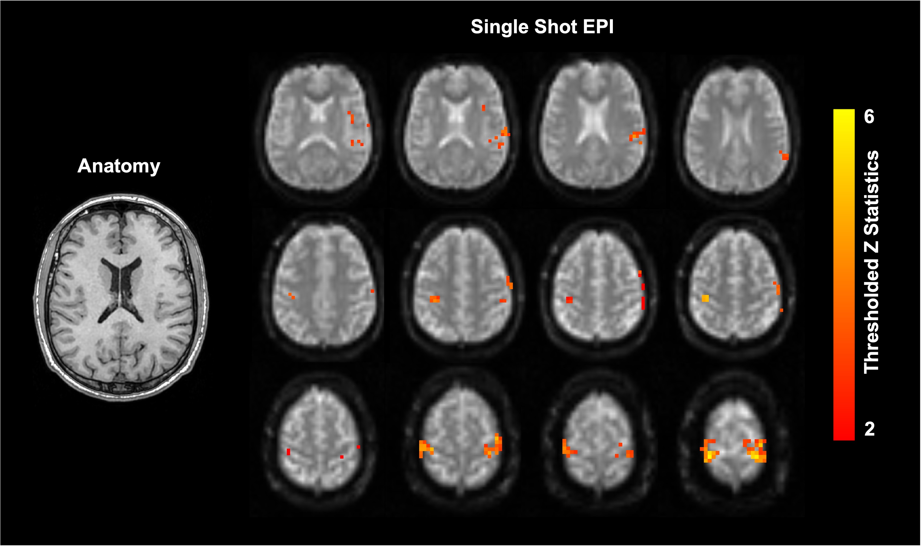
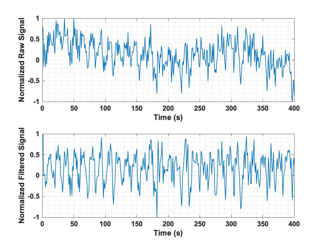
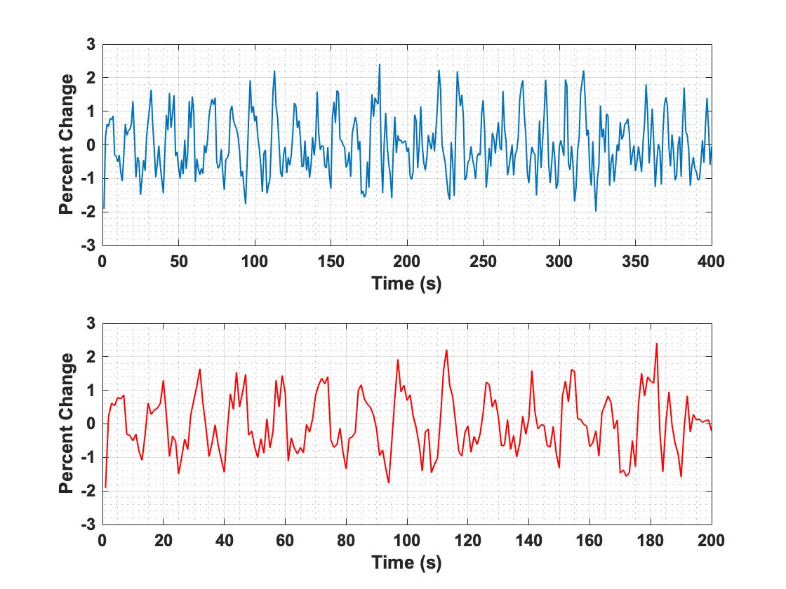
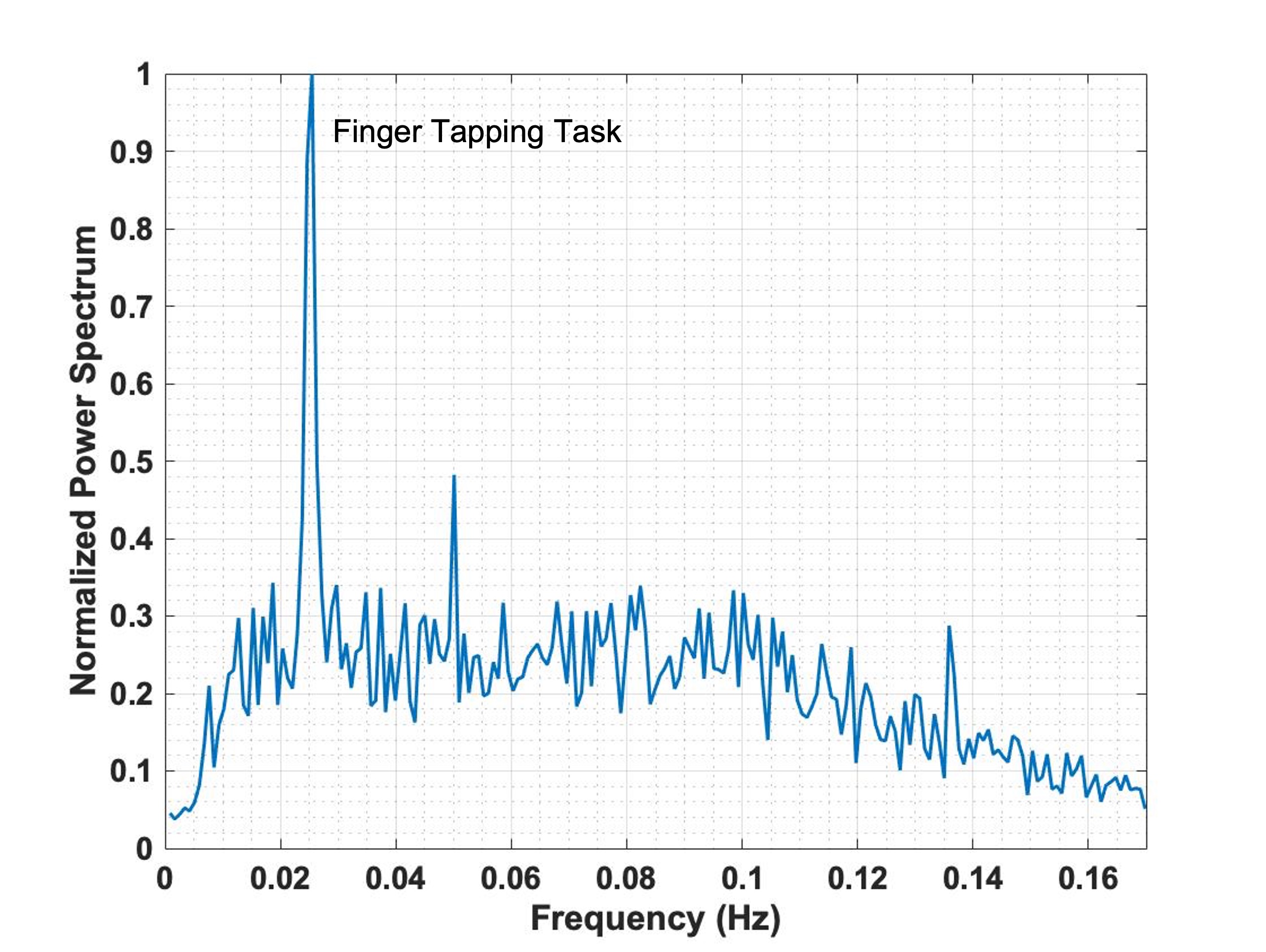
Fig 1. shows the anatomical image of a representative slice along with the thresholded Z statistic images superimposed on top of the raw EPI data. The Z statistic images were thresholded using clusters determined by Z > 2 and a cluster significance threshold of P = 0.05. The activated regions seen in this figure are visually consistent with the expected behaviour in the bilateral primary motor cortex in terms of spatial pattern. The number of voxels activated in the right side of the brain was 54 with a maximum Z value of 6.3 whereas the number of voxels activated in the left side of the brain was 44 with a maximum Z value of 6.7. Fig 2. shows the time series behaviour of the normalized raw and filtered EPI data in the activated regions. The corresponding mean temporal SNR calculated over these regions was ~ 44. The percent change in signal of an averaged time course for a masked region on the right side of the brain consisting of 30 activated voxels is shown in Fig 3. The spectra of the averaged time courses are shown in Fig 4 which identifies the peak frequency to be ~ 0.025 Hz which corresponds to the task-related frequency.DISCUSSION
Mid-field scanners with reduced siting requirements can expand access to MRI [4]. The availability of imaging techniques such as fMRI in a point-of-care setting can provide neurological information which can be vital in cases of traumatic brain injury and acute ischemic stroke [5,6]. This preliminary study demonstrates the feasibility of functional imaging at 0.5 T with the standard technique irrespective of the reduced activity-induced magnetic susceptibility contrast. Detailed experiments will be conducted in the future with optimized parameters and greater population size. Previous studies (1) performed with visual stimuli show consistent activations with a percent signal change of ~2.5%. The resulting tSNR in the occipital lobe before filtering within an ROI of 106 voxels was ~ 36.4. This initial investigation with motor task shows a percent signal change of ~1.8 +/- 0.4 % and the tSNR before filtering with an ROI of 30 voxels was ~ 44. The current EPI protocol uses a TE of 70 ms, which results in the elongation of TR and dead time prior to the echo train. In future work, we intend to investigate and characterize the relationship between BOLD sensitivity and scan efficiency at 0.5T. Even so, any dead time should be intelligently filled to increase SNR, for instance by asymmetric acquisition [7]. At the same time, simultaneous multi-slice (SMS) acquisition [8] can be utilized to reduce the TR further.EPI at low fields offers reduced susceptibility-induced geometric distortions and signal loss. This is especially important for structures near the skull base, where it has been shown that the increased signal dropout from field inhomogeneities at high field overwhelms any advantage of increased voxel activation [9]. Furthermore, the increased safety profile of 0.5T offers the possibility of expanding the eligible population able to undergo fMRI exams, for instance, post-implantation of therapeutic devices such as deep brain stimulation (DBS) leads [10].
CONCLUSIONS
This preliminary work is intended to show the feasibility of performing task based BOLD fMRI experiments on a mid-field 0.5 T head-only scanner using standard EPI sequence.Acknowledgements
The authors would like to thank the research and financial support received from Natural Sciences and Engineering Research Council (NSERC) of Canada.References
1. Wang Y., Gelderen P. V., Zwart J. A. D., Campbell-Washburn A. E., Duyn J. H., FMRI based on transition-band balanced SSFP in comparison with EPI on a high-performance 0.55 T scanner. Magnetic Resonance in Medicine. 2021;85(6):3196–3210.
2. Panther A., Thevathasen G., et al., A dedicated head-only MRI scanner for point-of-care imaging. . In: Proc. Intl. Soc. Mag. Reson. Med. 27, Montreal, Canada, 2019.
3. Woolrich MW, Ripley BD, Brady M, Smith SM. Temporal Autocorrelation in Univariate Linear Modeling of FMRI Data. NeuroImage 2001;14:1370–1386 doi: 10.1006/nimg.2001.0931.
4. Campbell-Washburn AE, Ramasawmy R, Restivo MC, et al. Opportunities in interventional and diagnostic imaging by using high-performance low-field-strength MRI. Radiology. 2019;293: 384-393.
5. O’Neill TJ, et al. Applications of rs-fMRI to Traumatic Brain Injury. Neuroimaging Clin N Am. 2017;27(4):685-696.
6. Chi NF, et al. Cerebral Motor Functional Connectivity at the Acute Stage: An Outcome Predictor of Ischemic Stroke. Nature: Sci Rep. 2018;8:16803
7. Jung KJ and Zhao T, Parallel imaging with asymmetric acceleration to reduce Gibbs artifacts and to increase signal-to-noise ratio of the gradient echo echo-planar imaging sequence for functional MRI. MRM. 2011; 67(2):419-427.
8. Todd N, et al., Functional Sensitivity of 2D Simultaneous Multi-Slice Echo-Planar Imaging: Effects of Acceleration on g-factor and Physiological Noise. Front. Neurosci. 2017; 11(158):1-14.
9. Wardlaw JM, et al., A systematic review of the utility of 1.5 versus 3 Tesla magnetic resonance brain imaging in clinical practice and research. Eur Radiol. 2012; 22:2295-2303.
10. Connell I and Panther A. Increasing MRI Safety for Patients with Implanted Medical Devices: Comparisons of a 0.5T Head-Only MRI to 1.5T and 3T. Proc. ASNR. 2019; #3569
Figures