3660
Investigation of 4D Flow CMR technique for left ventricular hemodynamics in heart failure patients
Guo Jiaxuan1, Yue Xiuzheng2, Huang shan2, and Zhu Li3
1Ningxia medical university, Yinchuan, China, 2Philips Healthcare, Beijing, China, 3General Hospital of Ningxia Medical University, Yinchuan, China
1Ningxia medical university, Yinchuan, China, 2Philips Healthcare, Beijing, China, 3General Hospital of Ningxia Medical University, Yinchuan, China
Synopsis
Keywords: Heart, Cardiovascular, Heart Failure
The number of patients with heart failure is increasing. Our study is based on the 4D flow technique of cardiac magnetic resonance and attempts to find cardiac functional parameters that precede structural changes in the heart at the level of the ventricular inflow and outflow tracts. We have found easier methods for visualizing blood flow and have the potential to further advance clinical diagnosis.Introduction
Heart failure (HF) is a clinical syndrome resulting from structural and/or functional abnormalities of the heart, leading to increased intracardiac pressure and/or inadequate cardiac output at rest and/or during exercise[1]. The alteration in hemodynamics is an important part of the pathophysiology of HF. However, proper assessment of hemodynamics remains challenging[2]. The present study was performed to investigate the left ventricular hemodynamic pattern in patients with HF by using four-dimensional flow (4D flow) cardiovascular magnetic resonance technique.
Methods
125 patients with HF with reduced ejection fraction (HFrEF, n=65), HF with mid-range ejection fraction (HFmrEF, n=30), or HF with preserved ejection fraction (HFpEF, n=30) and 30 healthy controls underwent cardiac magnetic resonance on a 3.0T scanner (Ingenia 3T;Phillips) were recruited for this study. Left ventricular inflow-outflow tract 4D flow data was collected via short axis cine sequence . The images were imported into CVI42 post-processing software. Left ventricular cardiac function parameters including LVEF and LVESV and peak velocity and pressure gradient of the aortic valve during systole were measured. Flow patterns were then observed and recorded for each patient. Statistical analysis was performed using SPSS software for Windows (version 26.0, SPSS, Chicago Illinois, US). Quantitative data for peak velocity and pressure gradient of the aortic valve was displayed as mean±SD. Correlation coefficient r was calculated to show the association between peak systolic velocity and pressure gradient across the aortic valve and cardiac function measures. R value >0.4 but <0.6 was considered as moderate association. A p value ≤0.05 was considered statistically significant.
Results
In the 4D flow pattern at the level of the heart's three chambers, patients with heart failure with reduced ejection fraction have significantly reduced and irregular blood flow.Peak systolic velocity across the aortic valve in HFrEF (118±20) was significantly lower than that of HFmrEF (145±32, p=0.002), HFpEF (140±19, p=0.005) and healthy controls (147±25, p<0.001). In patients with HF, peak systolic supra-aortic velocity showed a moderate association with LVEF (r=0.58, p<0.001).
Discussion
The main findings of this study are as follows. First, both peak and mean aortic valve pressure gradients during ventricular systole were significantly lower in patients with HF compared with healthy controls, indicating the presence of factors affecting pressure gradients in the ventricular outflow tract. Second, outflow tract pressure gradients were correlated with cardiac function parameters. Finally, the aortic valve systolic velocity in patients with HF is a more intuitive alteration of cardiac function parameters. Our results suggest that outflow tract velocity and pressure gradient are correlates of LV systolic function in patients with heart failure.
Conclusion
Peak supra-systolic aortic velocity in patients with HFrEF, which is obtained on a three-chamber heart 4D flow sequence, can be used as an adjunctive measure of cardiac function. It might also provide additional diagnostic information for patients with heart failure.
Acknowledgements
There are no other conflicts of interest in this study.References
[1] McDonagh TA, Metra M, Adamo M, Gardner RS, Baumbach A, Bohm M, Burri H, Butler J, Celutkiene J, Chioncel O et al: 2021 ESC Guidelines for the diagnosis and treatment of acute and chronic heart failure. Eur Heart J 2021, 42(36):3599-3726.
[2] Jain CC, Borlaug BA: Hemodynamic assessment in heart failure. Catheter Cardiovasc Interv 2020, 95(3):420-428.
Figures
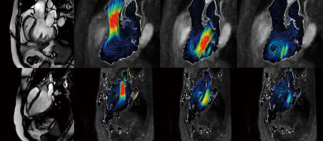
Figure 1 shows the three-chamber heart CMR images with 4Dflow images of heart failure patients and healthy volunteers.The upper row shows the three-chamber CMR images and 4Dflow images of heart failure patients with reduced ejection fraction in systole, early diastole, and late diastole, respectively; the lower row shows the corresponding images of healthy volunteers.
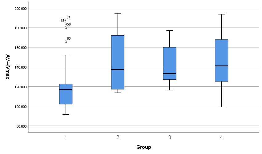
Figure 2 shows the different groups' Peak supra-systolic aortic velocity.
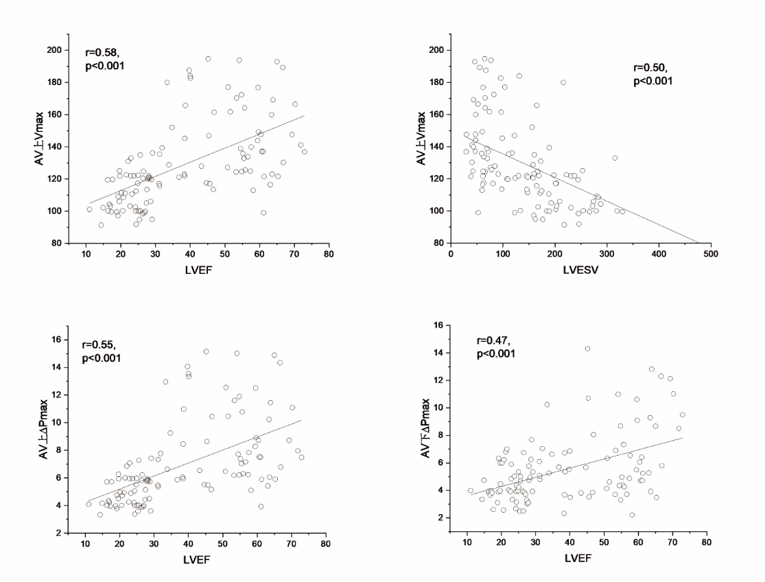
Figure 3 shows the linear correlation statistics.
DOI: https://doi.org/10.58530/2023/3660