3658
Radial phase-contrast FLASH acquisiton with SSA-FARY utilizing a Hadarmad-encoding sheme
Vanessa Thiesen1, Ansgar Adler1, and Martin Uecker1,2,3
1Institute of Biomedical Imaging, Graz, Austria, 2Institute for Diagnostic and Interventional Radiology of the University Center Göttingen, Göttingen, Germany, 3DZHK (German Center for Cardiovascular Research), Partner Site Göttingen, Göttingen, Germany
1Institute of Biomedical Imaging, Graz, Austria, 2Institute for Diagnostic and Interventional Radiology of the University Center Göttingen, Göttingen, Germany, 3DZHK (German Center for Cardiovascular Research), Partner Site Göttingen, Göttingen, Germany
Synopsis
Keywords: Flow, Velocity & Flow
The assessment of flow within a heart cycle places high demands on the temporal resolution of the measurement protocol. With self-gating we can exploit the quasi-periodicity of cardiac and respiratory motion to meet this challenge. We combined a Hadamard flow-encoding scheme together with SSA-FARY to further elaborate on the idea of a flow measurement with a high temporal and spatial resolution. The approach delivered promising results in 2D and can be seen as a basis for further developement towards 3D flow MRI.Introduction
Cardiac and respiratory self-gating in radial MRI using an adapted singular spectrum analysis (SSA-FARY) [1], is associated with advantages when considering patient comfort from the standpoint of preparations to be made and the respective time required. More precisely, SSA-FARY is a free-breathing technique which does not need trigger devices. Here, we combine SSA-FARY with a three dimensional flow-encoded radial FLASH sequence. Therefore, a high spatial resolution can be obtained and free-breathing is possible.Theory
After the formation of the AC-region, the basic steps performed by SSA-FARY include 1) Zero-padding, 2) Hankelization and 3) Decomposition of the signal using a singular value decomposition (SVD) [1]. After application of a sliding window to the time series data for every coil channel (step 2), a temporally localized principle component analysis (PCA) is performed. The resulting empirical orthogonal functions, encode the principal cardiac and respiratory motion over time and allow for subsequent binning. A detailed mathematical concept and schematic illustrations of SSA-FARY can be found in [1]. With the aim of three dimensional flow-encoding it becomes necessary to measure each spoke 4 times. Instead of a simple one-sided scheme, we utilized Hadamard encoding to gather more information within the repetitively measured spokes leading to overall smaller gradient moments [2]. Therefore a decreased gradient duration and shorter echo times are reachable. The additional reconstruction effort is low and negligible besides the potentially faster acquisition.Methods
A spoiled phase-contrast radial FLASH sequence, with combined flow gradients was used to collect images of the aortic arch from a transversal plane. Slices of 5mm thickness were measured at a bandwidth of 900Hz. Acquired spokes got recorded 4 times with respective flow gradients to enable 3d-encoding with a VENC of 150. The measurement was performed with a repetition time of 3.4ms and an echo time of 2.73ms. The extension to flow led to an adapted AC-region with dimensions [Partitions=1 x Flow-enodings=4 x Coils=10] in comparison to [1]. A sliding window of 45 and a moving average filter of 3 * w were set to determine the EOF's properly. In total we separated 25 cardiac phases and 5 different respiratory states. The collected data is binned according to the EOF's found by the SVD, resulting in an unequal distribution of spokes across cardiac and respiratory states. To quantify the presented flow measurement we extracted circular ROI's within the ascending aorta and calculated the mean velocity for each of the bins.Results
Figure 1 shows the measured through plane flow of an aorta for multiple cardiac phases. The negative flow is colored in blue and the positive in red.Figure 2 depicts the cross-sectional mean flow velocity over a full cardiac cycle (as visualized in Figure 1) extracted with a circular ROI from the ascending aorta in the transversal plane. We note that the cardiac phases cannot easily be translated to actual time steps because of the significantly varying number of spokes used for reconstruction of each phase.
Figure 3 demonstrates the concept of three dimensional flow encoding for a given cardiac and respiratory state. Here the colored arrows indicate the different flow encoding directions.
Discussion and Outlook
In this work we presented a proof of principal study combining the Hadamard flow encoding and the SSA-FARY self-gating technique to create a free breathing flow encoded acquisition. With a maximum value of about 120 cm/s during a full heart cycle our initial evaluation results display a physiologically consistent aortic flow. The width of the distribution in this representation is broader than expected. In our opinion this is due to the significantly varying number of spokes used for reconstructing the individual cardiac phase images. This inhomogeneity complicates the translation of cardiac states to time steps. A comparison with literature where the mean velocity distributions over time is typically shown is thus complicated.In the future, we will investigate a volumetric Stack-of-Stars approach to unlock the full potential of the presented method. Besides the fact that further development towards a 3D sequence will naturally prolong the time necessary for data acquisition, the additional 4 flow-encodings per spoke underline the importance of a time efficient measurement. An inadequate temporal resolution could reinforce the issue of a low spoke number per bin. With the already incorporated Hadamard-encoding a step has already been taken to meet this challenge, additionally an undersampling pattern together with a temporal regularization along partition direction will be a potentially beneficial strategies.
Acknowledgements
No acknowledgement found.References
[1] Rosenzweig S., Scholand N., Holme H. C., Uecker M.. (2018) Cardiac and Respiratory Self-Gating in Radial MRI using an Adapted Singular Spectrum Analysis (SSA-FARY). IEEE Transactions on Medical Imaging 2020;39:3029-3041
[2] Kollmeier, J.M. "Multi-Directional Phase-Contrast Flow MRI in Real Time." PhD diss., Georg-August-Universität Göttingen, 2020.[3] Dyverfeldt, P., Bissell, M., Barker, A.J. et al. (2015) 4D flow cardiovascular magnetic resonance consensus statement. J Cardiovasc Magn Reson 17, 72
[3] Garcia, J. et al, Distribution of blood flow velocity in the normal aorta: Effect of age and gender. J Magn Resonance Imaging. (2018)
Figures
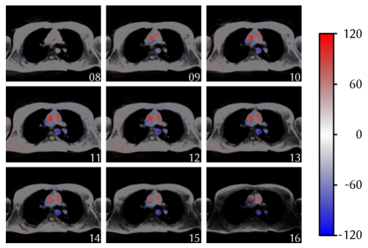
Figure 1: Color coded representation of flow in z-direction for different cardiac phases. The flow in z-direction is plotted for cardiac phases 8-16 (respiratory phase 0).
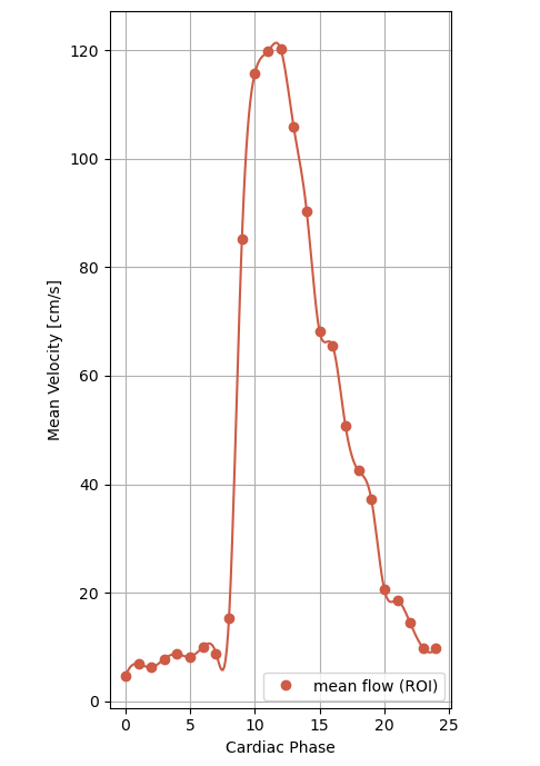
Figure 2: Mean flow velocity in the ascending aorta over a full heard cycle.
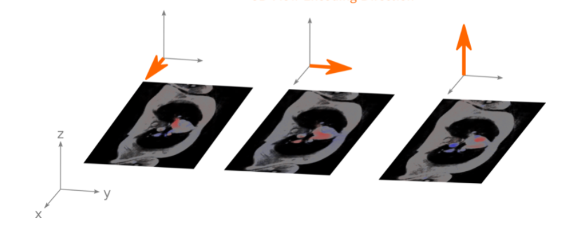
Figure 3: Flow encodings for the three spatial directions x, y, z.
DOI: https://doi.org/10.58530/2023/3658