3655
View-Sharing and KWIC Filtering Deblur 64-fold Accelerated Real-Time Phase-Contrast MRI Obtained with Radial k-space Sampling
Huili Yang1,2, Andrine Larsen3, Joshua D Robinson4, KyungPyo Hong1, Cynthia K Rigsby5, and Daniel Kim1,2
1Radiology, Northwestern University, Chicago, IL, United States, 2Biomedical Engineering, Northwestern University, Evanston, IL, United States, 3Biomedical Engineering, Lehigh University, Bethlehem, PA, United States, 4Pediatrics, Ann & Robert H. Lurie Children’s Hospital, Chicago, IL, United States, 5Medical Imaging, Ann & Robert H. Lurie Children’s Hospital, Chicago, IL, United States
1Radiology, Northwestern University, Chicago, IL, United States, 2Biomedical Engineering, Northwestern University, Evanston, IL, United States, 3Biomedical Engineering, Lehigh University, Bethlehem, PA, United States, 4Pediatrics, Ann & Robert H. Lurie Children’s Hospital, Chicago, IL, United States, 5Medical Imaging, Ann & Robert H. Lurie Children’s Hospital, Chicago, IL, United States
Synopsis
Keywords: Flow, Cardiovascular, compressed sensing, real-time imaging, phase-contrast MRI
Highly-accelerated real-time phase-contrast (rt-PC) using compressed sensing may underestimate (~17%) velocity measurements due to over regularization. We sought to address this underestimation by incorporating view-sharing (VS) and k-space weighted image contrast (KWIC) filtering. This is particularly important for pediatric patients who have faster heart rates and smaller hearts than adults, it is particularly important to achieve high temporal resolution. The proposed reconstruction achieves 25 ms temporal resolution without significant temporal blurring artifacts.Introduction
Real-time phase-contrast (rt-PC) MRI has several advantages (faster, free-breathing, insensitive to arrhythmia) over clinical standard ECG-gated segmented PC MRI, which may be performed with breath-holding or free-breathing with averaging. In pediatric patients with fast heart rates and smaller hearts than adults, it is particularly important to achieve high spatio-temporal resolution. We had recently developed a 38.4-fold accelerated rt-PC MRI pulse sequence with 41.7ms temporal resolution using radial k-space sampling and compressed sensing (CS), which produced 17% underestimation of peak velocity in pediatric patients compared with clinical standard PC MRI1. This underestimation is due to over-regularization. We sought to achieve 64-fold acceleration – resulting in 25 ms temporal resolution - without significant loss in accuracy by incorporating view sharing (VS)2 and k-space weighted image contrast (KWIC) filtering3 to augment high spatial frequency components in our CS framework. In this study, we compare hemodynamic metrics between default CS alone and CS plus VS and KWIC in pediatric patients.Methods
Data acquisition: This is a retrospective study using existing raw k-space of 12 pediatric patients with congenital heart disease (8 males and 4 females, mean age = 11.0±3.2 years), who had undergone an rt-PC MRI at four locations (aortic valve, pulmonic valve, left pulmonary artery, right pulmonary artery; N=48 planes in total). Relevant imaging parameters are summarized in Figure 1.Image reconstruction: We compared the two image reconstruction methods: (i) CS alone with 3 radial spokes per frame (64-fold accelerated), (ii) CS with 3 native radial spokes per frame (64-fold accelerated) boosted with 10 shared k-space lines and KWIC filtering, which is necessary to eliminate temporal blurring from shared lines. Figure 2 illustrates the view-sharing and KWIC filtering schemes. CS reconstruction was performed with temporal total variation, as previously described1.
Post-processing: (Step 1) Background phase correction. We used the correction methods described in4. (Step 2) Regions of interest (ROI) were manually delineated using custom software. The same ROI masks were used for CS alone and CS + VS + KWIC for fairness.
Analysis: (Image quality) We computed the blur metric5 (0 [best] to 1 [worst]) to evaluate image sharpness, which is important for identifying the boundary of ROI. The blur metrics were compared using a two-tailed, paired t-test. (Accuracy in velocity measurements) We measured the peak velocity and mean velocity to evaluate the impact of deblurring using VS and KWIC filtering.
Results
Figure 3 shows the representative magnitude and phase images of a patient, and flow measurements. CS with 3 radial spokes per frame boosted by VS and KWIC filtering produced sharper images and better time-resolved flow curves with minimal underestimation of peak velocity. Summarizing the results over all 12 patients, the mean blur metric was significantly lower for CS + VS + KWIC than CS alone (0.39±0.05 vs. 0.50±0.05, p <0.001); the peak and mean velocity at end-systole were significantly higher for CS+VS+KWIC than CS alone (peak: 109.2±38.2 cm/s vs. 94.7±32.9, p < 0.01; mean: 64.0±24.9 cm/s vs. 60.3±23.4, p<0.01).Conclusion
This study demonstrates that the incorporation of VS and KWIC filtering into CS deblurs 64-fold accelerated rt-PC MR images with 25 ms temporal resolution. Our new approach minimizes underestimation compared with CS alone, which is a common consequence of over-regularization. The future study includes more extensive testing in a larger cohort of pediatric patients with congenital heart disease.Acknowledgements
This work was supported in part by the following grants: National Institutes of Health (R01HL116895, R01HL151079, R21EB030806A1) and American Heart Association (19IPLOI34760317, 949899).References
1. Haji‐Valizadeh, Hassan, et al. "Highly accelerated, real‐time phase‐contrast MRI using radial k‐space sampling and GROG‐GRASP reconstruction: a feasibility study in pediatric patients with congenital heart disease." NMR in Biomedicine 33.5 (2020): e4240.2. Markl, Michael, and Jürgen Hennig. "Phase contrast MRI with improved temporal resolution by view sharing: k-space related velocity mapping properties." Magnetic resonance imaging 19.5 (2001): 669-676.
3. Song, Hee Kwon, and Lawrence Dougherty. "k‐Space weighted image contrast (KWIC) for contrast manipulation in projection reconstruction MRI." Magnetic Resonance in Medicine: An Official Journal of the International Society for Magnetic Resonance in Medicine 44.6 (2000): 825-832.
4. Stalder, Aurélien F., et al. "Quantitative 2D and 3D phase contrast MRI: optimized analysis of blood flow and vessel wall parameters." Magnetic Resonance in Medicine: An Official Journal of the International Society for Magnetic Resonance in Medicine 60.5 (2008): 1218-1231.
5. Crete, Frederique, et al. "The blur effect: perception and estimation with a new no-reference perceptual blur metric." Human vision and electronic imaging XII. Vol. 6492. SPIE, 2007.
Figures

Figure 1. Relevant imaging parameters.
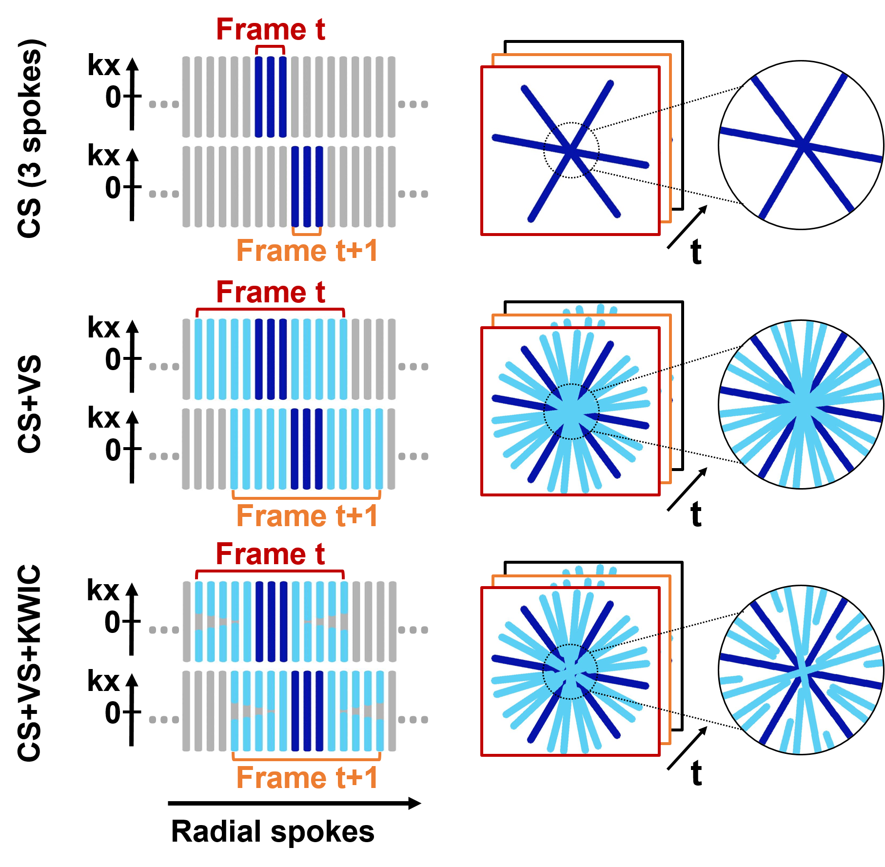
Figure 2. View sharing and KWIC filtering schemes with 3 native spokes per frame. The left column shows the shared spokes and KWIC shape; the right column shows the resultant k-space trajectory. We used a trapezoid-shaped KWIC filter because the shared spokes closer to native ones provide more similar information with native spokes than those farther away from the native spokes. Dark blue: native spokes; light blue: shared spokes.
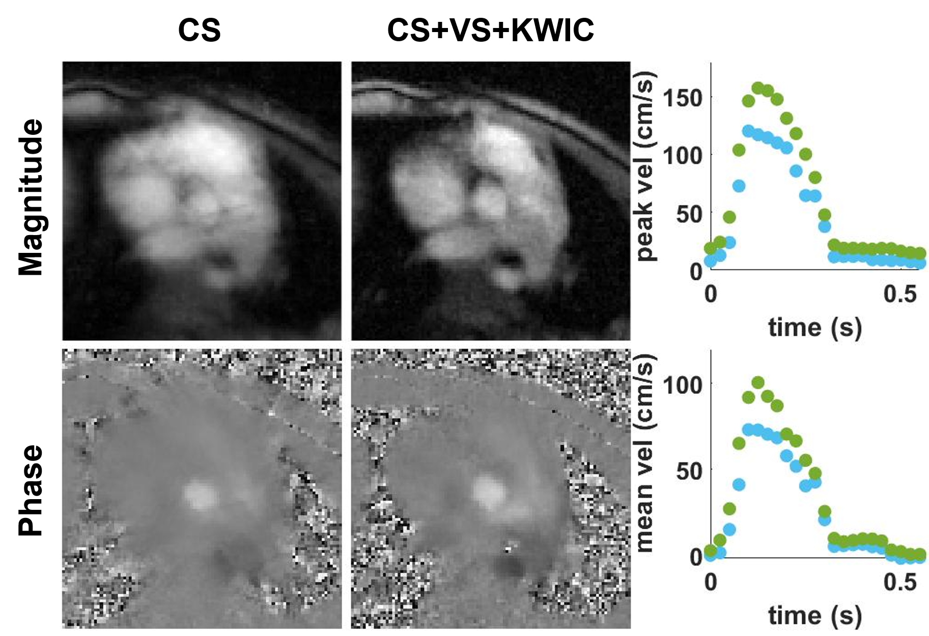
Figure 3. Representative aortic valve magnitude (top row) and phase (bottom row) images from CS alone (left column) and CS+VS+KWIC (middle column), and the resulting peak (right column, top) and mean (right column, bottom) velocity measurements (blue: CS, green: CS+VS+KWIC). CS with 3 radial spokes per frame boosted by VS and KWIC filtering produced better image quality and time-resolved velocity curves. AV: aortic valve.
DOI: https://doi.org/10.58530/2023/3655