3647
In-Plane Balanced Phase-Contrast Steady-State Free Precession (PC-SSFP) for All-in-One Diastolic Function Evaluation1Department of Biomedical Engineering, Yale University, New Haven, CT, United States, 2Department of Radiology and Biomedical Imaging, Yale University, New Haven, CT, United States
Synopsis
Keywords: Flow, Quantitative Imaging
Diastolic function evaluation requires estimates of early and late diastolic mitral valve flow velocities (E and A), and mitral annulus tissue velocity (e’). Our goal was to develop a bSSFP phase-contrast (PC) sequence (PC-SSFP) for in-plane flow-encoding, for simultaneous recording of E and A, and estimation of e’ based on tracking mitral valve motion on cine magnitude images, in a single breath-hold. Phantom and in vivo experiments showed agreement of PC-SSFP with velocities on GRE-based PC providing similar velocity curves, while achieving the high SNR and contrast of bSSFP cine images.Introduction
The blood flow velocity during filling of the left ventricular (LV) has peaks in early diastole (E wave) and late diastole (caused by atrial contraction (A wave)). E/A is a marker of the LV diastolic function1. Additionally, the peak velocity of the mitral valve in diastole (e’) plays a critical role in evaluating its diastolic function and E/e’ is a surrogate for LV end-diastolic pressure2. Though clinically assessed by transthoracic echocardiography (TTE), these diastolic parameters can be measured with good reproducibility by cardiovascular MRI and validated to have strong relationship with TTE3. The blood flow and valve plane velocity are normally obtained by two MR acquisitions, GRE based phase contrast (PC-GRE)4 with a high VENC (for blood) and low VENC (for mitral tissue). However, e’ can also be evaluated by tracking the valve displacements on bSSFP cine, using semi-automated5 or automated methods6. To have an all-in-one diastology scan, which can provide E, A and e’ in a single breath-hold, we developed a bSSFP phase-contrast method (PC-SSFP) to simultaneously measure E and A, using its phase-information, and e’ using automated processing of the magnitude images. To our knowledge only a few reports on PC-SSFP7,8 exist, with no study reporting in-plane flow evaluation.Methods
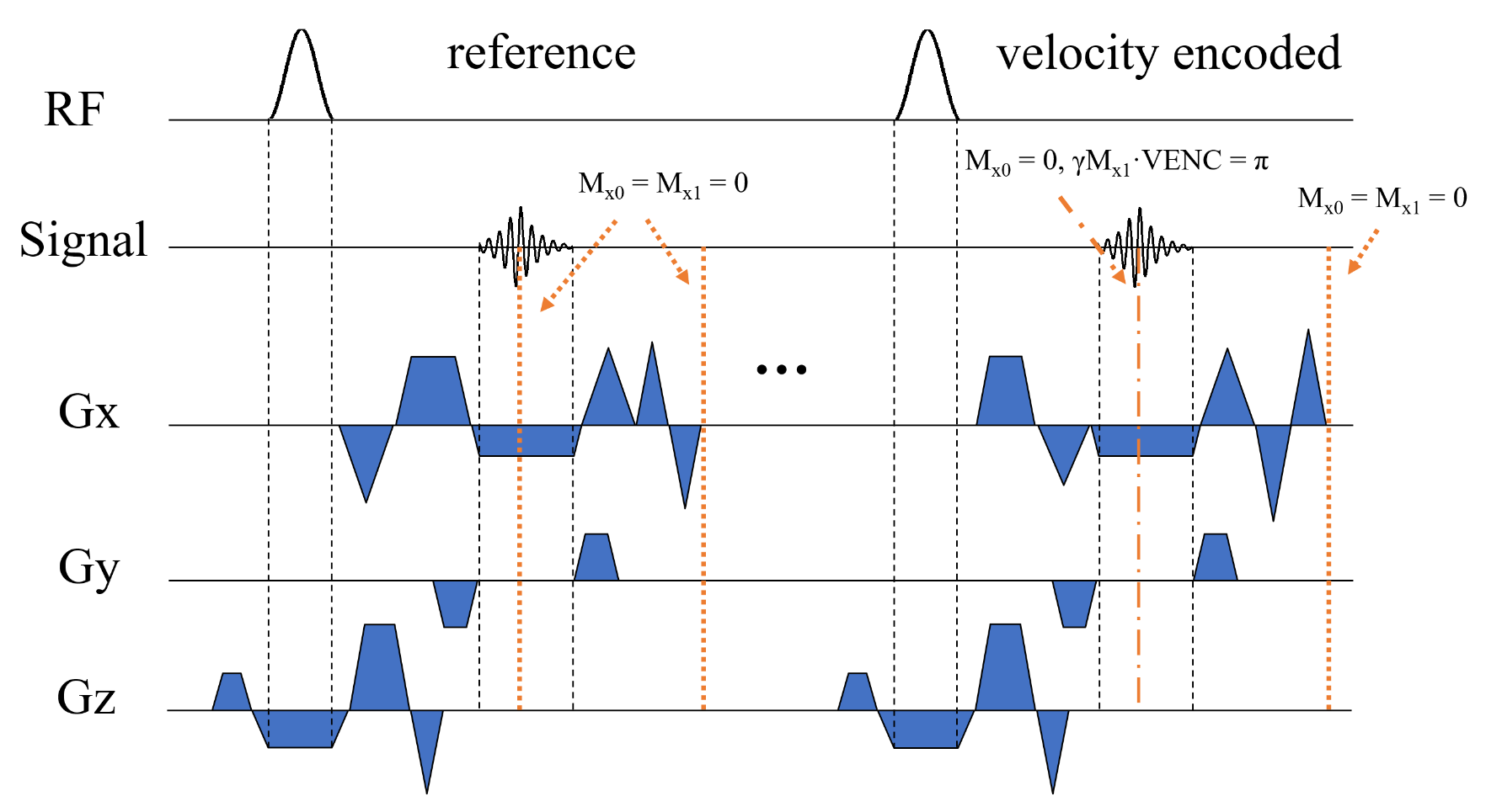
Figure 1 illustrates the PC-SSFP sequence we have developed for in-plane (readout) velocity-encoding. The 0th gradient moments are always balanced at TE and the end of each TR, to have the bSSFP image contrast and high SNR. Reference and velocity encoded acquisitions are executed sequentially to maintain their respective steady states. Both acquisitions use bipolar gradients to either null the first moment at the TE of the reference or encode the velocity for the specified VENC. Importantly, bipolar gradients are also added at the end of the readout, to null the 0th and first moment in the flow-encoding direction.All studies were performed on a Siemens 3T scanner. Scan parameters (for phantoms and in vivo) were: 2D Cartesian cine with ECG retrospective-gated, TR/TE/θ = 3.9ms/1.9ms/35°, 160 matrix, 380mm FOV, 75% phase, 8mm slice thickness, 4 views per segment, 150cm/s VENC, GRAPPA with acceleration factor R = 2, 24 reference lines, 18 heart-beat breath-hold, strong asymmetric echo. The same parameters for PC-GRE except TR/ TE/θ = 4.7ms/2.5ms/15°.
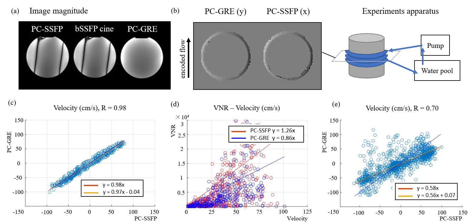
We tested the proposed method on phantoms first. The bSSFP contrast was confirmed by comparing banding patterns of PC-SSFP and bSSFP. A tube with constant flow inside was wrapped around a cylindrical phantom, to generate a wide range of velocities in the velocity encoded direction. We compared PC-GRE and PC-SSFP from two acquisitions on a pixel-by pixel basis for the same constant flow and VENC. Velocity noise ratio (VNR = Vel / Noise, using the mean velocity and variance across time at each pixel as the noise) were also measured.
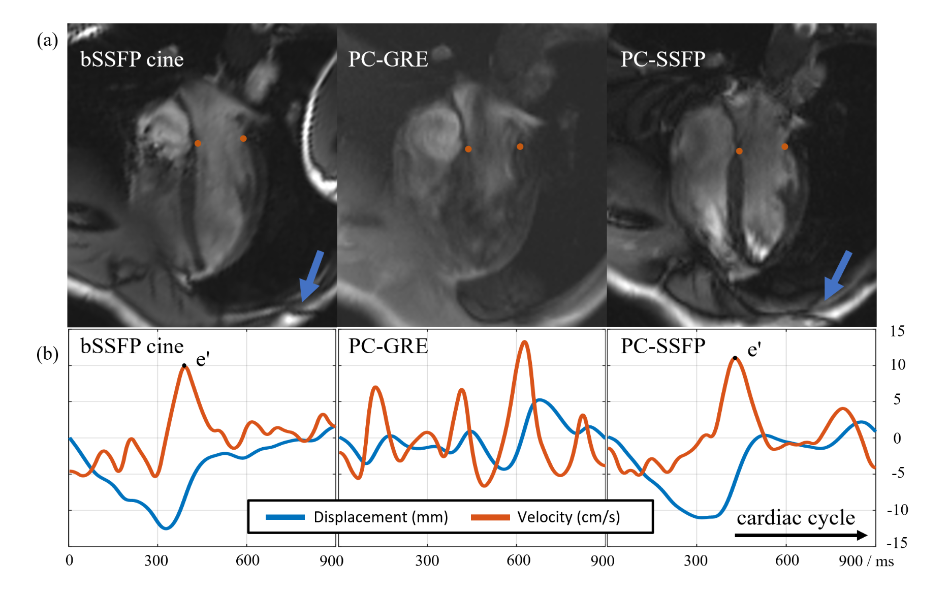
We then performed four-chamber PC-SSFP on healthy subjects, with all studies approved by our IRB and all subjects providing informed written consent. Blood flow curves and corresponding E/A by PC-SSFP were compared to those obtained from PC-GRE. MVnet6 was used for automatic mitral valve tracking of the PC-SSFP magnitude images. The valve velocity was calculated by taking the derivative of the smoothed displacement curve and compared to e’ obtained by tracking standard bSSFP cine.
Results
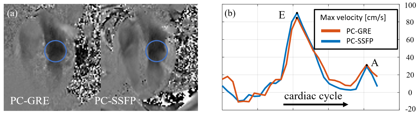
Figure 2 showed the image magnitude and phase in phantom experiments. PC-SSFP had the same banding pattern as bSSFP cine. The flow experiment apparatus is shown in Figure 2b. To compare the measured phase velocity, the velocity of pixels within the tube were plotted. PC-GRE and PC-SSFP were highly correlated but had noisy or wrapped pixels (see abnormal dark or bright point in figure 2b). After removing outliers, velocity measurement by PC-SSFP and PC-GRE agreed strongly (see Figure 2c). In addition, PC-SSFP demonstrated higher VNR than PC-GRE in this experiment (Figure 2d). Importantly, we also found that it was essential to null the first moment in the velocity encoding direction at the end of the TR, for both reference and velocity encoding TRs. If not nulled, the effective VENC changed and measured flow decreased by a protocol-dependent factor (Figure 2e).Figure 3 showed a phase image in early diastole and plotted blood flow velocity across the cardiac cycle, measured with PC-GRE and PC-SSFP. The peak velocities at E wave and A wave both agreed well between these two acquisitions. At the same time, the high SNR and strong blood-myocardium contrast of PC-SSFP enabled processing with network-based automatic valve tracking (figure 4) to obtain e’, which failed to follow the valve displacement in PC-GRE.
Discussion
We explored the potential of in-plane PC-SSFP for all-in-one left ventricular diastolic function evaluation, simultaneously obtaining E, A, e’ in a single breath-hold. Results of phantom and in vivo experiments were promising. One challenge is that the additional bipolar gradients at the middle and end of each TR further increased TR, resulting in worse susceptibility to off-resonance bSSFP banding artifacts, and also compromised temporal resolution7. Future work will focus on TR reduction and more experiments for optimization in both acquisition and reconstruction. This all-in-one-diastology method could be used to rapidly estimate pressure (e.g. using E/e’) during physiological testing, such as hyperventilation or cold pressor test9,10.Acknowledgements
No acknowledgement found.References
[1] Galderisi M. Diastolic dysfunction and diastolic heart failure: diagnostic, prognostic and therapeutic aspects. Cardiovasc Ultrasound. 2005;3:9. Published 2005 Apr 4.
[2] Wang M, Yip GW, Wang AY, et al. Peak early diastolic mitral annulus velocity by tissue Doppler imaging adds independent and incremental prognostic value. J Am Coll Cardiol. 2003;41(5):820-826.
[3] Seemann F, Baldassarre LA, Llanos-Chea F, et al. Assessment of diastolic function and atrial remodeling by MRI - validation and correlation with echocardiography and filling pressure. Physiol Rep. 2018;6(17):e13828.
[4] Bollache E, Redheuil A, Clément-Guinaudeau S, et al. Automated left ventricular diastolic function evaluation from phase-contrast cardiovascular magnetic resonance and comparison with Doppler echocardiography. J Cardiovasc Magn Reson. 2010;12(1):63. Published 2010 Nov 9.
[5] Thavendiranathan P, Guetter C, da Silveira JS, et al. Mitral annular velocity measurement with cardiac magnetic resonance imaging using a novel annular tracking algorithm: Validation against echocardiography. Magn Reson Imaging. 2019;55:72-80.
[6] Gonzales RA, Seemann F, Lamy J, et al. MVnet: automated time-resolved tracking of the mitral valve plane in CMR long-axis cine images with residual neural networks: a multi-center, multi-vendor study. J Cardiovasc Magn Reson. 2021;23(1):137.
[7] Markl M, Alley MT, Pelc NJ. Balanced phase-contrast steady-state free precession (PC-SSFP): a novel technique for velocity encoding by gradient inversion. Magn Reson Med. 2003;49(5):945-952.
[8] Rolf MP, Hofman MB, Kuijer JP, et al. Extrinsic multiecho phase-contrast SSFP: evaluation on cardiac output measurements. Magn Reson Imaging. 2009;27(3):385-392.
Figures