3640
Preliminary exploration of 3D-APTw imaging in renal tumor applications1Department of Radiology, Xi'an GaoXin Hospital, Xi'an, China, 2Philips Healthcare, Beijing, China, 3Department of Urology Surgery, Xi'an GaoXin Hospital,, Xi'an, China
Synopsis
Keywords: Kidney, CEST & MT
APTw imaging focuses on cranial tumor studies with significant clinical value, with fewer studies in mid-upper abdomen, because of the movement of organs and B0 fields inhomogeneous. Previously used showed that intermittent breath-hold (IBH) could to improve the image quality of 3D-APTw imaging in healthy adults. In this study, we used the IBH respiratory compensation mode to initially explore the feasibility of 3D-APTw imaging of renal tumors. The findings suggested that 3D-APTw imaging with IBH can be not only used for imaging renal tumors, but also showed potential values in renal tumors diagnosis and differential diagnosis.Introduction
Renal clear cell carcinoma (RCCC) and renal angiomyolipoma (RAML) are the most common malignant and benign tumors of the kidney, respectively. Typical RAML tends to contain varying proportions of mature adipose tissue and is easy to diagnose, while the lack of adipose RAML is difficult to differentiate from RCCC. As a novel molecular imaging technique, amide proton transfer-weighted (APTw) imaging has mainly focused on tumors, which can be used for diagnosis, histological grading, and assessment of efficacy [1]. However, there is a lack of relevant studies in the kidney due to respiratory motion and inhomogeneity of the B0 field [2,3]. Intermittent breath-hold (IBH) modality in healthy adult kidney 3D-APT imaging improved the image quality of 3D APT imaging in the previous study [4]. In this study, we used IBH respiratory compensation mode was used to initially explore the feasibility of APT imaging in renal tumors.Method
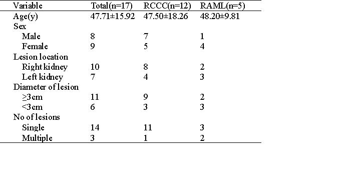
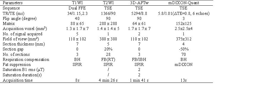
Seventeen patients with renal tumors were retrospectively collected with complete clinical data and MRI sequences from November 2021 to October 2022, of which 11 cases of renal clear cell carcinoma (RCCC) and 1 case of renal angiomyolipoma (RAML) were confirmed by histopathology, and other 4 RAML diagnosed by MR images. All MR images were obtained on a 3.0 T MR scanner (Ingenia CX, Philips Healthcare) using a 32-channel phased-array abdominal coil. Routine clinical imaging, included axial T1WI and T2WI, and coronal T2WI sequences. Axial and coronal 3D-APTw with IBH, and axial mDIXON-Quant sequences were scanned (Table 1). 3D APTw imaging selected the largest level of the renal tumor for 3-layer imaging, and the other sequences covered bilateral kidneys. The quality of APTw images were evaluated by 2 senior radiologists, and the tumor maximum cross-sectional and normal renal tissue of APT values were measured by selecting the image with the best orientation and slice location. One-way analysis of variance (ANOVA) was used to analyze the differences of APT values among RCCC, RAML, and normal renal tissue.Result
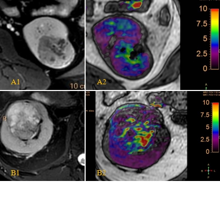
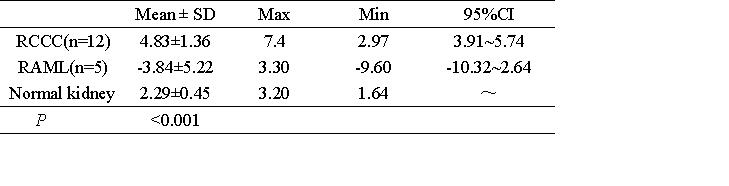
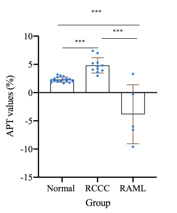
The APTw images of RCCC tumor tissue mainly showed high signal intensity and positive APT values; while RAML adipose tissue showed low signal intensity and mainly negative APT values (Fig. 1). There was a significant statistical difference among RCCC, RAML, and normal renal tissue (p<0.001). The mean APTw values of RCCC, normal renal tissue and RAML were 4.83±1.36%, 2.29±0.45% and -3.84±5.22% respectively from high to low order (Table 3, Fig. 2).Discussion
Preliminary findings of this study showed that 3D-APTw imaging with IBH could be used for kidney tumor imaging, and there were significant differences in the signal characteristics and APT values of 3D-APTw imaging in RCCC, RAML and normal kidney tissues. The RCCC tumor tissue had the highest APT values with predominantly increased signal, while the RAML tumor tissue had predominantly negative APT values with decreased signal.Conclusions
These findings showed that 3D-APTw imaging with IBH could be not only for imaging renal tumors, but also showed potential values in the diagnosis and differential diagnosis of common tumors.Acknowledgements
We sincerely thankful all participants of this study.References
[1] Zhou J, Zaiss M, Knutsson L, et al. Review and consensus recommendations on clinical APT- weighted imaging approaches at 3T: Application to brain tumors[J]. Magn Reson Med, 2022, 88: 546-574
[2] Chen Y, Dang X, Zhao B, et al. B0 Correction for 3T Amide Proton Transfer (APT) MRI Using a Simplified Two-Pool Lorentzian Model of Symmetric Water and Asymmetric Solutes. TOMOGRAPHY, 2022, 8(4):1974-1986.
[3] Togao O, Keupp J, Hiwatashi A, et al. Amide proton transfer imaging of brain tumors using a self-corrected 3D fast spin-echo Dixon method: comparison with separate B0 correction. Magn Reson Med, 2017, 77:2272-2279
[4] Xia Wang, Yu Jiang, Zeliu Du, et al. Exploring the reproducibility of APT imaging technology in healthy adult kidneys based on breathing patterns Proc. Iintl. Soc. Mag. Reson. Med. 30(2022).
Figures