3634
Preliminary study on the influence of renal motion amplitude on APTw image quality of normal kidney1Xi'an GaoXin Hospital, Xi'an, China, 2Philips Healthcare, Beijing, China
Synopsis
Keywords: Kidney, CEST & MT
Respiratory movement is the main reason for upper and middle abdominal organ movement, which significantly affects the quality of MR imaging.This study aims to analyze the influence of renal motion amplitude on renal APT imaging through free breathing (FB), and breath holding (BH) based on multi-phase balanced turbo fast field echo (B-TFE). The results showed that renal motion amplitude was negatively correlated with APTw image quality scores. The renal motion amplitude of bilateral kidneys of BH mode was significantly reduced compared with FB, and the APT image quality of BH mode was better.Introduction
In recent years, amide proton transfer-weighted (APTw) imaging,as a new MR imaging method at the molecular level[1], has received increasing attention for its application in the body. The technology was currently in the early stages of exploration, and significant clinical progress has been made in diagnosing, treating, and prognosis chronic kidney disease and renal tumors[2].However, APT imaging still lacked effective respiratory compensation technology. Our study has shown that respiratory motion has a significant effect on renal APTw image quality[4].The influence of renal motion amplitude on renal APT image quality is unknown. The aim of this study was to investigate the effect of the motion amplitude of bilateral kidneys in FB and BH modes on the quality of APTw images in healthy adults and to provide reliable support for the clinical application of renal APTw imaging.Method
Twenty healthy adult volunteers were selected in April-May 2022. All MR images were obtained on a 3.0 T MR scanner (Ingenia CX, Philips Healthcare) using a 32-channel phased-array abdominal coil. MR images of bilateral kidneys, including balanced turbo fast field echo (BTFE) and 3D-APTw imaging in the free-breathing (FB) and breath-holding (BH) modes. BTFE sequences acquired coronal and sagittal orientations, and 3D-APT imaging acquired axial and coronal orientations (Table 1, Figure 1). Two radiologists with more than 5 and 6 years of experience each independently measured the amplitude of motion of bilateral kidneys in FB and BH in the cranial-caudal(CC), right-left(RL), anterior-posterior(AP) direction using IntelliSpace Portal(Version 8, Philips Healthcare)) (Figure 1a)[3] . The APTw image quality was scored using a 5-point Likert scale. All statistical analyses were performed using SPSS 25.0.The intraclass correlation coefficient (ICC) was used to evaluate the consistency of the renal motion amplitude. The paired sample t-test was used to compare the difference in renal motion amplitude with different respiratory modes. Bivariate Spearman rank correlation was used to analyze the correlation between renal motion amplitude and APTw image quality scores.Result
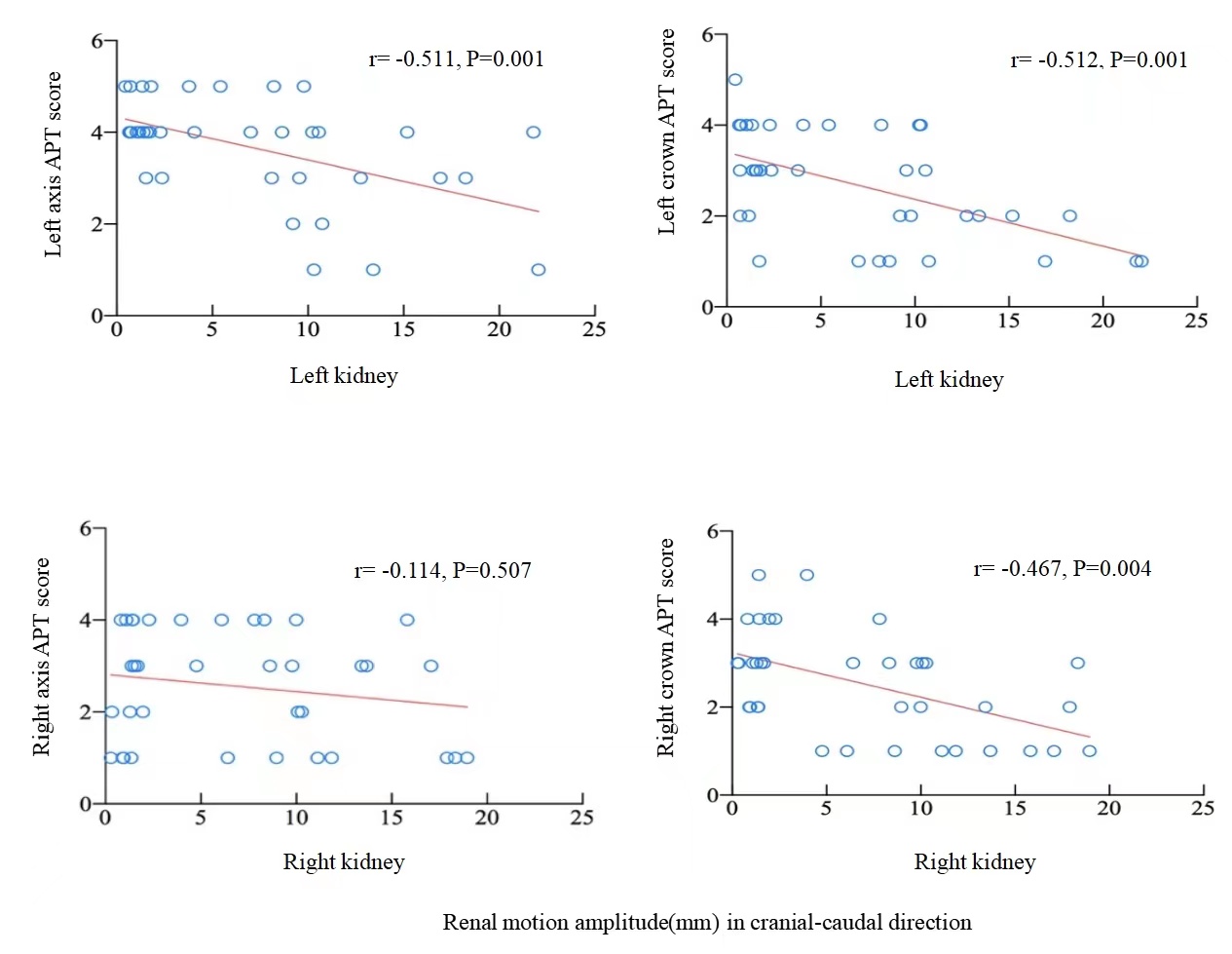
The consistency of renal motion amplitude measurement was as follows, high consistency in the cranial-caudal direction (ICC>0.9), moderate or high consistency in the right-left direction, anterior-posterior direction (0.55-0.69,0.70-0.84).The renal motion amplitudein BH was significantly smaller than which in FB (P<0.05).Except for the axial of the right kidney, the APTw image quality score of bilateral kidneys in BH was higher than that in FB (P<0.05)(Table 2, Figure 2).The left kidney motion amplitudes in all directions were negatively correlated with the axial and coronal APTw image scores of the left kidney(r=-0.338 ~ -0.563, P<0.05). The right kidney motion amplitudes in all directions were weakly negatively correlated with the coronal APTw image scores ofthe right kidney (r=-0.419 ~ -0.467, P<0.05).There was no significant correlation between the amplitude of motion of the right kidney in all directions with the axial APTw image scores of the right kidney(P>0.05) (Figure 3).Discussion
The preliminary analysis of this study showed that the amplitude of kidney motion was an important factor affecting the quality of renal APTw images, and the greater the amplitude of motion, the worse the image quality[4]. Therefore, the renal APTw study should adopt the IBH respiratory compensation mode.Our results suggest that effective respiratory compensation is very necessary and urgent for renal APTw imaging.Conclusion
The preliminary analysis of this study showed that the amplitude of kidney motion was an important factor affecting the quality of renal APTw images. The amplitude of motion of bilateral kidneys in BH (e.g.LCC:10.43(6.55) mm, RCC:12.12±4mm) were significantly lower than FB (e.g.LCC:1.46(1.56)mm, RCC1.39(1.11)mm).To improve the images success rate, axial APTw images in BH was more advantaged.Acknowledgements
We sincerely thank the participants of this study.References
[1] Lin Yue, Li Chunmei, Chen Min. Advances in the application of amide proton transfer imaging[J]. Radiology Practice, 2018, 33(5):4.
[2] Ye Ju, Ailian Liu, Yue Wang, et al. Amide proton transfer magnetic resonance imaging to evaluate renal impairment in patients with chronic kidney Magnetic Resonance Imaging, 2022, 87:177-182.
[3] Fan WJ, Long MIAO, Shen W, et al. Cine-MRI measurement of renal motion amplitude in healthy adults[J]. Chinese Journal of Medical Imaging, 2013, 05:380-382.
[4] Xia Wang, Yu Jiang, Zeliu Du, et al. Exploring the reproducibility of APT imaging technology in healthy adult kidneys based on breathing patternsProc. Iintl. Soc. Mag. Reson. Med. 30(2022).
Figures
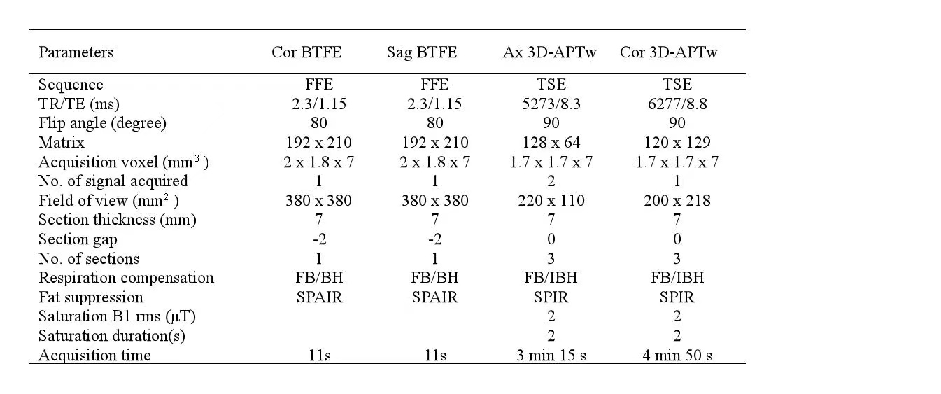
Table 1. The detail parameters of MRI pulse sequences.(FB: free breathing; BH: breath-hold; IBH: Intermittent breath-hold; Cor:coronal;Sag:sagittal;Ax:axis).
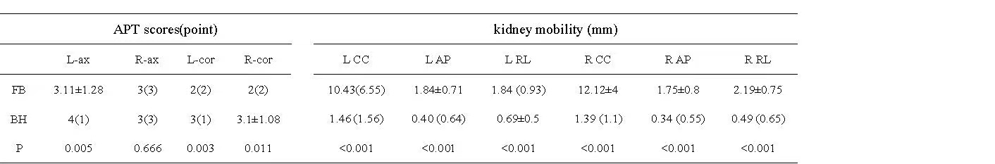
Table 2.APTscores and amplitude of kidney motioncomparative analysis in different breathing modes.(Cor:coronal;Sag:sagittal;Ax:axis; CC:cranial-caudal;AP:anterior-posterior;RL:right-left).