3632
Improved accuracy of triexponential diffusion analysis of the kidney using motion-corrected reconstruction for free-breathing DWI
Yuki Makino1, Naoki Ohno2, Johannes M Peeters3, Masami Yoneyama4, Yu Ueda4, Tosiaki Miyati2, Yukihiro Matsuura1, Toshifumi Gabata5, and Satoshi Kobayashi1,2,5
1Radiology Division, Kanazawa University Hospital, Ishikawa, Japan, 2Faculty of Health Sciences, Institute of Medical, Pharmaceutical and Health Sciences, Kanazawa University, Ishikawa, Japan, 3Philips Healthcare, Best, Netherlands, 4Philips Japan, Tokyo, Japan, 5Department of Radiology, Kanazawa University Hospital, Ishikawa, Japan
1Radiology Division, Kanazawa University Hospital, Ishikawa, Japan, 2Faculty of Health Sciences, Institute of Medical, Pharmaceutical and Health Sciences, Kanazawa University, Ishikawa, Japan, 3Philips Healthcare, Best, Netherlands, 4Philips Japan, Tokyo, Japan, 5Department of Radiology, Kanazawa University Hospital, Ishikawa, Japan
Synopsis
Keywords: Kidney, Diffusion/other diffusion imaging techniques
The clinical application of triexponential intravoxel incoherent motion analysis in the kidney has been limited because the blurring and misregistration of images caused by respiratory motion during acquisition. To solve this issue, we applied retrospective motion-corrected (MC) reconstruction to free-breathing (FB) DWI of the kidney for accurate triexponential diffusion analysis. Our results showed that triexponential analysis using MC FB-DWI can improve the measurement accuracy and repeatability of triexponential diffusion parameters of the kidney.INTRODUCTION
Intravoxel incoherent motion (IVIM) analysis with a triexponential model provides information on three diffusion components, including perfusion-related, fast-free, and slow-restricted diffusion.1,2 Since kidney possess complex tissue structures, including blood vessels, tubules, and connective tissues, it has been illustrated that the DWI signals are well compatible with the triexponential model.3 However, the clinical application of triexponential IVIM analysis of the kidney has been limited because the blurring and misregistration of images caused by respiratory motion during acquisition can deteriorate the measurement accuracy and repeatability.4 To solve this issue, we applied retrospective motion-corrected (MC) reconstruction5 to free-breathing (FB) DWI of the kidney for accurate triexponential diffusion analysis.METHODS
Single-shot diffusion echo-planar imaging with FB acquisition was performed on a 3.0 T MRI system to obtain coronal diffusion-weighted images of the kidney. Seven healthy volunteers (all men; mean age, 25.1 ± 5.7 years) were scanned twice to assess the repeatability of measurements. The imaging parameters were as follows: repetition time, 4000 ms; echo time, 62.9 ms; acquisition matrix, 128 × 128; b-values, 1, 10, 30, 50, 100, 200, 400, 600, 800, 1000, and 1200 s/mm2; separate diffusion measures in three orthogonal directions; field of view, 340 mm; slice thickness, 7 mm; number of signals averaged, 3; half-scan factor, 0.803; and sensitivity encoding factor, 2.1. All subjects were asked to fast for at least 4 hours before the scan. After the acquisition, we applied a fast elastic image registration algorithm5 to the DWI data to correct the blurring and misregistration of images caused by respiratory motion. Next, voxel-wise estimation of perfusion-related, fast-free, and slow-restricted diffusion coefficients (Dp, Df, and Ds, respectively), their corresponding fractions (Fp, Ff, and Fs, respectively), and multiplication of Dp and Fp (DpFp) were performed by fitting the triexponential model to the MC and motion-uncorrected (non-MC) DWI data. Normalized root-mean-square error (nRMSE) was calculated to evaluate the fitting accuracy. We placed region of interest (ROI) in the renal cortex and determined the triexponential diffusion parameters and nRMSE within the ROI. Repeatability coefficients (RC) were calculated from Bland-Altman plots to assess the repeatability of diffusion parameters. Additionally, to evaluate the respiratory motion-induced variation of diffusion parameters, coefficient of variation for each diffusion parameter in the ROI (CVROI) was computed as the standard deviation divided by the mean. Finally, these values were compared between the MC and non-MC schemes.RESULTS AND DISCUSSION
Representative images of triexponential diffusion parameters and nRMSE obtained with the MC and non-MC schemes are presented in Figure 1. Table 1 presents the diffusion parameters and nRMSE in the renal cortex compared between both schemes. The MC had significantly lower nRMSE compared with the non-MC, indicating that the fitting accuracy of the triexponential analysis can be improved by using the MC.5 In addition, the MC showed significantly lower Dp than the non-MC. This result indicates that compared with the other parameters, Dp is more susceptible to signal variations due to respiration-induced image blurring and misregistration.6 Table 2 shows RC of each diffusion parameter. The MC improved the repeatability of Dp, Fp, DpFp, Ff, and Fs. CVROI of diffusion parameters are shown in Table 3. CVROI for Dp, Fp, DpFp, Ff, Ds, and Fs in the MC were significantly lower than those with the non-MC, suggesting that the MC can improve the respiratory motion-induced variation of these diffusion parameters.CONCLUSION
The MC FB-DWI of the kidney improved the fitting accuracy and repeatability of triexponential diffusion parameters.Acknowledgements
No acknowledgement found.References
- Hayashi T, et al. Diffusion analysis with triexponential function in liver cirrhosis. J Magn Reson Imaging. 2013; 38: 148-153.
- Ohno N, et al. Modified triexponential analysis of intravoxel incoherent motion for brain perfusion and diffusion. J Magn Reson Imaging. 2016; 43: 818-823.
- Baalen S, et al. Intravoxel incoherent motion modeling in the kidneys: Comparison of mono-, bi-, and triexponential fit. J Magn Reson Imaging. 2017; 46: 228-239.
- Jerome NP, et al. Comparison of free-breathing with navigator-controlled acquisition regimes in abdominal diffusion-weighted magnetic resonance images: Effect on ADC and IVIM statistics. J Magn Reson Imaging. 2014; 39: 235-240.
- Buerger C, et al. Comparing nonrigid registration techniques for motion corrected MR prostate diffusion imaging. Med Phys. 2015; 42: 69-80.
- Meeus EM, et al. Evaluation of intravoxel incoherent motion fitting methods in low-perfused tissue. J Magn Reson Imaging 2017; 45: 1325-34.
Figures
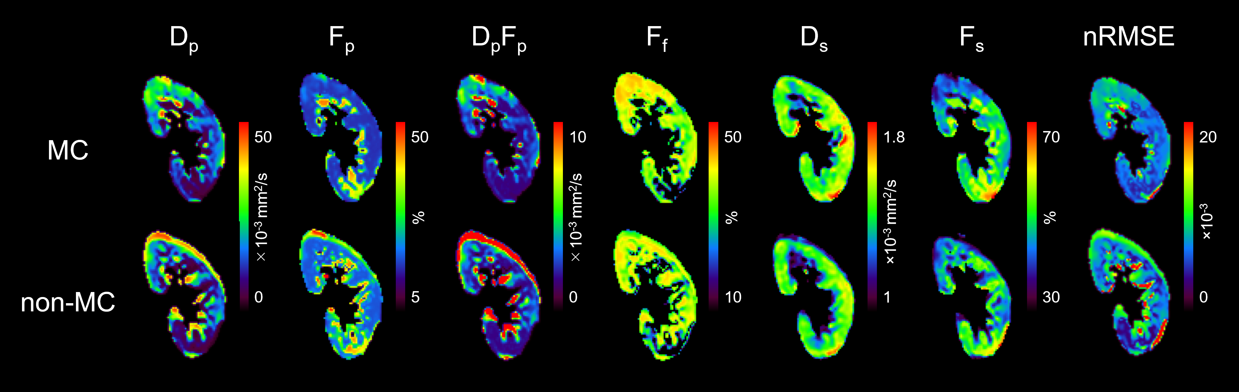
Figure 1. Representative
images of perfusion-related diffusion coefficient (Dp), the fraction (Fp), the
multiplication of Dp and Fp (DpFp), fast-free diffusion fraction (Ff), slow-restricted
diffusion coefficient (Ds), and the fraction (Fs), and normalized
root-mean-square error (nRMSE) with triexponential analysis in motion-corrected
(MC) and motion-uncorrected (nonMC) data.
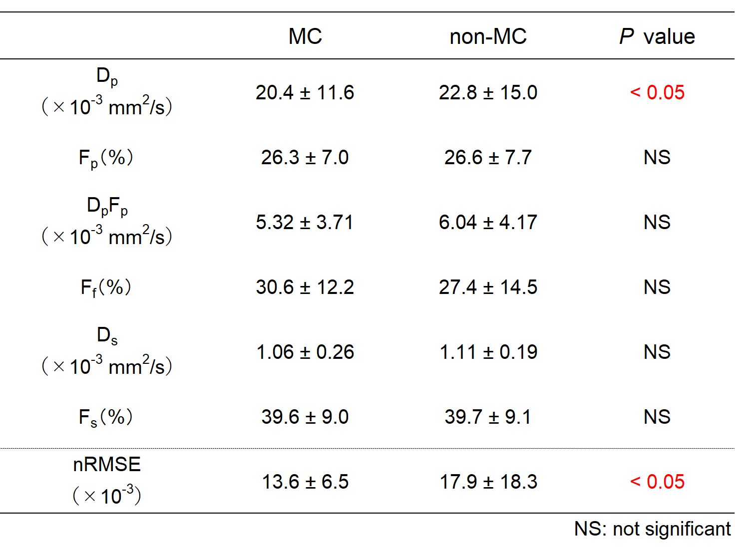
Table
1 Triexponential diffusion parameters and nRMSE in MC and non-MC
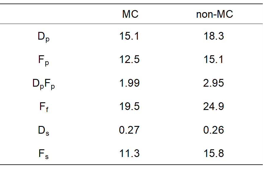
Table 2 Repeatability
coefficient (RC) of diffusion parameters in MC and
non-MC
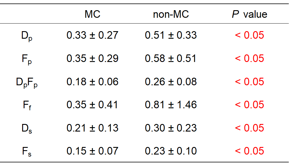
Table 3 Coefficient
of variation for each diffusion parameter in region of interest (CVROI)
in renal cortex
DOI: https://doi.org/10.58530/2023/3632