3610
Motion robust and high resolution DWI utilizing the combination of Multi-shot Reduced FOV imaging with motion-compensated diffusion gradients1Philips Healthcare, Shenzhen, Ltd., Shenzhen, China, 2Philips Health Technology, Suzhou, China, 3MR Clinical Science, Philips Healthcare, Mississauga, ON, Canada, 4BU MR Application, Philips Health Technology, Suzhou, China, 5BU MR R&D, Philips Health Technology, Suzhou, China, 6Philips Healthcare, Beijing, China
Synopsis
Keywords: Pulse Sequence Design, Diffusion/other diffusion imaging techniques, Diffusion, Motion compensated diffusion gradients, Reduced FOV imaging, Multi-shot DWI
Reduced FOV imaging (rFOV) and multi-shot DWI both are very useful techniques to improve spatial resolution for detection of pancreatic lesion. However, respiratory and cardiovascular motion will introduce severe artifacts and ADC bias. Motion compensated diffusion gradients (MOCO) could be used to improve the image quality. In this study, a new sequence named MOCO-rFOV IRIS was developed, which combines the advantages of rFOV, MOCO and image reconstruction using image-space sampling function based multi-shot DWI (IRIS). Results from in vivo data demonstrated that the proposed method could be used to realize motion robust and high resolution DWI for pancreasIntroduction
Diffusion imaging has showed great potential for the detection, staging, and treatment monitoring of pancreatic cancer. However, conventional diffusion imaging suffers from several challenges including low spatial resolution, distortions caused by B0 inhomogeneity, and signal loss caused by respiratory and cardiovascular motion1. Recently, several advanced techniques have been developed to solve these challenges. Reduced FOV imaging (rFOV)2,3 has been used to reduce distortion and increase resolution for pancreas. Multi-shot diffusion such as multiplexed sensitivity-encoding (MUSE)4 and image-space sampling function based multi-shot DWI (IRIS)5,could also be used to reduce the distortion for brain and other organ free from physiological motions. However, severe artifacts and signal voids can be introduced for both single -and multi-shot diffusion due to phase errors and signal loss from bulk motion, physiological motion (breathing, intestinal peristalsis, cardiac motion etc.), which have been investigated recently for brain6 and liver7. Ruiqi et al. showed that cardiovascular motion could introduce artifacts and ADC bias in pancreas DWI, and it could be addressed by motion-compensated diffusion gradients8.In this work, we developed a motion robust, reduced distortion and high resolution DWI by combining rFOV, IRIS and motion-compensated gradients.
Methods
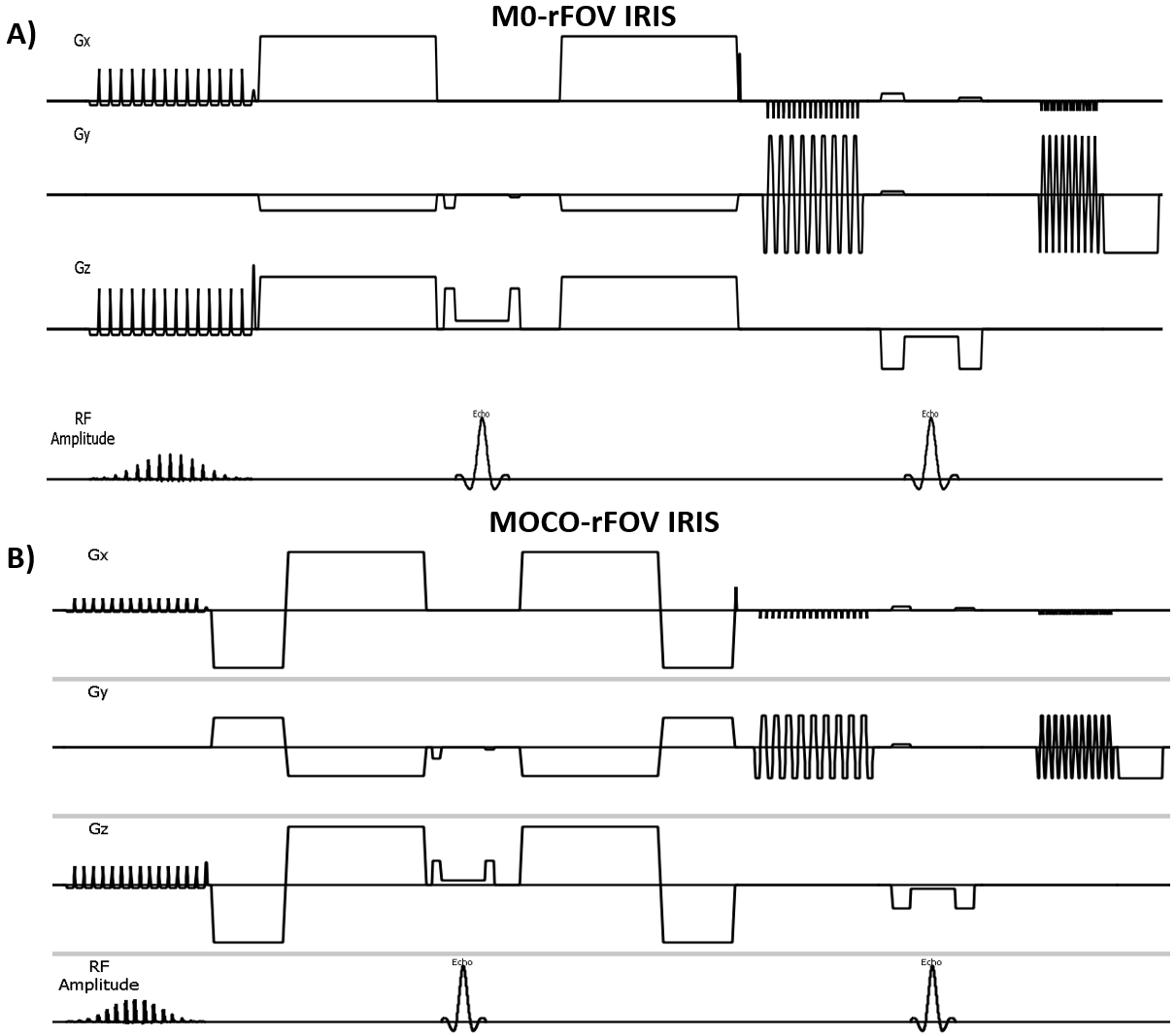
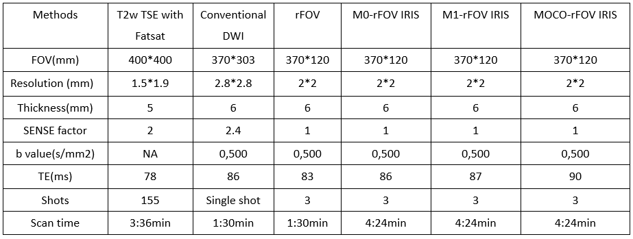
Pulse sequence: We combined reduced FOV imaging with IRIS 9, called rFOV IRIS. Fig. 1A shows the conventional rFOV IRIS scheme which acquires data using normal pulsed gradients without motion compensation. Fig. 1B shows our proposed scheme, it uses second order motion -diffusion gradients to reduce the phase errors for rFOV IRIS, it’s named as MOCO-rFOV IRIS, means the combination of motion-compensated diffusion gradients (MOCO), rFOV and IRIS.To evaluate performance of MOCO-rFOV IRIS, conventional DWI (ssDWI) and rFOV without motion compensated diffusion gradients were used as reference which is based on single shot EPI acquisition. Several other rFOV IRIS diffusion schemes were compared with ssDWI, which were rFOV IRIS with conventional pulsed gradients without motion compensation (M0-rFOV IRIS), rFOV IRIS with first-order motion-diffusion gradients (M1-rFOV IRIS), and our proposal MOCO-rFOV IRIS using second-order motion-compensated diffusion gradients. All scanning was performed on a Philips 3.0T Elition system (Philips Healthcare, Suzhou, China) , 24-ch torso & spine coil was used. The study was approved by the local IRB. All
Results
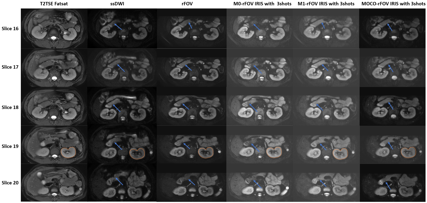
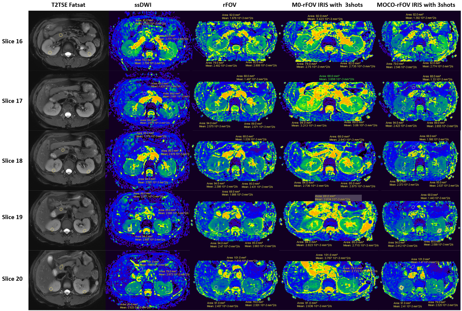
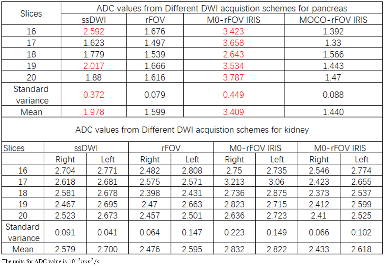
Compared to ssDWI and conventional rFOV, Fig. 2 shows that M0-rFOV IRIS will show severe artifacts which are introduced by phase errors between shots due to the motion from respiratory and cardiovascular, which is too large and can’t be corrected by the navigator echoes. The use of M1-rFOV IRIS with first-order motion compensated DWI reduces the artifacts, but it still shows some residual artifacts. Our proposal MOCO-rFOV IRIS with second-order motion-compensated diffusion gradients improves image quality dramatically with little residual artifacts. MOCO-rFOV IRIS with second-order motion shows smaller distortion and better spatial resolution than other schemes.To evaluate the performance of MOCO-rFOV IRIS further, Fig. 3 and Table 2 also compare the ADC values from different schemes for kidney and pancreas over 5 continuous slices; it shows that ADC values by MOCO-rFOV IRIS have better consistency between slices and smaller standard variance than observed for other schemes, and it also provides ADC values that are comparable to those from ssDWI and rFOV. The other schemes show some abnormal values, which may arise from respiratory or cardiovascular motion.For example, M0-rFOV IRIS shows a severe bias in ADC value compared to ssDWI and to other schemes.
Discussion
In this study, MOCO-rFOV IRIS was implemented from the combination of rFOV, IRIS and motion-compensated diffusion gradients. That approach was used to reduce the phase errors between shots, minimize motion related artifacts and could improve image quality dramatically. Preliminary results showed promising image quality with reduced ADC bias on kidney and pancreas and less overall distortion. That technique could be used for the assessment of metastases, for which high spatial resolution, high image quality and motion robustness is required. Further studies are required to test its performance in clinical settings, and it could be also combined with navigator free multi-shot DWI acquisition in the future.Conclusions
We investigated the feasibility of MOCO-rFOV IRIS which combines rFOV, IRIS and motion-compensated diffusion gradients for the first time. Results from in vivo data demonstrated that MOCO-rFOV IRIS shows better performance than other schemes for pancreas imaging. This technique holds potential for clinical applications that require motion robustness, high resolution and low distortion, such as pancreas, kidney etc.Acknowledgements
No.References
1.Diego H, et al. Quantitative diffusion MRI of the abdomen and pelvis. Med Phys. 2022;49(4):2774-2793.
2. Masahiro T, et al Reduced Field-of-View Diffusion-Weighted Magnetic Resonance Imaging of the Pancreas With Tilted Excitation Plane: A Preliminary Study. J Magn Reson Imaging. 2021;54(3):715-720. 3. Hyungjin K, et al. Reduced Field-of-View Diffusion-Weighted Magnetic Resonance Imaging of the Pancreas: Comparison with Conventional Single-Shot Echo-Planar Imaging. Korean J Radiol. 2015;16(6):1216-25.
4. Nan-Kuei C, et al. A robust multi-shot scan strategy for high-resolution diffusion weighted MRI enabled by multiplexed sensitivity-encoding (MUSE). Neuroimage. 2013 May 15;72:41-7.
5. Ha-Kyu J, et al. High-resolution human diffusion tensor imaging using 2-D navigated multishot SENSE EPI at 7T. Magn Reson Med. 2013;69(3):793-802.
6. Eric SM, et al. Multi-shot diffusion MRI of the human brain with motion-compensated oscillating gradients. Proc. Intl. Soc. Mag. Reson. Med. 29 (2021) 1324.
7. Yuxin Z, et al. Motion‐robust and blood‐suppressed M1‐optimized diffusion MR imaging of the liver. Magn Reson Med. 2019 Jul;82(1):302-311.
8. Ruiqi G, et al. Characterization and correction of cardiovascular motion artifacts in diffusion-weighted imaging of the pancreas. Magn Reson Med. 2021;86(4):1956-1969. 9. Wenxing F, et al. High-Resolution DWI using reduced FOV multi-shot EPI with IRIS reconstruction. Proc. Intl. Soc. Mag. Reson. Med. 24 (2016)3052.
Figures