3609
Impact of gradient spoiling for diffusion-weighted Double-Echo Steady-State sequences1Philips Research Europe, Hamburg, Germany
Synopsis
Keywords: Pulse Sequence Design, Diffusion/other diffusion imaging techniques
Double-Echo Steady-State (DESS) sequences are a promising candidate for diffusion weighted imaging (DWI) free of geometric distortions. While diffusion weighted DESS (dwDESS) sequences were originally introduced with a unipolar diffusion weighting gradient GD, a bipolar GD is required to obtain a motion robust fully balanced sequence. The inevitable banding artefacts occurring for bipolar GD can be handled via different techniques like gradient spoiling (thus deviating from the fully balanced sequence) or phase cycling. This study compares these techniques to optimize SNR for a given scan time and given diffusion weighting.Introduction
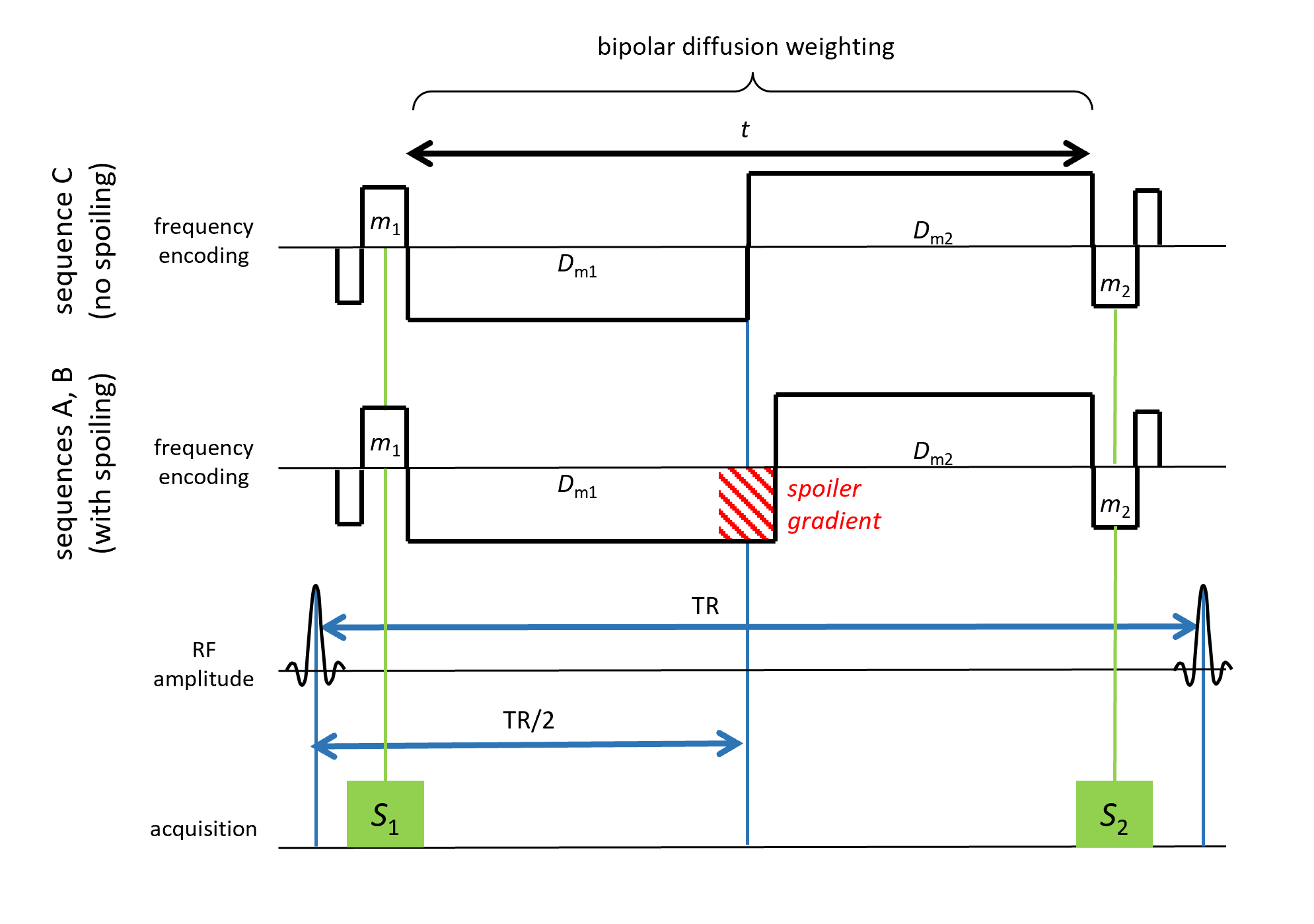
To avoid the pervasive geometric distortions of diffusion weighted imaging (DWI), the use of diffusion-weighted Double-Echo Steady-State (dwDESS) sequences was suggested 1. Such dwDESS sequences were originally introduced with a unipolar diffusion weighting gradient GD 2, but to achieve a motion robust fully balanced sequence, a bipolar GD is required 3, leading inevitably to banding artefacts 4. This study investigates two different techniques to remove these banding artefacts: (a) phase cycling is applied by repeating the dwDESS sequence with different phase offsets of the RF pulse, and suitable subsequent combination of the different images (see, e.g. 5), (b) a spoiler gradient is inserted between the two lobes of the bipolar diffusion gradient lobes, thus introducing a slight imbalance of GD (see, e.g. 6). These techniques are compared with respect to obtained SNR and required gradient moment. The comparison is performed for a fixed scan time and a fixed diffusion weighting, which implies that first an (effective) b-value for the different sequence types needs to be determined.Methods
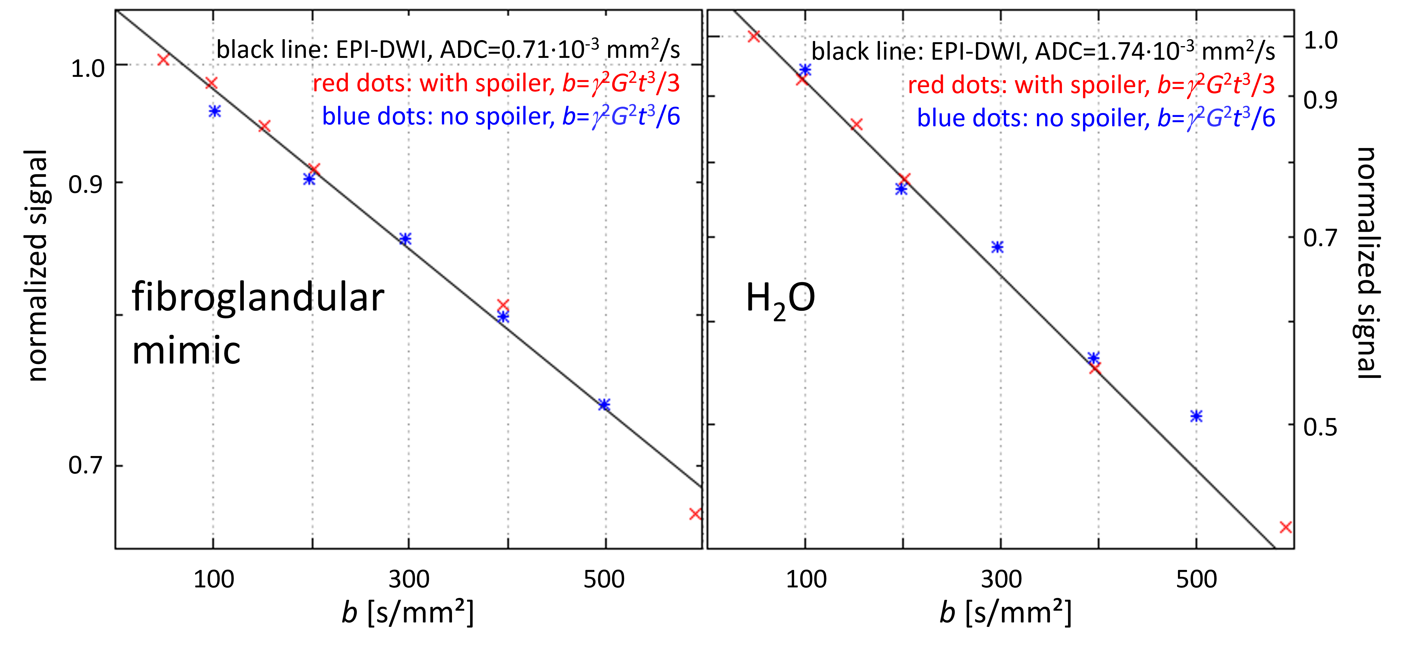
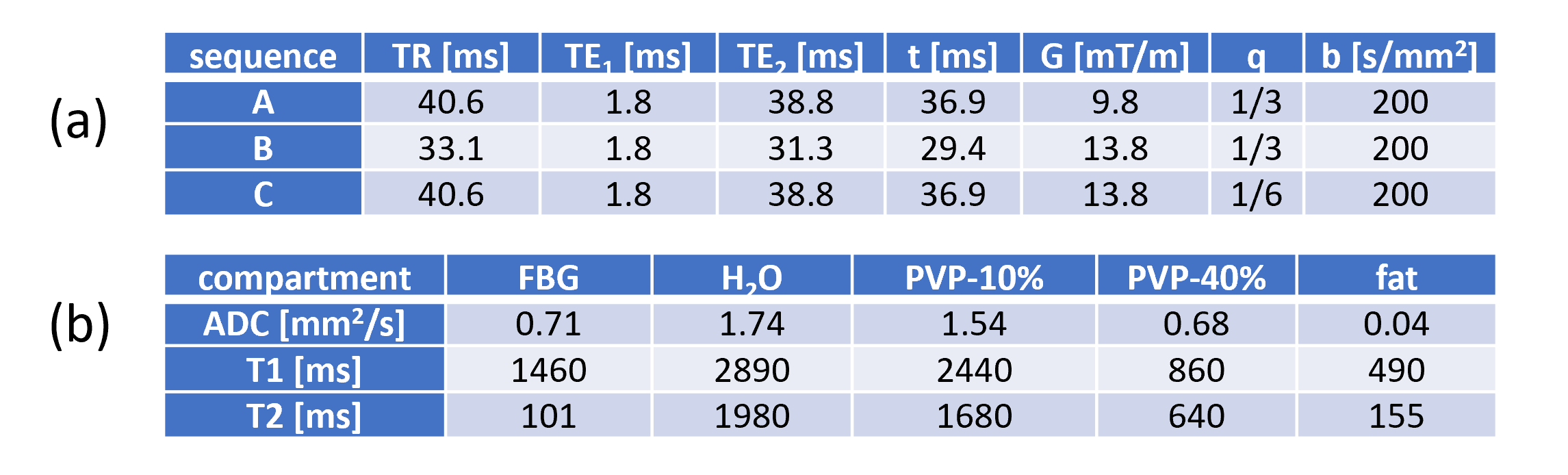
The phantom “Breast Standard Model 131” (CaliberMRI, Boulder, USA) was investigated with a clinical 1.5T MRI system (Ingenia, Philips Healthcare, Best, Netherlands) and a 32-channel anterior/posterior coil array.Determination of effective b-values: For standard, EPI-based DWI as well as for the dwDESS sequences investigated in this study (Fig. 1), the corresponding b-value is given by b=qγ2G2t3 with γ the gyromagnetic ratio, G the diffusion gradient strength, and t the total duration of the diffusion gradient (i.e. including both, positive and negative lobe, with no gap between the lobes assumed). The constant q depends on the type of sequence and was verified in a preparation experiment (Fig. 2) as q=1/3 for dwDESS with spoiling (in line with 7) and q=1/6 for dwDESS without spoiling (in line with 8). Thus, to achieve the same b as for dwDESS without spoiling, dwDESS with spoiling can be implemented by (a) reducing G by 21/2 maintaining t or (b) shortening t by 21/3 maintaining G (or a suitable mixture of both, which however was not further pursued in this study).
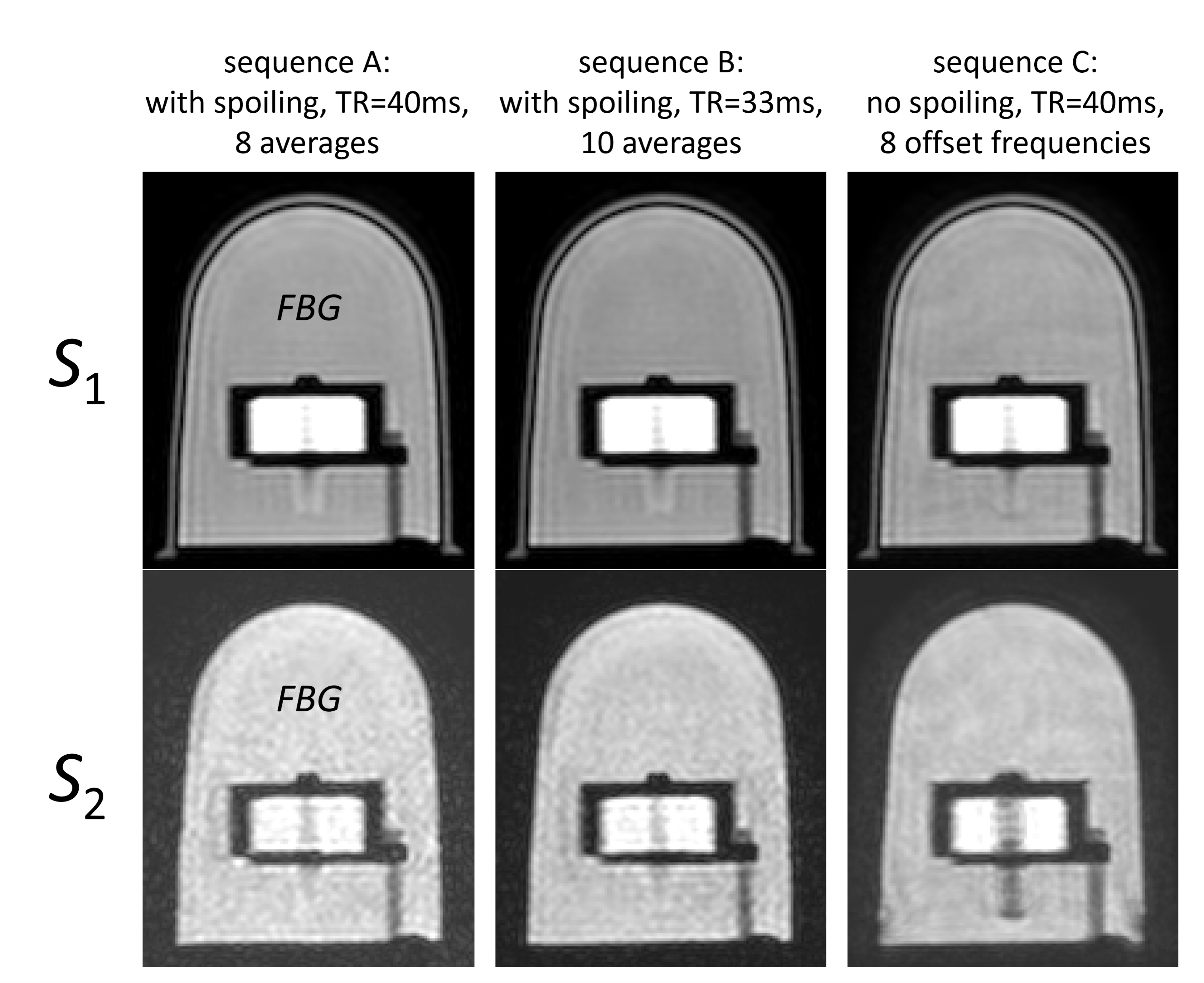
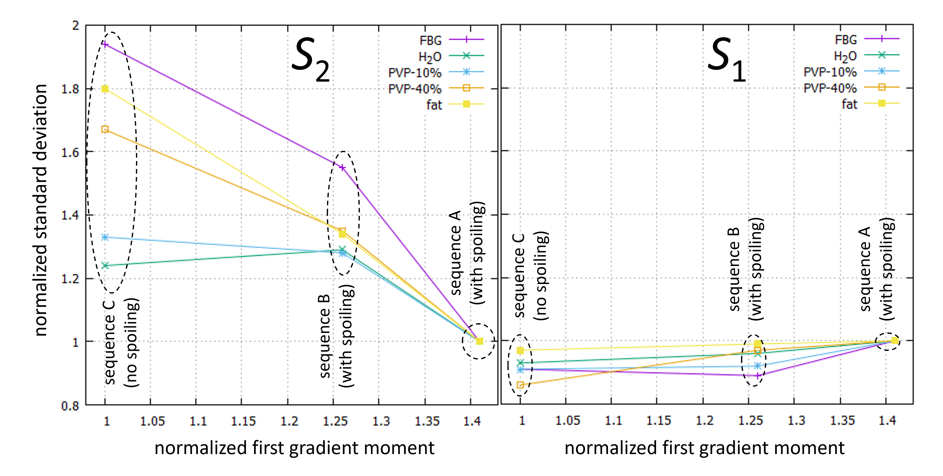
Sequences: All dwDESS sequences of this study used a voxel size of 1.8 × 1.8 × 2.0 mm³ and flip angle α=25°. Three different variants (A, B, C) of dwDESS were tested, sequences A and B with gradient spoiling and sequence C without gradient spoiling (parameters see Fig. 3a). As mentioned above, G was lowered for sequence A and t was lowered for sequence B such that both spoiled sequences yield the same b=200 s/mm2 as sequence C. For sequence C, phase cycling was performed by repeating the sequence with 8 different, equidistant phase offsets. Sequence A (having same TR as sequence C) was performed with 8 averages, and sequence B (having shorter TR than sequence C) with 10 averages, such that all sequences had a comparable total acquisition time of about 4:50 min. “Nonlinear Averaging” was applied to combine the 8 phase offsets of sequence C to the final image 5. From the two echoes S1 and S2, the Coefficient of Variation (CoV) was determined in 5 different phantom compartments specified in Fig. 3b, and plotted versus normalized gradient moment G×t.
Results
Examples images of the FBG compartment are shown in Fig. 4 to illustrate the obtained image quality of the two echoes. A trade-off between SNR (in terms of CoV) and gradient moments required for the different sequence types was found for S2, while SNR was almost constant for S1 (Fig. 5). The maximum SNR gain for S2 and FBG was ×1.95 (C vs A) and ×1.26 (C vs B). Substances with short T2 showed a larger impact of the sequence type than substances with long T2.Discussion and Conclusion
This phantom study exhibited a significant impact of the dwDESS sequence type on the obtained SNR of echo S2 for equivalent b-values, which thus is also obtained for the resulting diffusion weighted image given by S2/S1 1. The effect was exemplarily demonstrated for b=200 s/mm2, but applies accordingly for the whole range of b-values. It can be explained by higher configuration states evoked by the gradient spoiler, having lower signal but higher diffusion sensitivity 9. As long as phase-cycling techniques sufficiently remove banding artefacts of dwDESS without gradient spoiling, a significantly higher SNR can be expected for tissue types with short T2. For instance, the investigation of breast cancer shall benefit from applying a phase-cycled dwDESS without spoiling to obtain distortion-free, diffusion weighted breast images with high SNR.Acknowledgements
The authors cordially thank Peter Koken and Peter Vernickel for system maintenance.References
1. Freed DE et al., Steady-state free precession experiments and exact treatment of diffusion in a uniform gradient. J Chem Phys. 2001;115(9):4249-4258.
2. Wu EX et al., Effect of diffusion on the steady-state magnetization with pulsed field gradients. J Magn Reson. 1990;90(1):243-253.
3. Zur Y et al., A new diffusion SSFP imaging technique. Magn Reson Med. 1997;37(5):716–722.
4. Staroswiecki E et al., Simultaneous Estimation of T2 and ADC in Human Articular Cartilage In Vivo with a Modified 3D DESS Sequence at 3T. Magn Reson Med. 2012;67(4):1086-1096.
5. Elliott AM et al, Nonlinear averaging reconstruction method for phase-cycle SSFP. Magn Reson Imag. 2007; 25(3):359-364.
6. Keupp J et al., Simultaneous acquisition of diffusion weighted images and conductivity maps using a balanced double echo steady state sequence. Annual Meeting of ISMRM 2020;28:955.
7. Katscher U et al., B-value derivation for diffusion-weighted Double-Echo Steady-State (dwDESS) sequences. Annual Meeting of ISMRM 2021;29:1144.
8. Ding S et al., High Resolution Renal Diffusion Imaging Using a Modified Steady-State Free Precession Sequence. Magn Reson Med. 1995;34(4):586-595.
9. Weigel M, Extended Phase Graphs: Dephasing, RF Pulses, and Echoes - Pure and Simple. J Magn Reson Imag. 2015;41(2):266–295.
Figures