3576
Brain iron content increase and oxygen extraction fraction reduction after iron repletion in blood donation induced iron deficiency1Biomedical Engineering, Cornell University, Ithaca, NY, United States, 2Department of Radiology, Weill Medical College of Cornell University, New York, NY, United States, 3Weill Medical College of Cornell University, New York, NY, United States, 4Columbia University Medical Center, New York, NY, United States
Synopsis
Keywords: Hematologic, Neuro
We examined brain iron content using susceptibility source separation and oxygen extraction fraction (OEF) in adult healthy blood donors with blood donation-induced iron deficiency treated with intravenous iron repletion at the first donation and a second donation after approximately 5 months and compared it with a control group who was not treated after the first donation. Iron content increases in the parietal cortex, the caudate and the putamen were observed in the treatment group. OEF reduction was observed in the whole brain and multiple ROIs, suggesting iron repletion treatment helps reduce oxidative stress.
Motivation
Blood donation-induced iron deficiency has a high prevalence among regular donors.1 Previous studies have reported little effect on donor cognition and wellbeing under the current criteria of blood donation.2 The primary effect of iron deficiency and iron repletion on the brain still needs to be examined. In this study we examine the effect of intravenous iron repletion in subjects with blood donation-induced iron deficiency on brain iron content and oxygen metabolism using MRI based analysis methods.Materials and Methods
Adult iron-deficient blood donors (n = 59) were enrolled in this study. After a first standard blood donation, donors were randomized to intravenous iron repletion (intravenous one gram low molecular weight iron dextran) or placebo (intravenous saline). The randomization was stratified by sex and blinded to the research pharmacist and data analysis individuals. A second blood donation occurred approximately 5 months. Multiecho gradient echo (mGRE) data were acquired at both time points (TE1/∆TE/TR = 5.9/9.1/66.8ms) for cerebral susceptibility source separation and OEF mapping. Positive and negative susceptibility maps (χ+ and χ-) were reconstructed using a R2*-based magnetic susceptibility source separation method.3 χ+ maps served as a measure of cerebral iron content. OEF maps were reconstructed from quantitative susceptibility mapping (QSM) plus quantitative blood oxygen level-dependent magnitude (QSM + qBOLD = QQ)-based mapping.4 ROIs were identified using FreeSurfer5 for each subject. ROI values were compared longitudinally between the two time points and cross-sectionally between treatment and placebo groups using paired t-tests with the significance level set at p<0.05. Multiple comparison correction was performed using the Benjamini-Hochberg procedure to control false discovery rate method6.Results
We found that in the treatment group, the iron content indicated by χ+ increased in the parietal cortex (p = 0.0295) from 35.24±3.59ppb to 36.31±3.74ppb. χ+ also increased in the treatment group in the caudate (p = 0.0445) from 46.84±7.06ppb to 48.71±7.09ppb and in the putamen (p = 0.0065) from 52.96±7.83ppb to 54.87±8.09ppb. The whole-brain OEF was reduced in the treatment group at the second donation compared to at the first donation (p = 0.0187) from 35.46±7.36% to 32.17±5.42%. Regional OEF also showed similar reduction in the treatment group in the cortical gray matter (p = 0.0232) from 33.77±7.35% to 30.65±5.65%, in the caudate (p=0.031) from 34.36±8.1% to 31.79±5.39%, in the globus pallidus (p=0.0052) from 38.28±8.66% to 33.28±5.78%, in the putamen (p = 0.0052) from 39.51±7.96% to 35.2±5.22%, in the thalamus (p = 0.0205) from 36.87±7.72% to 34.00±5.27% and in the white matter (p = 0.0084) from 38.06±7.70% to 34.32±5.74%.Discussion and Conclusion
Our results show that the effect of iron repletion for blood donation-induced iron deficiency can be seen in the increase in the brain iron content measurement using the susceptibility source separation algorithm in several ROIs. The effect size is small and unlikely to affect brain iron homeostasis.7 Oxygen extraction fraction was reduced after the iron repletion in the whole brain and all ROIs. Although decreased, the OEF values are still in the normal range of healthy subjects using similar MR based OEF mapping methods.8, 9 Thus, the decreased OEF values may indicate that the subjects experience less oxidative stress after iron repletion.Acknowledgements
No acknowledgement found.References
1. Cable RG, Glynn SA, Kiss JE, et al. Iron deficiency in blood donors: the REDS-II Donor Iron Status Evaluation (RISE) study. Transfusion 2012; 52: 702-711. 2011/10/26. DOI: 10.1111/j.1537-2995.2011.03401.x.
2. Hod EA, Brittenham GM, Bitan ZC, et al. A randomized trial of blood donor iron repletion on red cell quality for transfusion and donor cognition and wellbeing. Blood 2022 2022/09/08. DOI: 10.1182/blood.2022017288.
3. Dimov AV, Nguyen TD, Gillen KM, et al. Susceptibility source separation from gradient echo data using magnitude decay modeling. Journal of Neuroimaging 2022; 32: 852-859. DOI: 10.1111/jon.13014.
4. Cho J, Spincemaille P, Nguyen TD, et al. Temporal clustering, tissue composition, and total variation for mapping oxygen extraction fraction using QSM and quantitative BOLD. Magnetic Resonance in Medicine 2021. DOI: 10.1002/mrm.28875.
5. Fischl B, Salat DH, Busa E, et al. Whole brain segmentation: automated labeling of neuroanatomical structures in the human brain. Neuron 2002; 33: 341-355. 2002/02/08. DOI: 10.1016/s0896-6273(02)00569-x.
6. Benjamini Y and Hochberg Y. Controlling the False Discovery Rate - a Practical and Powerful Approach to Multiple Testing. J R Stat Soc B 1995; 57: 289-300. DOI: DOI 10.1111/j.2517-6161.1995.tb02031.x.
7. Langkammer C, Schweser F, Krebs N, et al. Quantitative susceptibility mapping (QSM) as a means to measure brain iron? A post mortem validation study. Neuroimage 2012; 62: 1593-1599. DOI: 10.1016/j.neuroimage.2012.05.049.
8. Cho J, Lee J, An H, et al. Cerebral oxygen extraction fraction (OEF): Comparison of challenge-free gradient echo QSM+qBOLD (QQ) with (15)O PET in healthy adults. J Cereb Blood Flow Metab 2021; 41: 1658-1668. 2020/11/28. DOI: 10.1177/0271678X20973951.
9. Jiang D, Deng S, Franklin CG, et al. Validation of T2 -based oxygen extraction fraction measurement with (15) O positron emission tomography. Magn Reson Med 2021; 85: 290-297. 2020/07/10. DOI: 10.1002/mrm.28410.
Figures
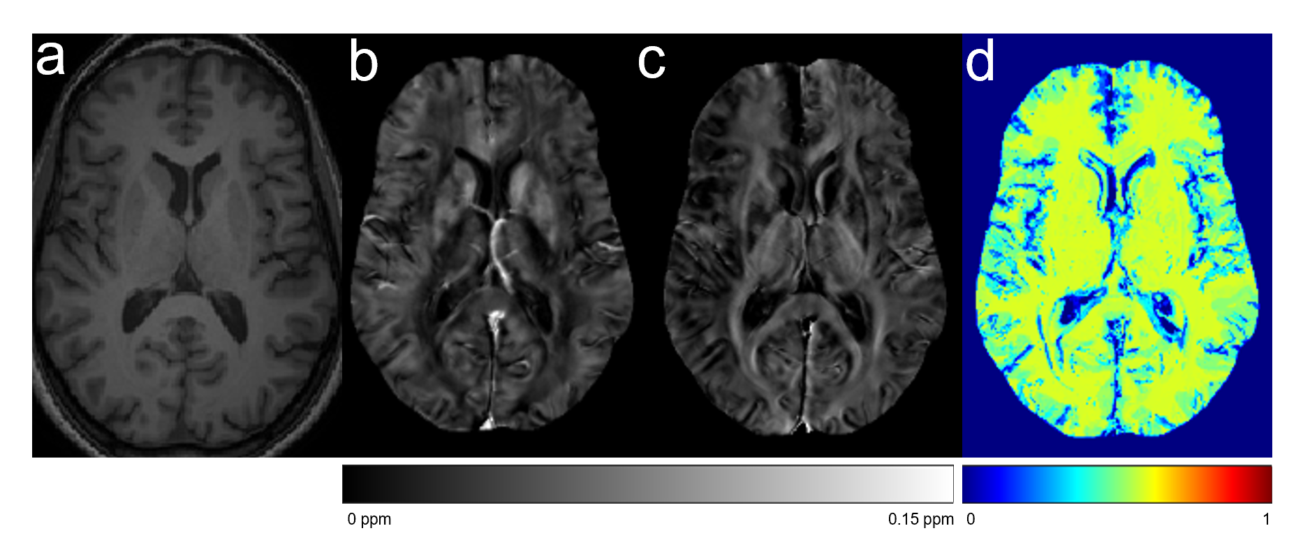
An example of the subject a.T1w image, b. χ+ map, c. χ- map, d. OEF map

Comparison of iron content in the treatment group between the two time points in select brain ROIs. In the parietal cortex, the caudate and the putamen, iron increase was observed.
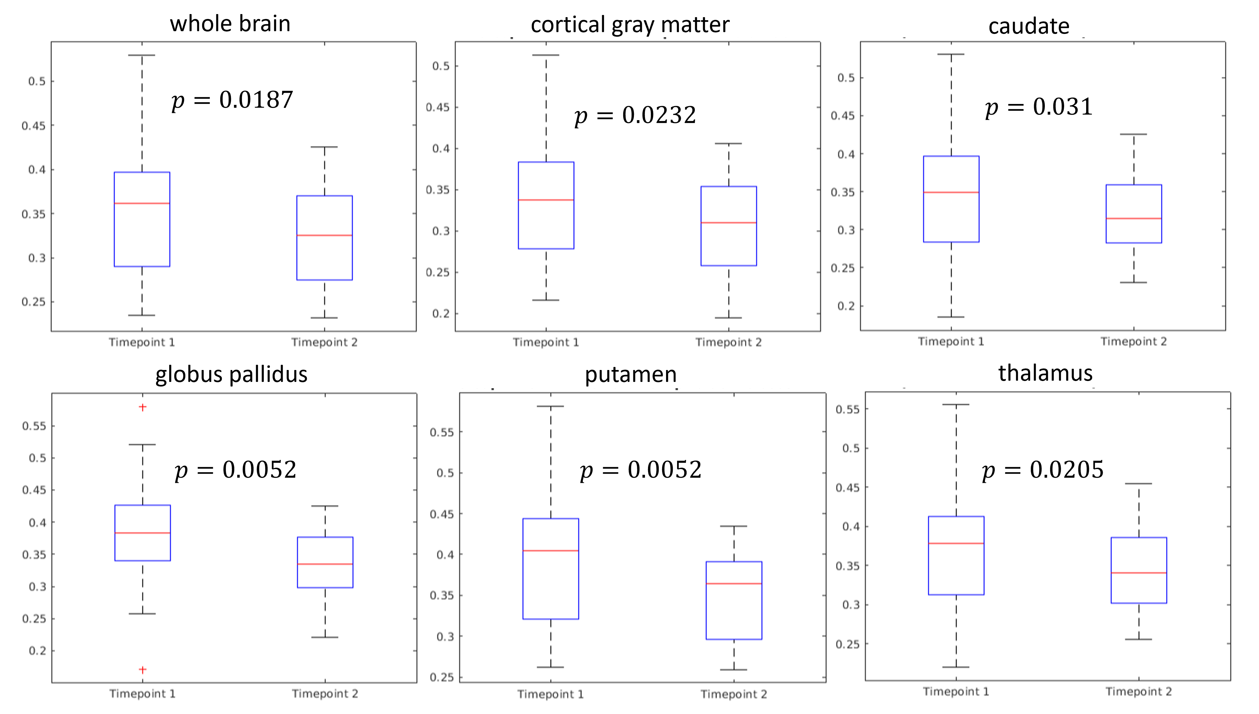
Comparison of OEF in the treatment group between the two time points in select brain ROIs. In all ROIs shown, OEF reduction can be detected approximately 5 months after iron repletion.