3550
Brain morphological alterations in patients with lung cancer after chemotherapy
Liu Renyuan1, Rong Ping1, Han Xiaowei1, and Zhang Bing1
1Radiology, Drum Tower hospital, Nanjing, China
1Radiology, Drum Tower hospital, Nanjing, China
Synopsis
Keywords: Gray Matter, Cancer
The pathophysiological and biochemical effects of lung cancer and chemotherapy would result in abnormal alterations of brain morphometry. In particular, chemotherapy could endanger critical cognition-related subcortical nucleus, such as amygdala.Introduction
Chemotherapy-related brain morphological alterations are frequent consequences in patients with lung cancer, especially after platinum chemotherapy1. Volumetric alterations and abnormalities might result in mental and psychological disorders, thus may compromise survivors’ quality of life2. Therefore, it is necessary to investigate the characteristics of brain morphology in patients with lung cancer after chemotherapy.Methods
Overall, 38 patients with chemotherapy (Ch+), 113 patients without chemotherapy (Ch-), and 40 healthy controls (HC) were retrospectively enrolled in this study. Inclusion criteria for patients: 1) diagnosed with lung cancer based on pathological results; 2) without brain metastasis; 3) with high resolution T1 weighted imaging data for structural analysis; 4) with or without platinum chemotherapy. Exclusion criteria: 1) patients with stroke, arterial aneurysm, cerebral hemorrhage, or other neuropsychiatric diseases; 2) patients with a duration after chemotherapy of < 30 days. This study was approved by Medical Ethics Committee of the Nanjing Drum Tower Hospital. Imaging data were acquired using a 3.0T MR scanner (Ingenia CX, Philips) with a 32-channel head coil. A three-dimensional fast-spoiled gradient-echo sequence with repetition time/echo time = 6.6/3.0 ms, flip angle = 8°, field of view = 250 × 250 × 180 mm3, matrix = 250 × 250 × 180, voxel size = 1.0 × 1.0 × 1.0 mm3 was used for T1 weighted imaging. FreeSurfer v7.2.0 was used for images segmentation and group-wise volumetric comparisons. Structural covariance network (SCN) was constructed and analyzed by brain connectivity toolbox (BCT). In this study, SCN analysis was performed separately on three volumetric properties: cortical thickness, volumes of subcortical structures, and volumes of hippocampal subfields and nuclei of amygdala. Structural regions of interest (ROIs) from the Freesurfer segmentation were defined as SCN nodes, while Pearson’s correlations between ROIs pairs were defined as SCN edges. Group-wise comparisons of network properties were performed by permutation test with significance level and number of permutations were set to p < 0.05 and 5000 times, respectively. Additional FDR corrections (p < 0.05) were used for comparisons of nodal properties.Results
No significant differences of volumetric properties, such as cortical thickness and volumes of subcortical nucleus, were found among the groups (vertex/voxel-wise p < 0.001, cluster-wise p < 0.05). However, cortical SCN based on the ROIs of Destrieux Atlas indicated an enhanced small-worldness connections of the Ch+ group (fig.1), while no significant differences of SCN properties were found with the ROIs of Desikan-Killiany Atlas. On subcortical structural covariance analysis, among the 17 subcortical structures selected in this study, reduced nodal degree (p = 0.0002) and nodal betweenness (p = 0.0008) were found in right amygdala of Ch+, in compared with HC (fig.2-3). No significant differences of nodal properties were found between the Ch- and HC. For global SCN characteristic, a trend of reduced gamma was found in both Ch+ and Ch- (both p value < 0.0001), in compared with HC. For the SCN analysis of subfields of hippocampal-amygdalar complex, reduced gamma (p = 0.0342) and sigma (p < 0.0001) were found in Ch-, in compared with HC (fig.4). No significant differences of hippocampal-amygdalar complex SCN properties were found between the Ch+ and HC.Discussion
In this study, we identified abnormal brain morphological alterations in patients with lung cancer, with or without chemotherapy. Reduced SCN nodal properties were found in the right amygdala of Ch+, in compared with HC. No such alterations were found between the Ch- and HC, suggesting the cognitive-related side effects of chemotherapy. However, reduced SCN properties of hippocampal-amygdalar complex were only observed in Ch-, indicate the neuropsychological effects of cancer biology. Thus, the relationship of brain morphological changes and cancer chemotherapy remain unclear. On the other hand, enhanced cortical small-worldness connections were observed in Ch+. Such topological alterations might be resulted from cortical edema and ROIs selection, indicating the necessity of further validations from different cortical atlas. The retrospective nature of this study indicated the lack of insights from follow-up research; secondly, without clinical assessments of cognitive performances, the associations of morphological alterations and cognitive decline remain unclear.Conclusion
The pathophysiological and biochemical effects of lung cancer and chemotherapy would result in abnormal alterations of brain morphometry. In particular, chemotherapy could endanger critical cognition-related subcortical nucleus, such as amygdala.Acknowledgements
This work was supported by the National Natural Science Foundation of China (82171908 XH, 81720108022 BZ, 81971596, XZ, 82001793, JL).References
1.Kathleen VD, Catherine MC, Laura P, et al. Identifying Cancer-Related Cognitive Impairment Using the FACT-Cog Perceived Cognitive Impairment. JNCI Cancer Spectr. 2019; 29; 4(1): pkz099.
2.Omar FK, Ellen C Soundouss R, et al. Immediate-term cognitive impairment following intravenous (IV) chemotherapy: a prospective pre-post design study. BMC Cancer. 2019; 19(1): 150.
Figures
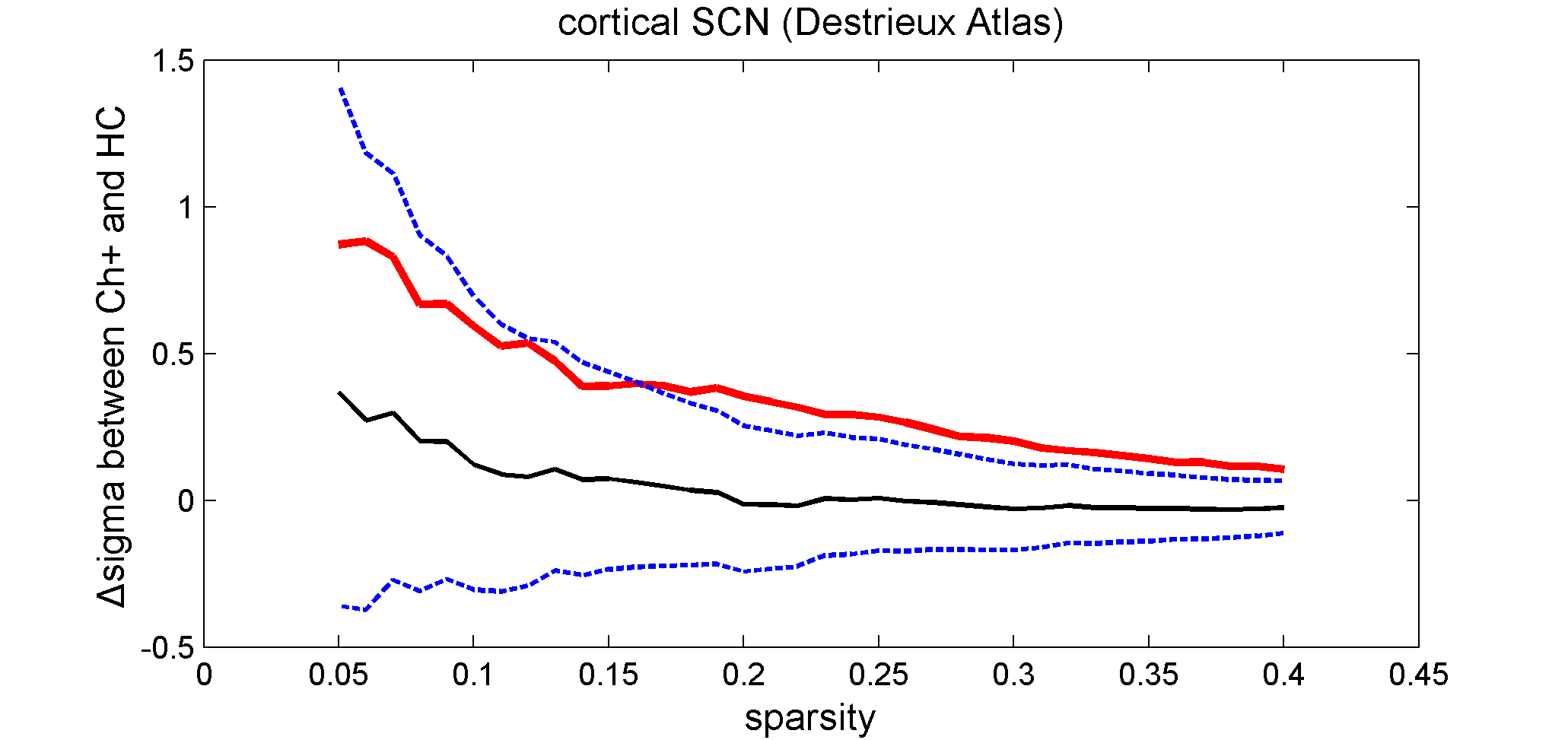
Enhanced sigma of Ch+,
in compared with HC. Red line: the differences between Ch+ and HC; black line:
the differences between Ch+ and HC from permutation test; blue line: 95% confidence
interval.
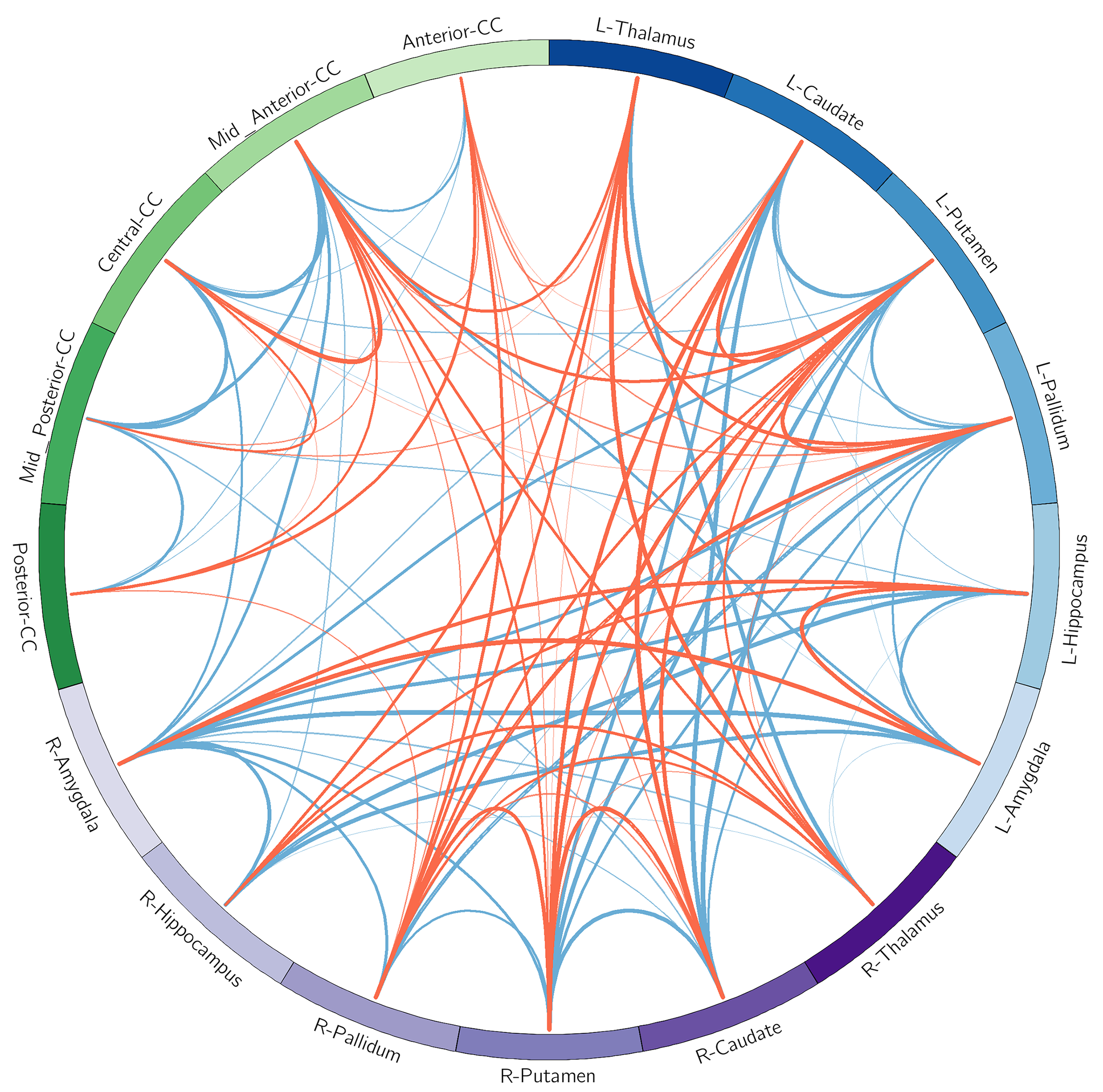
Reduced subcortical SCN
connections of Ch+, in compared with HC. One-sample t test (p<0.0001) of Ch+
(red line) and HC (blue line).
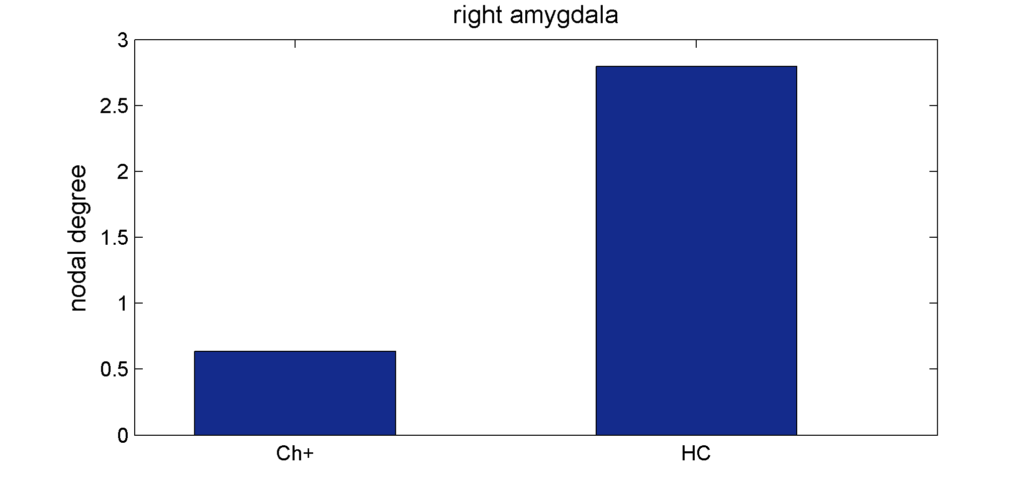
Reduced nodal degree in
right amygdala of Ch+, in compared with HC (FDR<0.05).
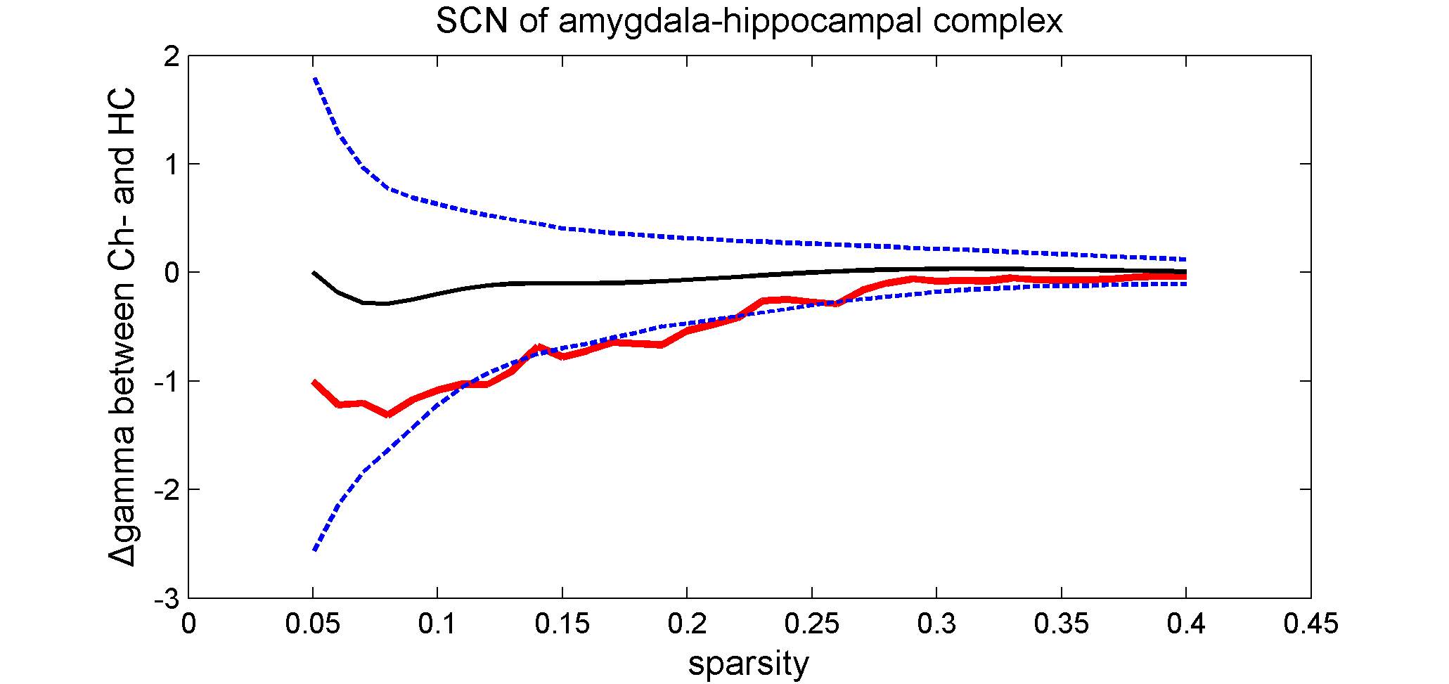
Reduced gamma of Ch-,
in compared with HC. Red line: the differences between Ch- and HC; black line:
the differences between Ch- and HC from permutation test; blue line: 95% confidence
interval.
DOI: https://doi.org/10.58530/2023/3550