3549
Mapping extraversion in brain activation patterns: a functional neuroimaging meta-analysis of resting state studies1Department of Interventional Therapy, National Cancer Center/National Clinical Research Center for Cancer/Cancer Hospital, Chinese Academy of Medical Sciences and Peking Union Medical College, Beijing, China, 2Research Unit of Psychoradiology, Chinese Academy of Medical Sciences, Chengdu, China, 3Huaxi MR Research Center (HMRRC), Department of Radiology, West China Hospital of Sichuan University, Chengdu, China, 4Functional & Molecular Imaging Key Laboratory of Sichuan Province, West China Hospital of Sichuan University, Chengdu, China, 5Department of Radiology, West China Xiamen Hospital of Sichuan University, Xiamen, China
Synopsis
Keywords: Gray Matter, Brain
Our meta-analysis reveals that extraversion was linked with resting-state brain activity differences widely distributed across cortical and subcortical regions involved in emotion and behavioral regulation. The meta-regression results suggest an effect of gender on the association between extraversion and neural activity in the right inferior frontal gyrus. Our findings support that extraversion could lead to neural activity changes, which may be correlated with behavioral differences between extraverts and introverts.PURPOSE
Extraversion is a fundamental personality dimension that contributes to individuals’ health and well-being. With the rapid development of functional brain imaging in the past two decades, a number of studies have examined the brain activation patterns of extraversion, but the results are extremely inconsistent and not integrated. Therefore, we conducted a quantitative meta-analysis of functional neuroimaging studies to obtain a convergent understanding of the functional brain basis at rest underlying extraversion.METHODS
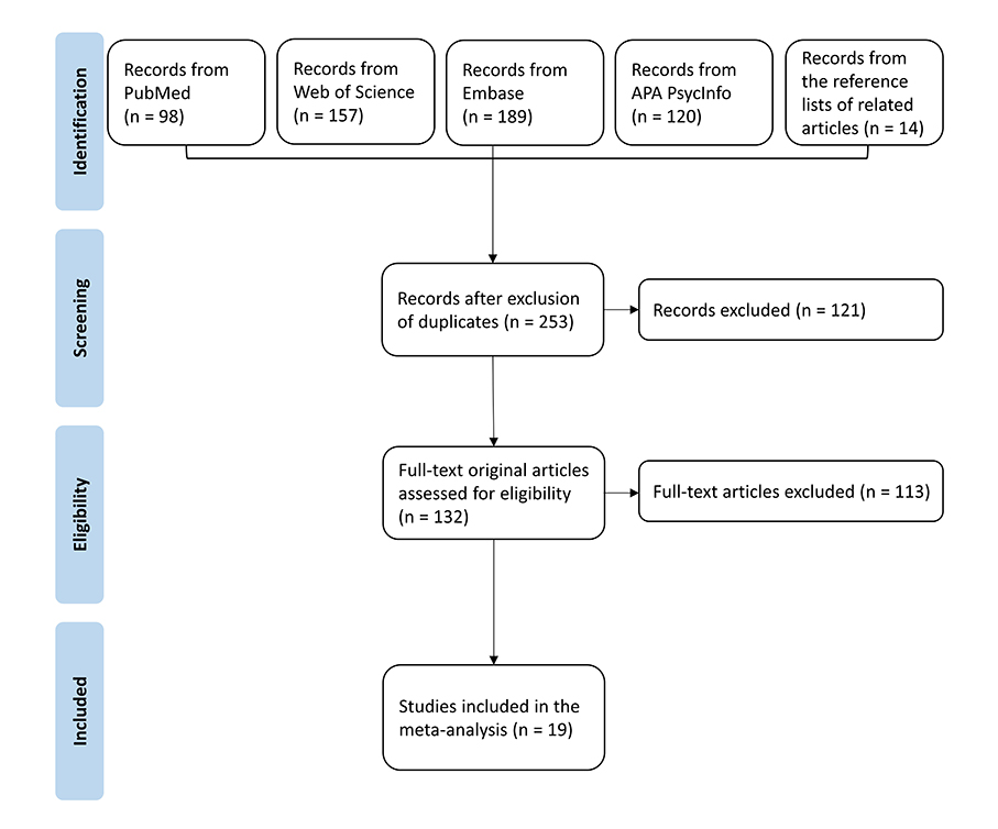
Based on the Preferred Reporting Items for Systematic Reviews and Meta-Analyses statement (PRISMA)1, we implemented a comprehensive online literature search to identify brain activation patterns of extraversion in resting-state neuroimaging studies. To identify precise and unbiased brain regions where neural activities at rest were significantly associated with extraversion, we performed a coordinate-based whole-brain meta-analysis using anisotropic effect-size seed-based d mapping (AES-SDM) software2. AES-SDM (https://www.sdmproject.com/software/) is a voxel-based meta-analytic approach by accommodating peak coordinates and their effect sizes. It has been validated for the conjunction of functionally alternated brain regions in many neuropsychiatric disorders and healthy controls3. Then we carried out jackknife sensitivity analyses to assess the reliability and replicability of our results. Meta-regression analysis was performed to explore the potential effects of gender and age on the association between extraversion and neural activity.RESULTS
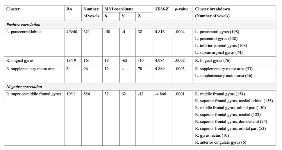
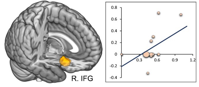
A total of 19 studies (18 datasets) meeting the criteria, which included 1,440 healthy subjects (648 females; 22.43 years old) and covered 116 peaks of stereotaxic coordinates, were used in the pooled meta-analysis. In resting-state neuroimaging studies with various analytical methods, extraverts showed increased functional activity compared to introverts in the left paracentral lobule (PCL) extending to the inferior parietal gyrus, right lingual gyrus (LING), and right supplementary motor area (SMA). A negative correlation between extraversion and neural activity was present in the right superior frontal gyrus (SFG) and middle frontal gyrus (MFG). The meta-regression also showed that the association in the right inferior frontal gyrus (IFG) were modulated by the gender ratio.DISCUSSION
To our knowledge, this is the first meta-analysis revealing the functional brain bases of extraversion which may contribute to the determination of a robust and study-invariant link between extraversion and brain function. Although previous neural functional research showed inconsistent brain regions, our review identified four core brain areas that are involved in emotion and behavioral regulation. These findings suggest that changes in spontaneous neural activity may reflect difficulties with interpersonal and affective regulation in introverts. Additionally, we found that gender modulated the association between extraversion and regional brain activity in the right IFG, which may be explained by interstudy heterogeneity. Overall, the current findings may help to elucidate the neurobiological mechanism underlying extraversion, and provide targeted brain regions for promising interventions on extraversion to decrease the risk of health problems and increase quality of life.CONCLUSION
In brief, our meta-analysis summarized the literature using AES-SDM and identified four core neurofunctional regions of extraverted personalities among healthy individuals. The results support that extraversion could lead to neural activity changes, which may be correlated with behavioral differences between extraverts and introverts. Our meta-analytic findings provide novel evidence on the neurobiological basis of extraversion, which may facilitate health interventions by helping identify individuals at risk of extraversion-related psychological/physical disorders.Acknowledgements
This study was supported by the National Natural Science Foundation of China (Grant Nos. 31800963, 81621003, 81761128023, 81820108018, 82027808).References
1. Knobloch, K., Yoon, U., & Vogt, P. M. Preferred reporting items for systematic reviews and meta-analyses (PRISMA) statement and publication bias. Journal of cranio-maxillo-facial surgery: official publication of the European Association for Cranio-Maxillo-Facial Surgery. 2011;39(2):91–92.
2. Radua, J., Mataix-Cols, D., Phillips, M. L., El-Hage, W., Kronhaus, D. M., Cardoner, N., & Surguladze, S. A new meta-analytic method for neuroimaging studies that combines reported peak coordinates and statistical parametric maps. European psychiatry: the journal of the Association of European Psychiatrists. 2012;27(8):605–611.
3. Liu, X., Lai, H., Li, J., Becker, B., Zhao, Y., Cheng, B., & Wang, S. Gray matter structures associated with neuroticism: A meta-analysis of whole-brain voxel-based morphometry studies. Human brain mapping. 2021;42(9):2706–2721.
Figures