3547
Cerebral Blood Flow quantification in anesthetized macaque using pseudo-continuous Arterial Spin Labeling at 3T1Radiology, Clínica Universidad de Navarra, Pamplona, Spain, 2IdiSNA,Instituto de Investigación Sanitaria de Navarra, Pamplona, Spain, 3Hepatology, CIMA Universidad de Navarra, Pamplona, Spain, 4School of Education and Psychology Universidad de Navarra, Pamplona, Spain, 5Siemens Healthcare, Madrid, Spain
Synopsis
Keywords: Gray Matter, Arterial spin labelling, Animal, Preclinical, White Matter
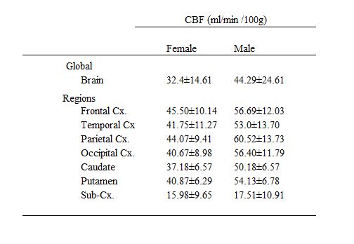
A pseudo-continuous arterial spin labeling (pCASL) technique was implemented on a Siemens 3T Skyra for quantitative cerebral blood flow (CBF) measurements in non-human primates (rhesus monkeys). Different regions were manually segmented based on T1 weighted images. ASL images were obtained in 10 min with 2.2-mm isotropic resolution. Whole brain CBF was 32.40±14.61 ml/100g/min (n=8) for female and 44.28±24.61 ml/100 g/min for male (n=4) rhesus monkeys under ketamine and midazolam anesthesia. Gender differences of CBF among brain regions will be address in this study.Introduction
Quantification of Cerebral Blood Flow (CBF) using MRI has been widely used to study different neurological disorders. Non-human primates (NHPs) resemble most aspects of humans in brain anatomy and physiology, especially macaque rhesus monkeys, which share 93.5% of human genome, and have ben used to translate relevant findings and treatments into clinical practice1. To date, isoflurane, ketamine, and propofol are the most widely used anesthetics in animal studies. Adverse effects of anesthetics may directly affect CBF values. Ketamine has been shown to have a strong vasodilation effect2. However, in this study, Ketamine has been combined with Midazolam to counteract this effect3. Arterial spin labeling (ASL) is a completely non-invasive MRI technique which allows measuring tissue perfusion using water from blood magnetically labelled as an endogenous tracer. Repeated measurements can be used to augment spatial resolution and/or signal-to-noise ratio. Most NHP studies have used continuous ASL modality, however, the pCASL technique provides a robust means to measure CBF in a conventional clinical scanner4. Thus, the main goal of this work was to evaluate the basal cerebral blood flow in non-human primates using 32-channel head-array coil with pCASL technique.Methods
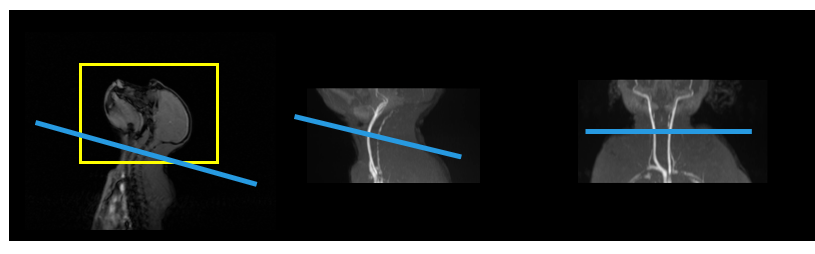
Subjects: Animal experiments were performed following a protocol previously approved by the Ethics and Biosafety Committee according to guidelines from the University of Navarra and government of Navarra. 12 rhesus monkeys (2-3 years old, mean weight ± standard deviation (SD)=2.432±0.169 Kg, 4 male) were employed in this study. The animals were initially anesthetized with 0.1 ml/kg of Ketamine (Ketamidor®, Ritcher Pharma,Wels, Austria) and 0.16 ml/kg Midazolam (Midazolam Normon®, Laboratorios Normon S.A, Madrid, Spain) administered intramuscularly, To keep them asleep, half doses of both anesthetics were administered every 30-40 minutes.MRI Protocol: Scans were performed on a 3T Skyra whole body scanner (Siemens Healthcare, Erlangen, Germany) using a 32-channel head-array coil. Pads were used for head stabilization. Animal was positioned in prone position and breathed on their own during the study. Pseudo-continuous ASL (pCASL) was implemented for CBF measurement with 2.2-mm isotropic resolution with a 3D GRASE readout and with background suppression. PCASL was unbalanced with average gradient=1,0 mT/m. Labeling duration was 2 s and PLD was 1 s. 24 pairs of control and labeling images were acquired. 3D time-of-flight (TOF) angiography was acquired to identify large vessels for placing the ASL labelling plane (Figure 1) using TR=22 ms, flip angle =18°, TE=3.67 ms, slice thickness=1 mm, FOV=200 × 150 mm, matrix= 384 × 269, 40 slices, and single average. T1 anatomical images were obtained using three-dimension (3D) magnetization-prepared rapid gradient-echo (MP-RAGE) sequence on the same imaging slices, FOV and matrix size= 112 mm x 112 mm, with TR=2300 ms, TE=3.69ms.
Image processing: Images were analysed using custom scripts in MATLAB (The MathWorks, Inc). CBF maps in ml/min/100g were computed using the single compartment model (Eq. 1) with the following simplified formula and parameters: $$CBF\ \left(\frac{\frac{ml}{min}}{100g\ }\right)= \frac{6000\lambda\ \left(SI_C\ {-\ SI}_L\right)\ {e\ }^{\left(\frac{PLD}{T1_b}\right)}}{2\ \alpha\ T1\ M0\ \left(1-e^{\left(\frac{-\tau}{T1_b}\right)}\right)}$$ where λ is the water brain–blood partition coefficient, α is the arterial spin-labeling efficiency, SIC and SIL are signal intensities of the non-labeled and labeled images, respectively, T1b is the longitudinal relaxation time (T1) of the arterial blood at 3T, PLD is the post-labeling delay and LD is the labeling duration. Briefly, α=0.60, λ=0.9 ml/g, T1b=1.66 s, LD=2 s, PLD=1 s. ASL pairs were corregistered to T1 image by the M0 image with Statistical Parametric Mapping version 12 (SPM12, The Wellcome Centre for Human Neuroimaging, UCL Queen Square Institute of Neurology, London, UK). Regional values for CBF were obtained in 28 circular ROIs of 5-mm in diameter based on T1 weighted images: two in frontal, three in temporal, two in parietal, two in occipital, two in caudate nucleus and two in putamen cortical regions, and three in subcortical regions; data were recorded for each hemisphere. A 2-way ANOVA was performed comparing CBF (ml/min/100g) between genders for frontal, temporal, parietal, occipital, caudate nucleus, putamen, and subcortical regions. Analysis were performed on RStudio (Posit Software, MA, USA).
Results
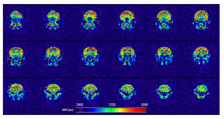
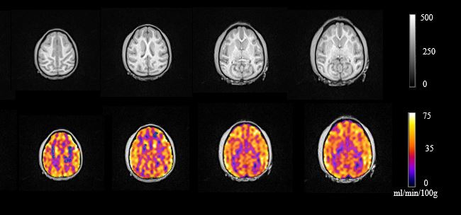
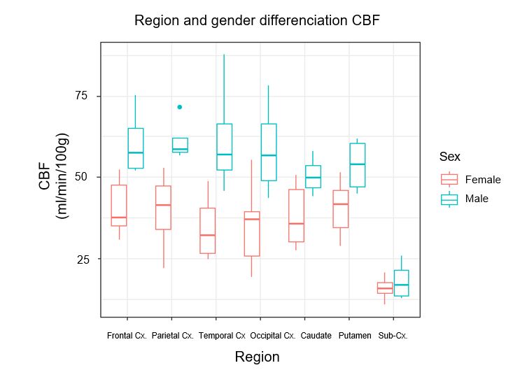
Clear visually differentiation between gray matter (GM) and white matter (WM) can be shown in the perfusion map along different slices (Figure 2) and in CBF map obtained with pCASL technique at 3T scanner (Figure 3). Caudate nucleus and putamen can be differentiated in T1-anatomical weighted image. Table 1 shows global and regionally CBF values for female and male rhesus monkeys, with GM/WM ratio of 2.7 and 3.2 for female and male respectively. Surprisingly, Male CBF values were higher than female ones in every single region (Figure 4).Discussion
Our present finding demonstrated significant CBFchanges across different cortical regions (p<0.001) and between gender (p<0.001) with higher GM and WM CBF values in male animals than female animals. Our CBF values (Table1), as well as GM/WM ratio matched previously reported values of and GM CBF value of 56–68 ml/100 g/min in the cortices and a WM CBF value of 34 ml/100 g/min in ketamine-anesthetized monkeys5.Conclusion
Although midazolam anesthetic has been scarcely used in NHP studies, the results of this study suggest that CBF measured using PCASL technique under ketamine and midazolam anesthesia could be optimal in future neurologic studies.Acknowledgements
Leyre Garcia-Ruiz received Ph.D. grant support from Government of Navarra.References
1. Zhang X. Effects of Anesthesia on Cerebral Blood Flow and Functional Connectivity of Nonhuman Primates. Vet Sci. 2022 Sep 22;9(10):516.
2. Zeiler FA, Sader N, Gillman LM, Teitelbaum J, West M, Kazina CJ. The Cerebrovascular Response to Ketamine: A Systematic Review of the Animal and Human Literature. J Neurosurg Anesthesiol. 2016 Apr;28(2):123-40.
3. Forster A, Juge 0, Morel D. 1982_Foster_Anesthesiology_Midazolam effects human brain. Anesthesiology. 1982;56:453–5.
4. Li CX, Patel S, Wang DJJ, Zhang X. Effect of high dose isoflurane on cerebral blood flow in macaque monkeys. Magn Reson Imaging. 2014;32(7):956–60.
5. Enlund M, Andersson J, Hartvig P, Valtysson J, Wiklund L. Cerebral normoxia in the rhesus monkey during isoflurane- or propofol- induced hypotension and hypocapnia, despite disparate blood-flow patterns. A positron emission tomography study. Acta Anaesthesiol Scand. 1997;41(8):1002–10.
Figures