3542
Inhomogeneous Magnetization Transfer (ihMT) of Peripheral Nerve in Patients with Chronic Inflammatory Demyelinating Polyradiculoneuropathy1Department of Radiology, Chiba university hospital, Chiba, Japan, 2Diagnostic Radiology and Radiation Oncology, Graduate School of Medicine, Chiba University, Chiba, Japan, 3Philips Japan, Tokyo, Japan, 4Philips Canada, Mississauga, ON, Canada
Synopsis
Keywords: Neurodegeneration, CEST & MT
Chronic inflammatory demyelinating polyradiculoneuropathy (CIDP) is an immune-mediated demyelinating disease. The purpose of this study is to evaluate the usefulness of ihMT in the diagnosis of CIDP by measuring myelin levels using ihMT imaging. ihMTR of the sciatic nerve of CIDP patients was significantly lower than that of the normal subjects. Our finding was compatible with previously reported pathological changes. Myelin imaging by ihMT has the potential to be used as a new biomarker in the diagnosis of CIDP.Introduction
Chronic inflammatory demyelinating polyradiculoneuropathy (CIDP) is an immune-mediated disease demyelinating1-3. The pathologic hallmark of CIDP is loss of the myelin sheath of the peripheral nerves4. In the diagnosis of CIDP, the electrophysiology study (EPS) is essential. However, EPS indirectly reflects demyelination and cannot evaluate the proximal nerves. Nerve biopsy, which allows direct observation of demyelination, has very limited indications and is not always sensitive to demyelination. On the other hand, nerve enlargement, increased signal, and gadolinium enhancement have been reported on MRI. MRI is considered a supportive criterion for the European Federation of Neurological Societies/Peripheral Neurological Society (EFNS/PNS) diagnosis5,6. The specificity of the current diagnostic criteria for CIDP is low. This is due to the lack of specific biological biomarkers for CIDP that can be used as a definitive diagnosis. Therefore, we focused on myelin imaging as a method to evaluate demyelination. Myelin imaging includes inhomogeneous magnetization transfer (ihMT) combined sequencing, a new endogenous imaging technique with high specificity for myelin7, which has been reported to be useful in evaluating the pathophysiology of multiple sclerosis8,9. Multiple sclerosis is a disease that causes demyelination as well as CIDP. Therefore, we thought it could be applied to CIDP. Therefore, in the present study, we evaluated the usefulness of ihMT in diagnosing CIDP by measuring myelin levels using ihMT imaging.Methods
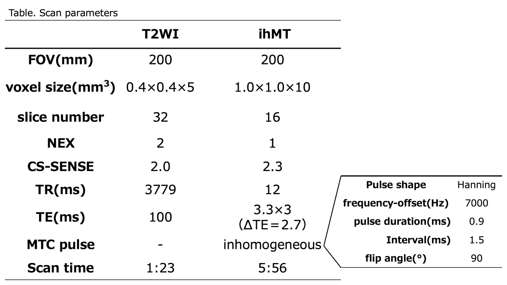
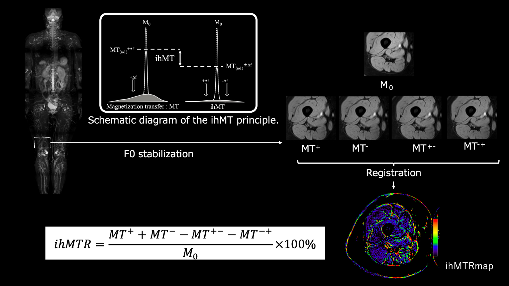
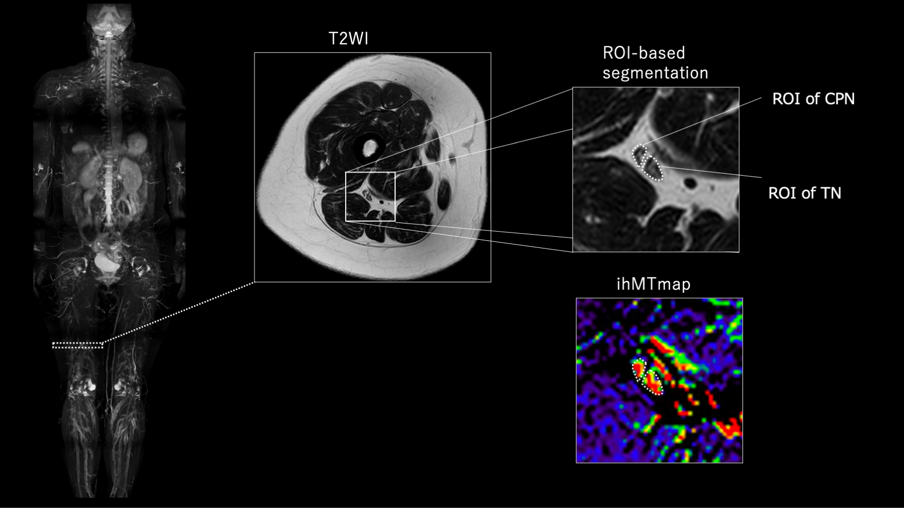
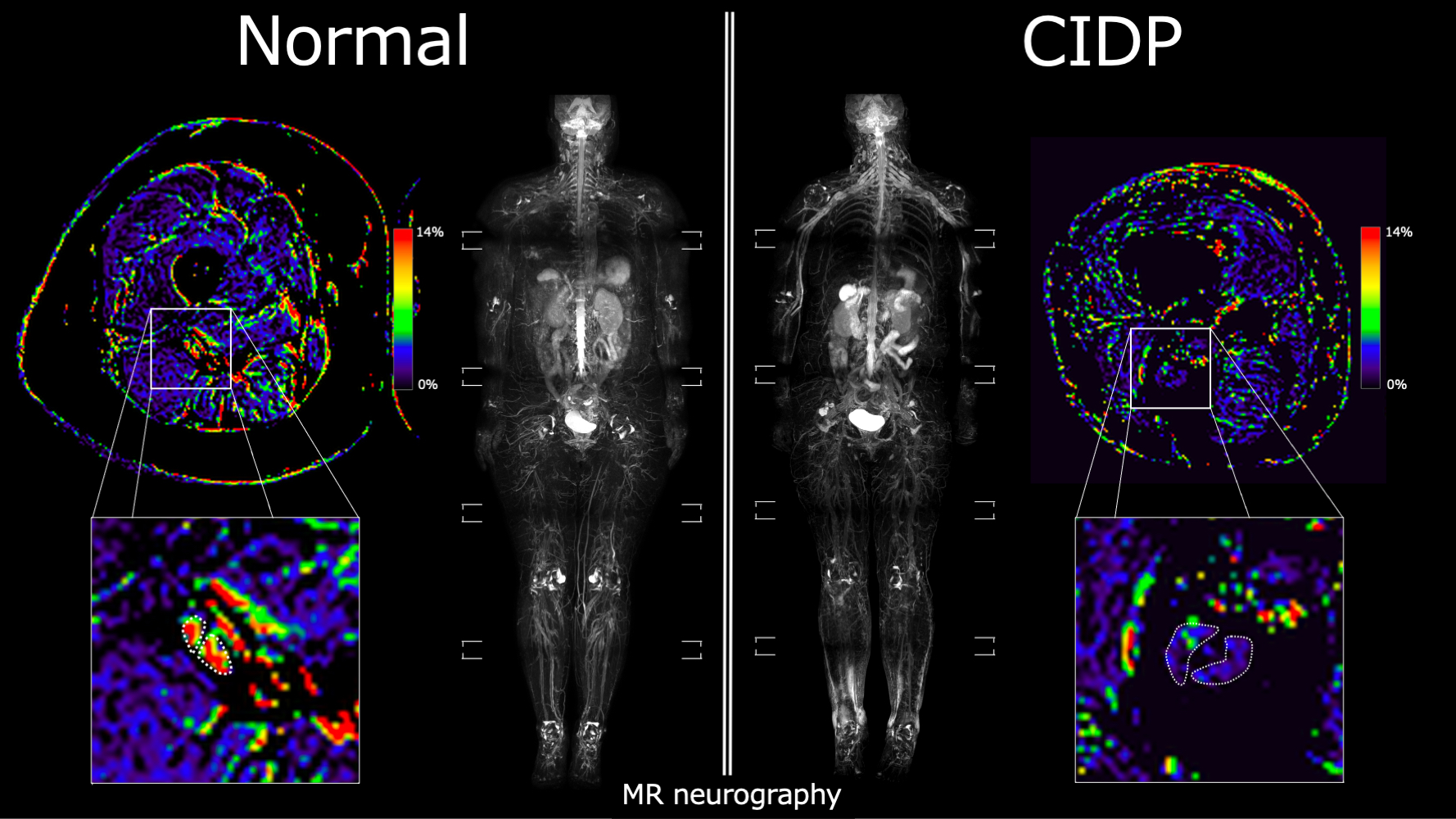
Four patients ( 2 females and 2 males; median age, 58; range 54-78) with CIDP and five normal subjects ( 2 females and 3 males; median age, 32; range 30-51) were included in this study. CIDP was diagnosed according to the guideline of PNS5. There were three cases of typical CIDP and one case of atypical CIDP (distal CIDP). MRI was performed on a 3.0-Tesla MR system (Philips Ingenia). ihMT images and T2-weighted images (T2WI) for position matching were acquired of the distal right thigh (up to 16 cm proximal to the patella) using a torso coil. The scanning parameters are summarized in Figure 1. To improve the reliability of the quantitative values, ihMT was subjected to a combination of stabilization and non-rigid registration. Stabilization corrects the center frequency (F0) during scanning. Non-rigid registration corrects retrospectively for moving objects (Figure 2). The quantitative value of ihMT was calculated by ihMT ratio (ihMTR) defined in Figure 2. Measurements were performed by a board-certified radiologist. An irregularly shaped region of interest (ROI) was placed to match the contours of the tibial and common peroneal nerves from the slice in which the nerve is most clearly delineated with reference to T2WI (Figure 3). ihMTR was the average of the two measurements of the tibial and common peroneal nerves. The statistical biases were defined as significant with P-values of less than 0.05 of a Mann-Whitney U test.Results
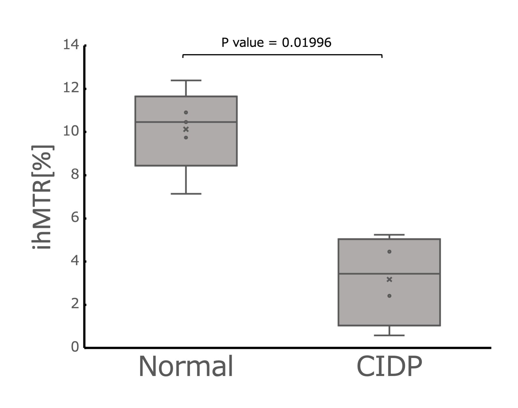
The results of ihMTR in the sciatic nerve of normal subjects and CIDP patients are shown in Figures 4 and 5. ihMTR of the sciatic nerve of CIDP patients was significantly lower than that of the normal subjects. (median 3.44% [IQR 1.04-5.04%] vs. median 10.46% [IQR 8.44-11.65%], P=0.01996)Discussion
Our finding that ihMTR was decreased in the sciatic nerves of CIDP patients is compatible with previously reported pathological changes. Multifocal inflammatory and demyelinating processes involving the peripheral nerve in a diffuse manner are causative factors in the development of CIDP, and demyelination and remyelination are pathological hallmarks of CIDP4. Therefore, imaging methods with high specificity for myelin may be useful for the diagnosis of CIDP. Prior studies have reported the utility of quantitative assessment in diffusion tensor imaging (DTI) of the tibial nerve in CIDP patients and controls10-12. DTI can assess changes in the microstructure of nerve fibers by various parameters. In particular, fractional anisotropy values are said to be significantly lower in the nerves of CIDP patients than in controls13. Although, DTI assesses axonal degeneration induced by chronic demyelinating lesions and does not directly reflect myelin status. On the other hand, ihMTR can directly assess myelin levels and may be able to assess conditions before axonal degeneration.Determining the efficacy of treatment for CIDP has a significant impact on treatment decisions, and it is recommended that decisions be based on quantification. An example of a quantitative assessment method is a clinical rating scale the Overall Neuropathy Limitation Scale. However, this measures the degree of physical activity and cannot assess demyelinating changes. EPS is also considered useful, but the reproducibility of the tests is not always good and that is difficult to assess the proximal nerve trunk14. ihMT has the potential to overcome the weaknesses of conventional methods of assessing CIDP and can be used as a new biomarker for diagnosing CIDP.
Several limitations should be noted in this study. First, our study population was relatively small. Second, we could not directly correlate ihMTR results with nerve histology and EPS. Third, the ihMTR had a large variation in quantitative values due to noise contamination caused by slight movements. Further study is needed to acquire ihMT with a higher signal-to-noise ratio.
Conclusion
Myelin imaging by ihMT has the potential to be used as a new biomarker in the diagnosis of CIDP.Acknowledgements
No acknowledgement found.References
1. Nobile-Orazio E, et al: Chronic inflammatory demyelinating polyradiculoneuropathy and variants: where we are and where we should go. J Peripher Nerv Syst 2014;19:2–13.
2. Dimachkie MM, et al: Chronic inflammatory demyelinating polyneuropathy. Curr Treat Options Neurol 2013;15:350–366.
3. Vedeler CA, et al: Chronic inflammatory demyelinating polyneuropathy (CIDP). Acta Neurol Scand Suppl. 2013;(196):48-51.
4. Vallat JM, et al: Chronic inflammatory demyelinating polyradiculoneuropathy: diagnostic and therapeutic challenges for a treatable condition. Lancet Neurol 2010;9:402–412.
5. Van den Bergh PY, et al; European Federation of Neurological Societies/Peripheral Nerve Society guideline on management of chronic inflammatory demyelinating polyradiculoneuropathy: report of a joint task force of the European Federation of Neurological Societies and the Peripheral Nerve Society - first revision. Eur J Neurol. 2010; 17: 356–363.
6. Joint Task Force of the EFNS and the PNS. European Federation of Neurological Societies/Peripheral Nerve Society Guideline on management of chronic inflammatory demyelinating polyradiculoneuropathy. Report of a joint task force of the European Federation of Neurological Societies and the Peripheral Nerve Society. J Peripher Nerv Syst 2005;10:220e8.
7. Varma G, et al: Magnetization transfer from inhomogeneously broadened lines: A potential marker for myelin. Magn. Reson. Med. 73, 614–622.
8. Van Obberghen E, et al: Evaluation of the Sensitivity of Inhomogeneous Magnetization Transfer (ihMT) MRI for Multiple Sclerosis. AJNR Am J Neuroradiol. 2018 Apr;39(4):634-641.
9. Van Obberghen E, et al: Inhomogeneous Magnetization Transfer (ihMT) in normal-appearing tissue correlates with disability of multiple sclerosis patients. Neurology.2017 Apr; 88 (16 Supplement) P4.360
10. Guggenberger R, Markovic D, Eppenberger P, et al. Assessment of median nerve with MR neurography by using diffusion-tensor imaging: normative and pathologic diffusion values. Radiology 2012; 265:194–203.
11. Mathys C, Aissa J, et al: Peripheral neuropathy: assessment of proximal nerve integrity by diffusion tensor imaging. Muscle Nerve 2013; 48:889–896.
12. Lehmann HC, et al: Diffusion tensor imaging to assess axonal regeneration in peripheral nerves. Exp Neurol 2010; 223:238–244.
13. Kronlage M, et al: Diffusion tensor imaging in chronic inflammatory demyelinating polyneuropathy. Invest Radiol 2017; 52:701–707.
14. Eftimov, Filip, et al: Diagnostic Challenges in Chronic Inflammatory Demyelinating Polyradiculoneuropathy. Brain 143, no. 11 (2021): 3214–24.
Figures