3529
Association of peripheral inflammation with disrupted brain functional network topology in bipolar disorder1First Affiliated Hospital of Jinan University, Guangzhou, China, 2MR Research, GE Healthcare, Beijing, China
Synopsis
Keywords: Neuroinflammation, fMRI (resting state)
This study provided preliminary evidence of the association between disrupted brain functional network topology and neuroinflammation in BD. The current study demonstrated disrupted topological organization in the whole brain and regional connectivity was associated with inflammatory cytokines of the IL-4, IL-8 and IL-10 levels in BD. Moreover, our results indicated that higher IL-4 levels and impaired regional connectivity in the temporal pole may be associated with the severer depressive symptoms in BD.Background
Recent studies have indicated that inflammation may contribute to the etiology and progression of BD [1, 2]. Although evolving structural and functional neuroimaging studies have reported abnormalities in specific brain regions and connections in BD, the neuroimmunology of BD is not fully understood. Graph theoretical methods have been extensively applied to resting-state functional magnetic resonance imaging (rs-fMRI) to study the topological organization of functional brain networks [3]. However, until now, there is no study concentrating on the correlation between functional brain network topology and peripheral inflammation in BD. In this study, we tried to explore the disrupted brain functional network topology, peripheral cytokines and their correlations to demonstrate the role of inflammation in brain functional network topology in BD.Methods
In this study, 53 BD patients and 47 healthy controls (HCs) underwent resting-state magnetic resonance imaging scans. Graph theory analysis was performed to investigate the topological properties of whole-brain functional connectome at both global and nodal levels. Serum levels of interleukin-4 (IL-4), interleukin-6 (IL-6) interleukin-8 (IL-8), interleukin-10 (IL-10), and tumor necrosis factor-α (TNF-α) were measured in all participants. Correlations between topological properties, clinical variables and cytokines levels in BD were calculated. Furthermore, mediation analysis was employed to determine whether inflammatory cytokines affect depressive symptoms by affecting brain network properties.Results
Global and nodal parameters of the brain functional networkFigure 1 shows the global parameters (the AUC of Cp, Lp, λ, γ, σ , Eglo, and Eloc) for the BD and HCs group. Statistical analyses revealed that BD group showed significantly greater AUC of Lp (t = 2.130, p = 0.036, FDR-p = 0.084) and λ (t = 2.919, p = 0.004, FDR-p = 0.028), and lower AUC of Eglo (t = -2.125, p = 0.036, FDR-p = 0.084) when compared with HCs group. Figure 2 shows the significantly different brain regions in nodal parameters (the AUC of Degi and Enodal) between BD group and HCs group (p < 0.0086). Of note, we found lower Degi in the bilateral lingual, lower Enodal in the right superior frontal gyrus (SFG), left middle temporal pole (MTP), right postcentral gyrus, and bilateral lingual in BD group when compared with HCs group.
Inflammatory cytokines levels between two groups
The BD group showed greater levels of IL-4 , IL-6, IL-8, IL-10 when compared with the HCs group.
Correlation analyses
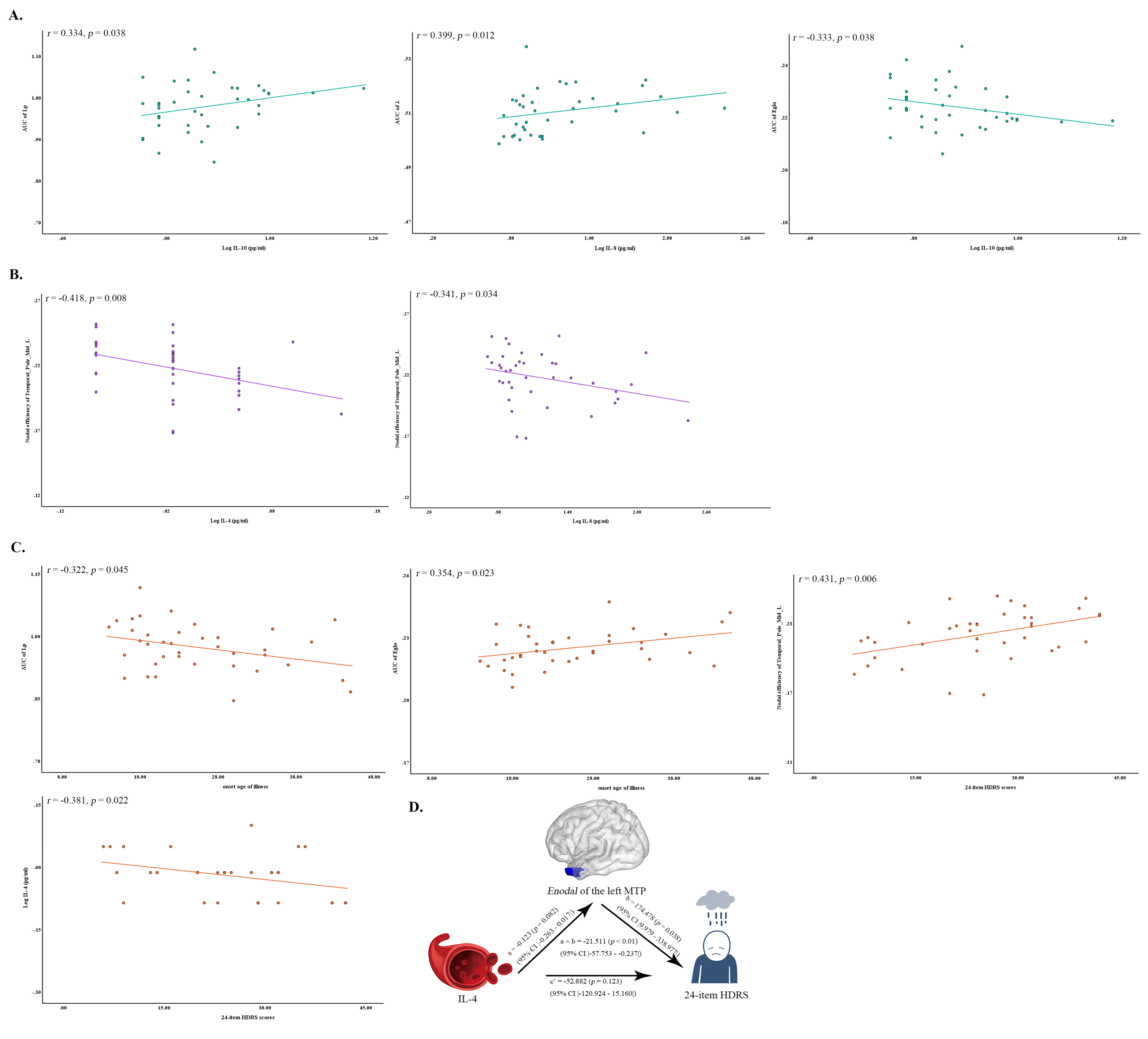
For the correlations between abnormal global parameters and inflammatory cytokines levels, in BD group, the AUC of Lp was significantly positively correlated with IL-10 level (r =0.334, p = 0.038), and the AUC of λ was significantly positively correlated with IL-8 level (r = 0.399, p = 0.012), and the AUC of Eglo was significantly inversely correlated with IL-10 (r = -0.333, p = 0.038) level (Figure 3A).
For the correlation between abnormal nodal parameters and inflammatory cytokines levels, in BD group, the AUC of Enodal of the left MTP was significantly negatively correlated with IL-4 level (r = -0.418, p = 0.008) and IL-8 level (r = -0.341, p = 0.034) (Figure 3B).
For the correlation between abnormal network parameters and clinical variable, the AUC of Lp was significantly inversely correlated with onset age of illness (r = -0.322, p = 0.045), and the AUC of Eglo was significantly positive with onset age of illness (r = 0.364, p = 0.023), and the AUC of Enodal of the left MTP was significantly positively correlated with 24-item HDRS scores (r = 0.431, p = 0.006) (Figure 3C).
For the correlation between abnormal inflammatory cytokines levels and clinical variable, IL-4 level was significantly negatively correlated with 24-item HDRS scores (r = -0.381, p = 0.022) (Figure 3C).
“Inflammatory-network-depression” mediation pathway analysis
Mediation analysis reported that the IL-4 level was associated with 24-item HDRS scores (total effect: β = -74.393, t = -2.2213, p = 0.034, 95% CI: [-142.776, -6.010]), and there was significant full mediation effect by Enodal of the left MTP (indirect effect: p < 0.01, β= -21.511, 95%CI: [-57.753, -0.237]) for the association between IL-4 level and on 24-item HDRS scores. After considering the significant effect via Enodal of the left MTP, the direct effect of IL-4 level on 24-item HDRS scores did not significant (β = -52.882, t = -1.583, p = 0.123, 95% CI: [-120.924, 15.160]) (Figure 3D).
Conclusion
In conclusion, our study provided preliminary evidence of the association between disrupted brain functional network topology and neuroinflammation in BD. The current study demonstrated disrupted topological organization in the whole brain and regional connectivity were associated with inflammatory cytokines of the IL-10, IL-8 and IL-4 levels in BD. Moreover, our results indicated that higher IL-4 levels and impaired regional connectivity in the temporal pole may be associated with the severer depressive symptoms in BD.Acknowledgements
The study was supported by grants from the National Natural Science Foundation of China (81671670, 81971597, and 82172530); National Key Research and Development Project (2020YFC2005700); Key-Area Research and Development Program of Guangdong Province (2020B1111100001). The funding organizations play no further role in study design, data collection, analysis and interpretation and paper writing.
References
1. Berk, M., et al., So depression is an inflammatory disease, but where does the inflammation come from? BMC Med, 2013. 11: p. 200.
2. de Melo, L.G.P., et al., Shared metabolic and immune-inflammatory, oxidative and nitrosative stress pathways in the metabolic syndrome and mood disorders. Prog Neuropsychopharmacol Biol Psychiatry, 2017. 78: p. 34-50.
3. Bullmore, E. and O. Sporns, Complex brain networks: graph theoretical analysis of structural and functional systems. Nat Rev Neurosci, 2009. 10(3): p. 186-98.
4. Yan, C.G., et al., DPABI: Data Processing & Analysis for (Resting-State) Brain Imaging. Neuroinformatics, 2016. 14(3): p. 339-51.
5. Zhang, J., et al., Disrupted brain connectivity networks in drug-naive, first-episode major depressive disorder. Biol Psychiatry, 2011. 70(4): p. 334-42.
Figures
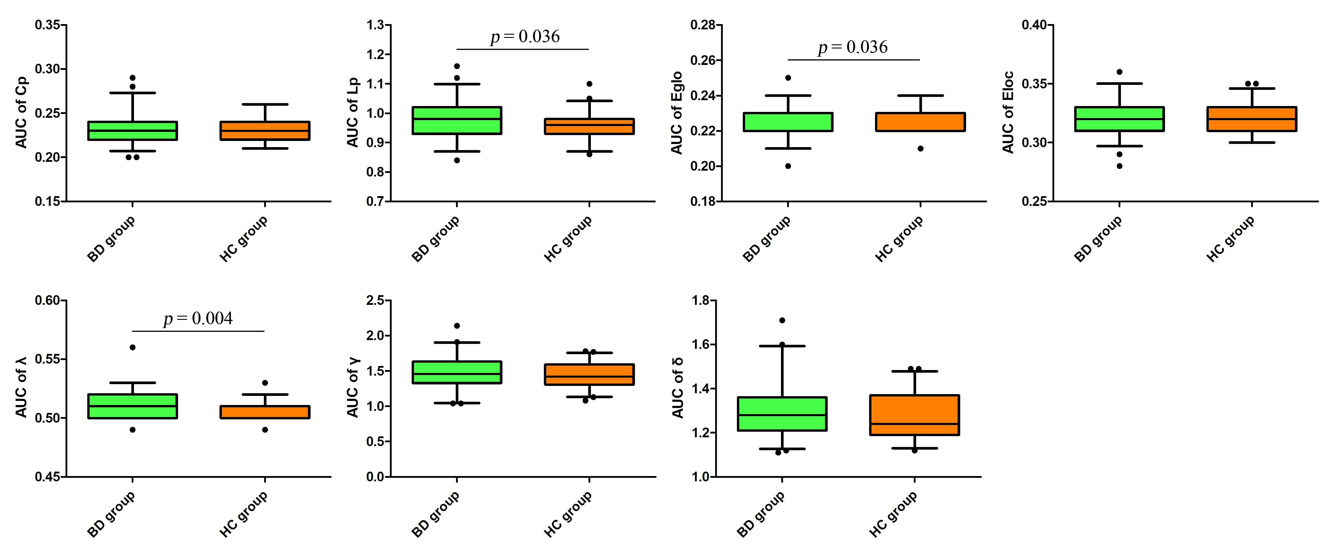
Fig 1. The global parameters (the AUC of Cp, Lp, λ, γ, σ, Eglo, and Eloc) for the BD and HCs group. BD group showed significantly increased AUC of Lp and λ, and decreased AUC of Eglo when compared with HCs group (p < 0.05).
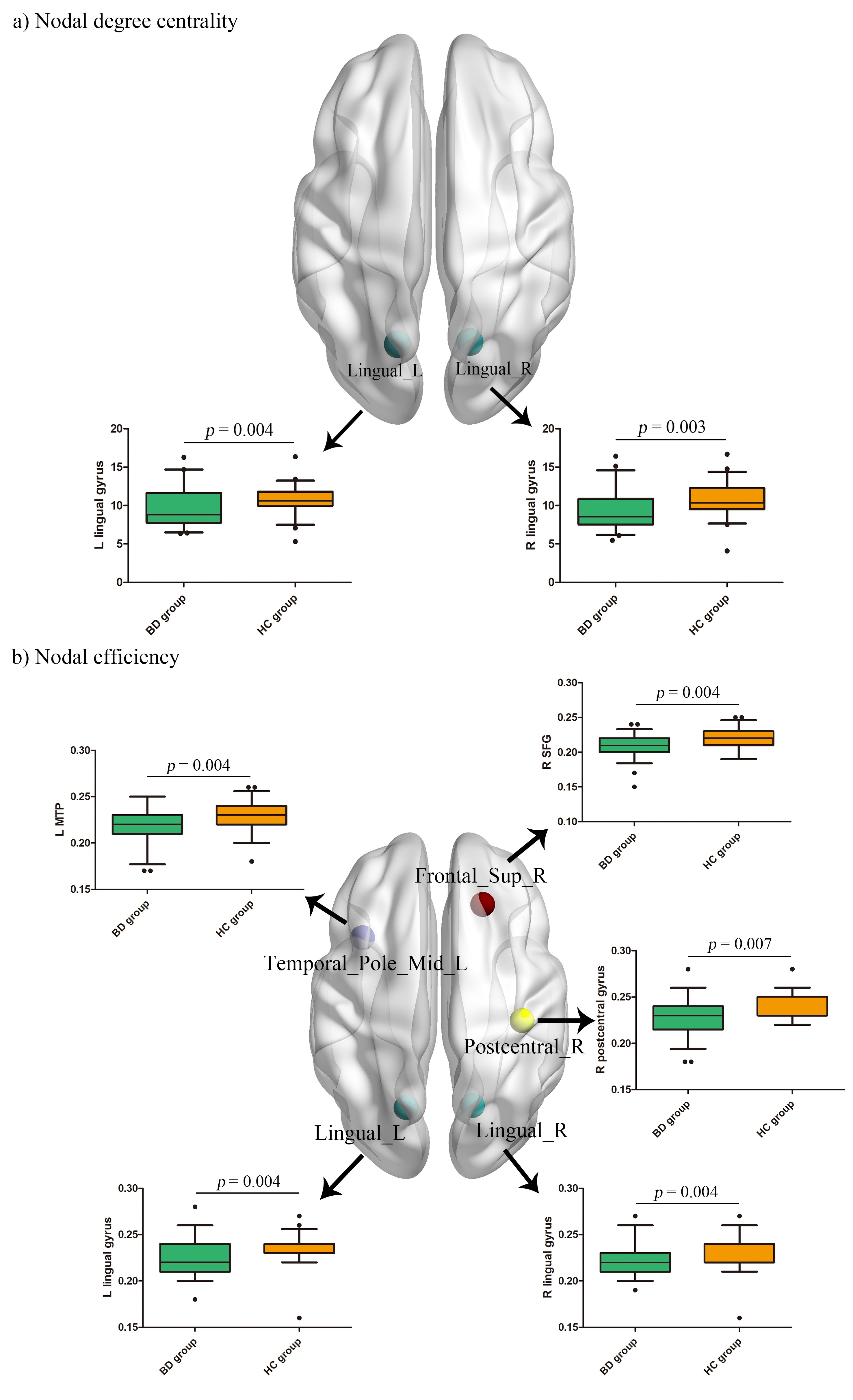
Fig 2. The significantly different brain regions in nodal parameters (the AUC of Degi and Enodal) between BD group and HCs group (p < 0.0086). The results showed decreased Degi in the bilateral lingual, decreased Enodal in the right superior frontal gyrus (SFG), left middle temporal pole (MTP), right postcentral gyrus, and bilateral lingual in BD group when compared with HCs group. L (R), left (right) hemisphere.