3527
Hippocampal metabolites in patients with amnestic mild cognitive impairment1Shandong Provincial Hospital Affiliated to Shandong First Medical University, Jinan, China, 2Philips Healthcare, Shanghai, China
Synopsis
Keywords: Alzheimer's Disease, Alzheimer's Disease
Amnestic mild cognitive impairment (aMCI) is a precursor to Alzheimer's disease (AD). The neurometabolic changes especially the neurotransmitters in the hippocampus contributed the course of AD. In-vivo magnetic resonance spectroscopy (MRS) can be used to measure brain metabolites noninvasively. In this study, we aim to explore the changes of hippocampal metabolic changes in aMCI patients using PRESS and MEGA-PRESS. aMCI patients exhibited decreased hippocampal Glx levels from MEGA-PRESS, having low concordance with PRESS.Introduction
Amnestic mild cognitive impairment (aMCI) is a precursor to Alzheimer's disease (AD). The neurometabolic changes especially the neurotransmitters in the hippocampus contributed the course of AD. In-vivo magnetic resonance spectroscopy (MRS) can be used to measure brain metabolites noninvasively. In this study, we aim to explore the changes of hippocampal metabolic changes in aMCI patients using PRESS and MEGA-PRESS.Methods
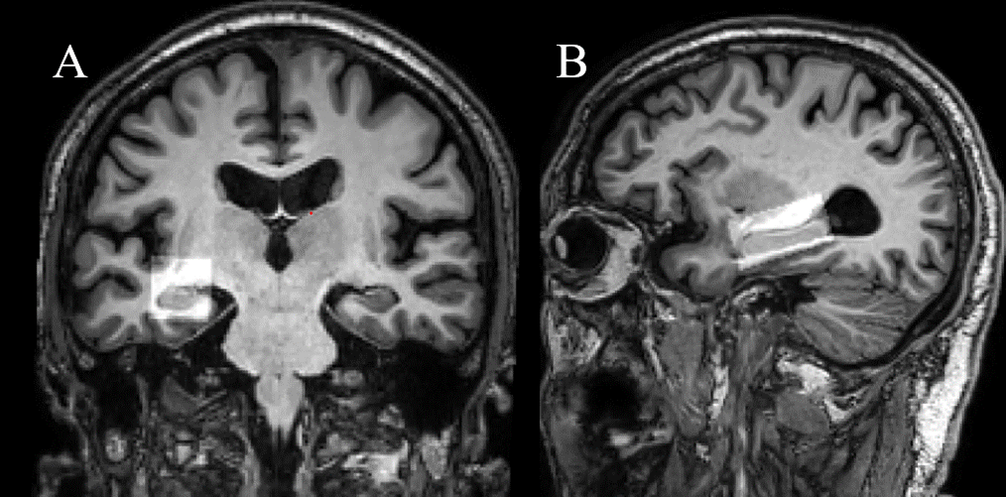
Twelve aMCI patients and 12 healthy participants were recruited. Metabolite spectra in the right hippocampus were acquired using PRESS and MEGA-PRESS on a 3 T MR scanner prospectively. The hippocampus neurometabolic levels (GABA+, Glu, Glx, Cr, GSH, NAA and tNAA) of aMCI patients were compared with healthy participants using Mann–Whitney U test.Results
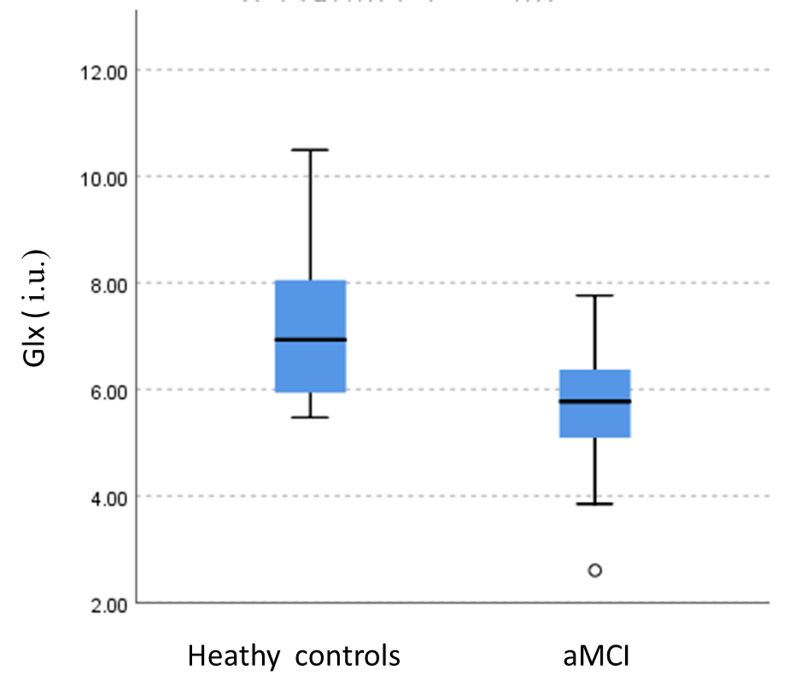
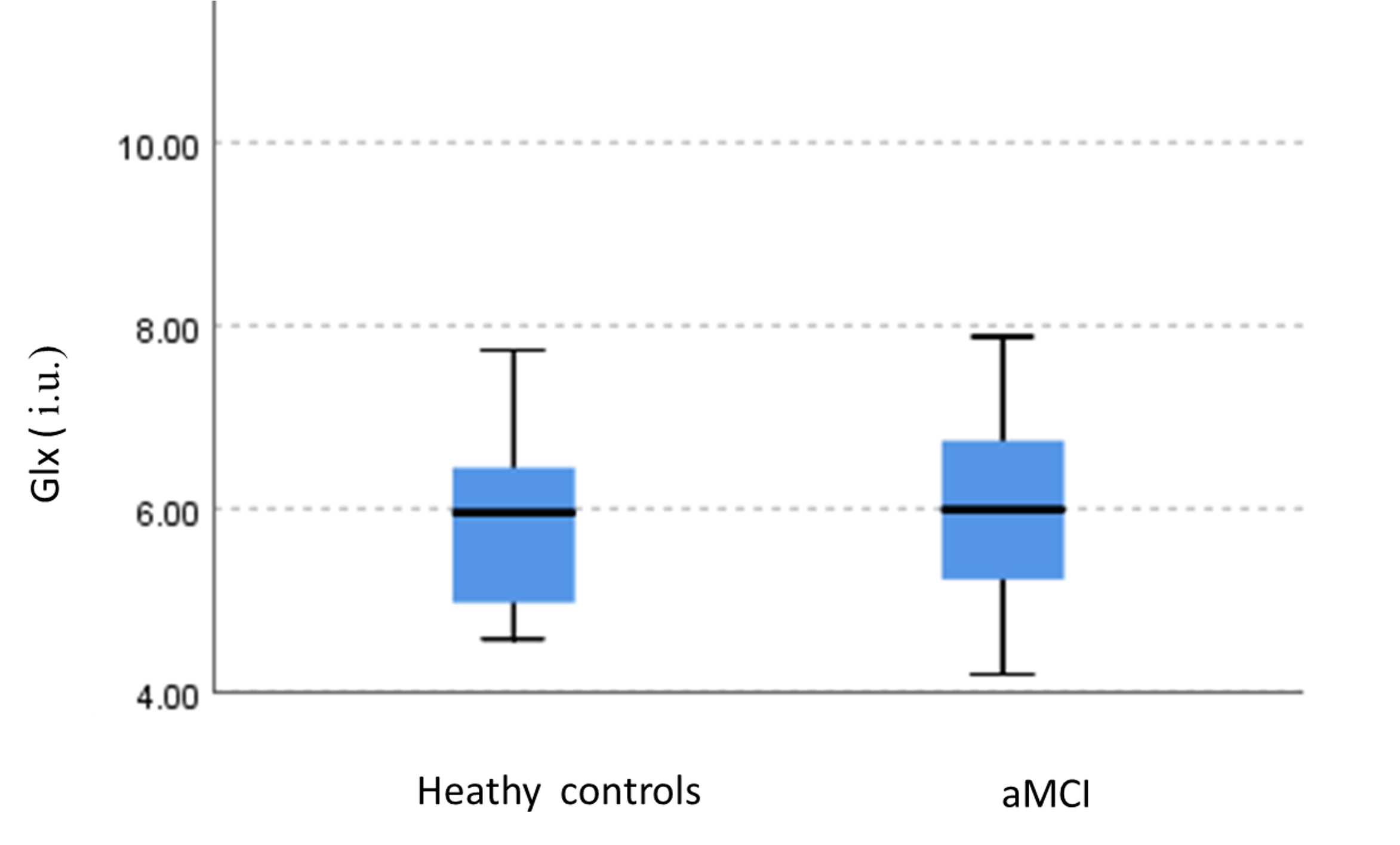
Glx (the combined signals of Glu and Gln) levels obtained using MEGA-PRESS were statistically significantly lower in right hippocampus of aMCI patients compared to heathy participants (6.95 ± 1.52 i.u. vs. 5.44 ± 1.43 i.u., P=0.022). However, Glx levels obtained using PRESS have no significant decreasing trends (6.14 ±0.94 i.u. vs. 5.78 ±1.08 i.u., P=0.285). The concordance between PRESS and MEGA-PRESS sequences was low (r=0.122, p=0.625). No significant difference of GABA, Glu, Cr, GSH, NAA and tNAA levels were observed between aMCI patients and healthy participants (P= 0.585, 0.709, 0.796, 0.546, 0.625, 0.323).Conclusion
aMCI patients exhibited decreased hippocampal Glx levels from MEGA-PRESS, having low concordance with PRESS.Discussion
According to the previous studies, glutamatergic dysfunction in the hippocampal drives the pathogenesis of AD and may be prominently affected in MCI stage. The concordance between PRESS and MEGA-PRESS sequences was low, which was consistent with Tamar’s study. However, in our study, the sample size was small, only 120 aMCI patients were enrolled in this study, so further large-scaled prospective trials are still warranted to verify this result.Acknowledgements
NoneReferences
1. Oltra-Cucarella J, Ferrer-Cascales R, Alegret M, Gasparini R, Diaz-Ortiz LM, Rios R, et al. Risk of progression to Alzheimer's disease for different neuropsychological Mild Cognitive Impairment subtypes: A hierarchical meta-analysis of longitudinal studies. Psychol Aging. 2018;33(7):1007-21.
2. Nilsen LH, Melo TM, Saether O, Witter MP, Sonnewald U. Altered neurochemical profile in the McGill-R-Thy1-APP rat model of Alzheimer's disease: a longitudinal in vivo 1 H MRS study. J Neurochem. 2012;123(4):532-41.
3. Harris AD, Puts NA, Edden RA. Tissue correction for GABA-edited MRS: Considerations of voxel composition, tissue segmentation, and tissue relaxations. J Magn Reson Imaging. 2015;42(5):1431-40.
4. an Veenendaal, T. M., Backes, W. H., van Bussel, F., Edden, R., Puts, N., Aldenkamp, A. P., & Jansen, J. (2018). Glutamate quantification by PRESS or MEGA-PRESS: Validation, repeatability, and concordance. Magnetic resonance imaging, 48, 107–114.
Figures