3521
Measurement of blood-brain barrier leakage in AD transgenic mice with aging: Improving sensitivity with capillary input function1Vilcek Institute of Graduate Biomedical Science, NYU School of Medicine, New York, NY, United States, 2Center for Advanced Imaging Innovation and Research, Radiology, NYU School of Medicine, New York, NY, United States, 3Radiology, Weill Cornell Medical College, New York, NY, United States, 4Neurology, NYU Langone Health, New York, NY, United States, 5Weill Cornell Medical College, New York, NY, United States
Synopsis
Keywords: Alzheimer's Disease, Aging
Recent studies have suggested that the increase in blood-brain barrier (BBB) permeability is associated with both aging and the progression of the Alzheimer’s disease (AD). However, the association of the amyloid pathology with the increased BBB leakage at different disease progression is still poorly understood. In this study, we performed a cross-sectional study to investigate the BBB permeability changes in AD transgenic mice with aging. We also propose the network-aided analysis allows the scan time reduction without compromising the accuracy of the detection of the subtle permeabilityPurpose
Recent studies have shown that increased blood-brain barrier (BBB) permeability is associated with aging (1), as well as the Alzheimer’s disease (AD) (2). However, these human studies are limited by challenges in identifying the causes of the vascular changes, because of the difficulties of conducting longitudinal studies and obtaining tissue samples. Animal models for the Alzheimer’s disease allow to segregate the different aspect of disease and its effect in pathological development. Although several studies identify the physiological changes associated with aging (3, 4), these imaging studies are conducted on relatively aged mice, which hinders the understanding of pathological development with aging. In this study, we conduct a cross-sectional study at a wide range of ages and investigate how the progression of the amyloid pathology contributes to the vascular changes along different ages. In addition, we adopt the previously proposed deep-learning approach(5) to demonstrate the improved sensitivity in detecting subtle BBB permeability changes with the clinically relevant scan time.Methods
AnimalsGroups of 5xFAD mice (n=6, Female/male (n = 3/3)) and the wild-type littermate (n=4, Female/male (n = 1/3)) were included in this study. 5xFAD mice express human Amyloid Precursor Protein (APP) and PSEN1 transgene, which drive an aggressive amyloid pathology(6). The age for the mice ranged between 4 to 16 months at the time of scan.
MRI experiment
The dynamic contrast-enhanced (DCE) MRI study using a 3D UTE pulse sequence with 3D golden angle projections were performed on a Bruker 7T micro-MRI system with a cryo-coil. (TR=5ms, TE=0.028ms, Flip-angle = 10 deg, Image Matrix=128x128x128, FOV=17x17x17mm3). The total scan time was 30min, while a bolus of gadolinium (Gadavist) contrast agent was injected at 2min into the scan. The acquired dynamic images were reconstructed at 5s temporal resolution using iterative GRASP reconstruction(7).
Pharmacokinetic Modelling analysis (PKM)
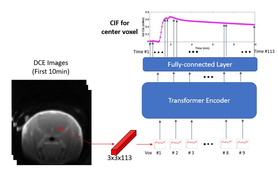
The PKM analysis was conducted on a single slice that contains the hippocampal region. The analysis was conducted on the whole-brain ROI. The arterial input function (AIF) was sampled using a DCE toolkit known as ROCKETSHIP(8). The capillary-input function (CIF) was estimated from the vision transformer-based deep learning network, which was trained on the simulated contrast dynamics from human studies. The CIF network receives an input of a patch of contrast dynamics, as shown in Figure 1, and predicts the local input function to that patch, as elucidated in our previous work(5). The graphical Patlak model (9) was used to assess the BBB permeability. The analysis was performed using AIF on 30-min data and truncated 10-min data. The same analysis was repeated on the 10-min data with the network-predicted CIF. The estimated permeability was compared among AIF-30min data (Ca-30min), AIF-10min data (Ca-10min) and CIF-10min data (Cp-10min).
Result
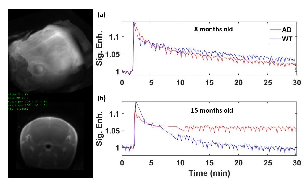
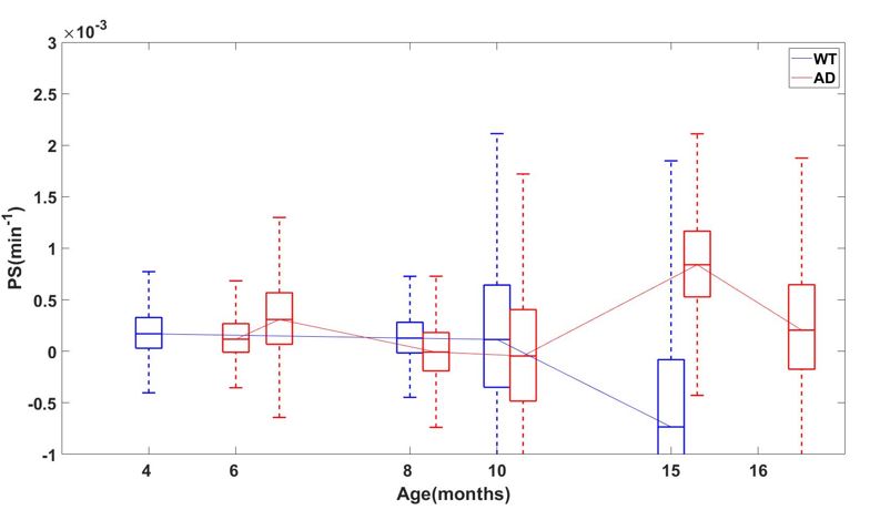
BBB permeability with agingFigure 2 shows example signal dynamics in WT and AD transgenic mice at different ages. As shown, the 8 month old mice show only subtle difference in contrast dynamics, while the contrast dynamics in more aged mice show remarkable difference between WT and AD. Figure 3 shows the estimated permeability with aging spectrum, which exhibits the increased trend in AD mice associated with aging, while the WT mice does not show increased permeability.
Improved sensitivity with CIF network
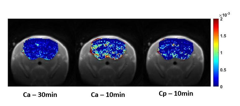
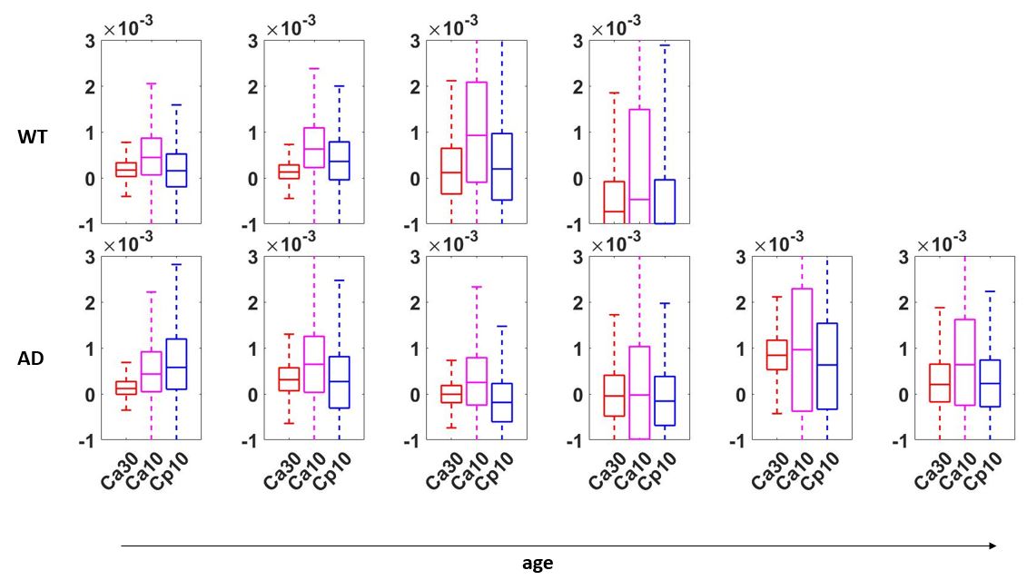
Figure 4 shows the estimated permeability maps from the 30-min data, 10-min data using AIF and 10-min data using CIF. When the scan time is reduced, the conventional approach with AIF results in overestimation of the permeability. However, when the local CIF is used, the overestimation is substantially reduced. Figure 5 shows the whisker-box plot for each measure for AD and WT mice along the age spectrum. With the scan time reduction, the estimates using CIF demonstrates better accuracy and precision as compared to the estimates using AIF.
Discussion & Conclusion
The preliminary result from our study demonstrates increased BBB permeability in AD transgenic mouse after 15 months, while the WT mouse does not exhibit vascular changes that are detectable with DCE-MRI study. In addition, our analysis suggests that when the scan time is reduced, the conventional approach may over-estimate the permeability estimates, as suggested by the previous study (10). However, the network-predicted CIF allows the scan-time reduction without compromising the accuracy of the subtle permeability estimates. Our future work aims to include more mice at different ages to investigate the longitudinal changes across a broader age spectrum.Acknowledgements
R01CA160620References
1. Montagne A, Barnes SR, Sweeney MD, Halliday MR, Sagare AP, Zhao Z, et al. Blood-brain barrier breakdown in the aging human hippocampus. Neuron. 2015;85(2):296-302.
2. Backes WH, van Buchem MA. Blood-Brain Barrier Leakage in Patients with Early Alzheimer Disease. Radiology. 2016;281:527-35.
3. Minogue AM, Jones RS, Kelly RJ, McDonald CL, Connor TJ, Lynch MA. Age-associated dysregulation of microglial activation is coupled with enhanced blood-brain barrier permeability and pathology in APP/PS1 mice. Neurobiology of aging. 2014;35(6):1442-52.
4. Blockx I, Einstein S, Guns P-J, Van Audekerke J, Guglielmetti C, Zago W, et al. Monitoring blood-brain barrier integrity following amyloid-β immunotherapy using gadolinium-enhanced MRI in a PDAPP mouse model. Journal of Alzheimer's Disease. 2016;54(2):723-35.
5. Bae J, Huang Z, Knoll F, Geras K, Pandit Sood T, Feng L, et al. Estimation of the capillary level input function for dynamic contrast‐enhanced MRI of the breast using a deep learning approach. Magnetic Resonance in Medicine. 2022;87(5):2536-50.
6. Devi L, Ohno M. Genetic reductions of β‐site amyloid precursor protein‐cleaving enzyme 1 and amyloid‐β ameliorate impairment of conditioned taste aversion memory in 5XFAD Alzheimer’s disease model mice. European Journal of Neuroscience. 2010;31(1):110-8.
7. Feng L, Grimm R, Block KT, Chandarana H, Kim S, Xu J, et al. Golden‐angle radial sparse parallel MRI: combination of compressed sensing, parallel imaging, and golden‐angle radial sampling for fast and flexible dynamic volumetric MRI. Magnetic resonance in medicine. 2014;72(3):707-17.
8. Barnes SR, Ng TS, Santa-Maria N, Montagne A, Zlokovic BV, Jacobs RE. ROCKETSHIP: a flexible and modular software tool for the planning, processing and analysis of dynamic MRI studies. BMC medical imaging. 2015;15(1):1-20.
9. Patlak CS, Blasberg RG, Fenstermacher JD. Graphical evaluation of blood-to-brain transfer constants from multiple-time uptake data. Journal of Cerebral Blood Flow & Metabolism. 1983;3(1):1-7.
10. Bae J, Zhang J, Wadghiri YZ, Minhas AS, Poptani H, Ge Y, et al. Measurement of blood‐brain barrier permeability using dynamic contrast‐enhanced magnetic resonance imaging with reduced scan time. Magnetic resonance in medicine. 2018;80(4):1686-96.
Figures