3520
Development of a high-resolution magnetic susceptibility template of the older adult brain in MIITRA space1Department of Biomedical Engineering, Illinois Institute of Technology, Chicago, IL, United States, 2Rush Alzheimer’s Disease Center, Rush University Medical Center, Chicago, IL, United States
Synopsis
Keywords: Alzheimer's Disease, Aging, Brain, Susceptibility, Atlas
Quantitative susceptibility mapping (QSM) is considered a promising tool for the detection and monitoring of disease processes that alter magnetic susceptibility in the brain of older adults. Voxel-wise and atlas-based QSM analyses require accurate spatial normalization of QSM data, which requires a high quality QSM template representative of the population. However, a QSM template of the older adult brain is not yet available. This study aimed to construct a high quality, high resolution QSM template of the older adult brain in the space of the MITRA atlas based on data from a large, diverse, community cohort of non-demented older adults.Introduction
Quantitative susceptibility mapping (QSM) provides important information about magnetic susceptibility throughout the brain and is sensitive to paramagnetic and diamagnetic elements that may be deposited in brain tissue as a result of aging or age-related neuropathologies [1]. For this reason, QSM is considered a promising tool for the detection and monitoring of disease processes in the brain of older adults. In neuroimaging, voxel-wise and atlas-based analyses require transformation of images from multiple individuals into a common space. Since QSM maps have a unique image contrast not present in other MR images, accurate spatial normalization of QSM data from multiple individuals requires a high quality QSM template representative of the population [2]. However, a QSM template of the older adult brain is not yet available. Therefore, the aim of this study was to construct a high quality, high resolution QSM template of the older adult brain in the space of the Multichannel Illinois Institute of Technology & Rush university Aging (MITRA) atlas [3] based on data from a large, diverse, community cohort of non-demented older adults.Methods
Data:Three dimensional T1w MPRAGE (1x1x1 mm3) and three dimensional 5-echo gradient echo (GRE) data (0.7x0.7x1.3 mm3) were collected on 3T MRI scanners for 400 non-demented older adults (50% male; 64.9-98.9 years of age; 54% white, 43% black) participating in the construction of the MIITRA atlas [4][5].
Template construction:
Step 1: A magnetic susceptibility map was generated for each participant using the Morphology Enabled Dipole Inversion (MEDI) method on the multi-echo GRE data, and was upsampled to 0.7x0.7x0.65 mm3 voxels using nonlocal upsampling [6].
Step 2: The images from the first echo of the GRE data of each participant were N4 bias field corrected, then upsampled to 0.7x0.7x0.65 mm3 using non-local upsampling [6], and converted to synthetic T1w images using mri_synthsr [7]. In addition, the original T1w data were upsampled to 0.5x0.5x0.5 mm3. Then the synthetic T1w images were linearly registered to the upsampled T1w data using ANTs [8].
Step 3: The transformations resulting from the previous step were used to forward-map magnetic susceptibility values from the native space of each participant to exact physical locations in the existing MIITRA space [9].
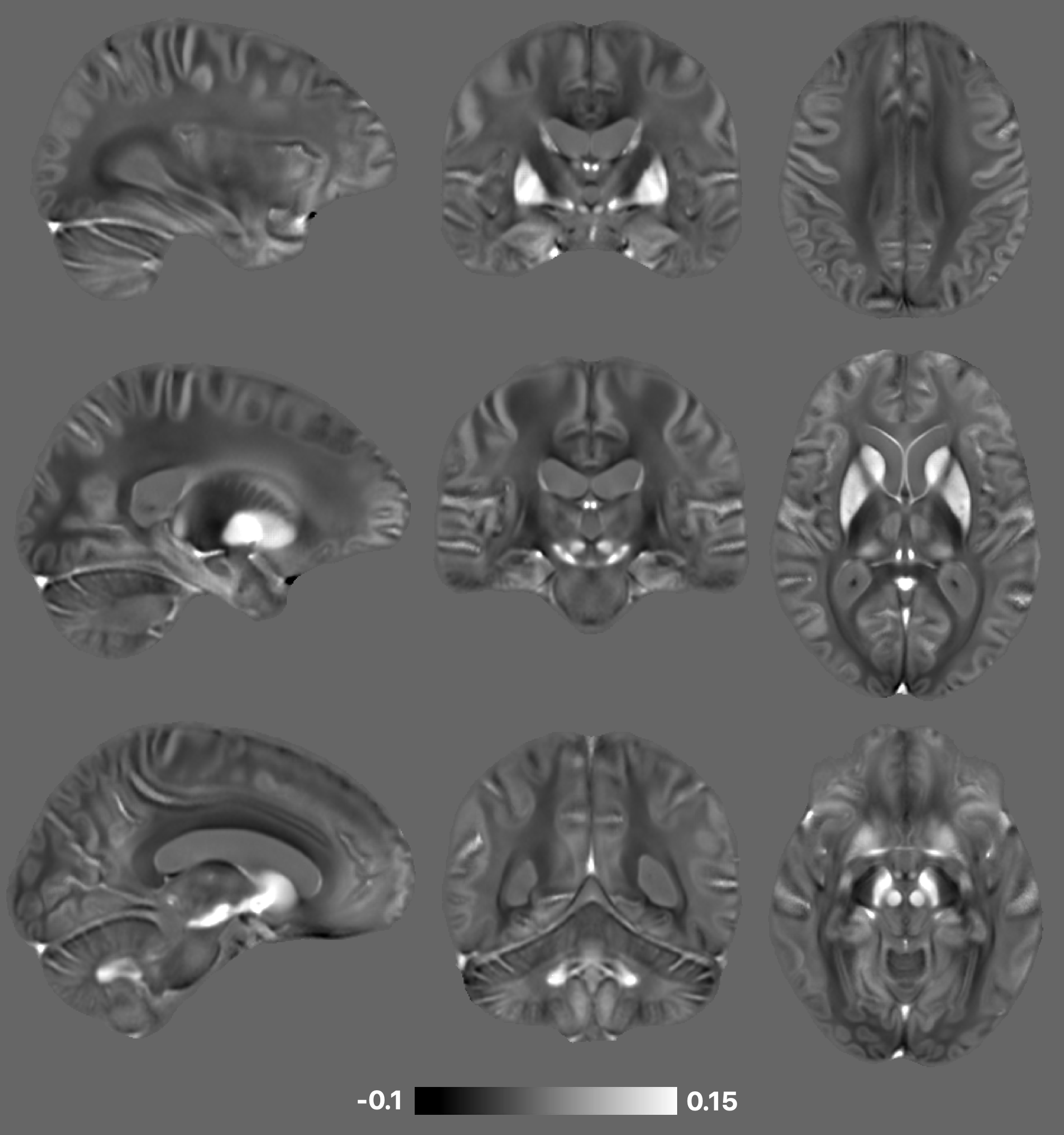
Step 4: A sparse representation based data-fusion approach [10] was used on the transformed signals from all participants to generate the final magnetic susceptibility template, denoted as MIITRA_QSM, at 0.5x0.5x0.5 mm3 resolution (Fig.1).
Evaluation:
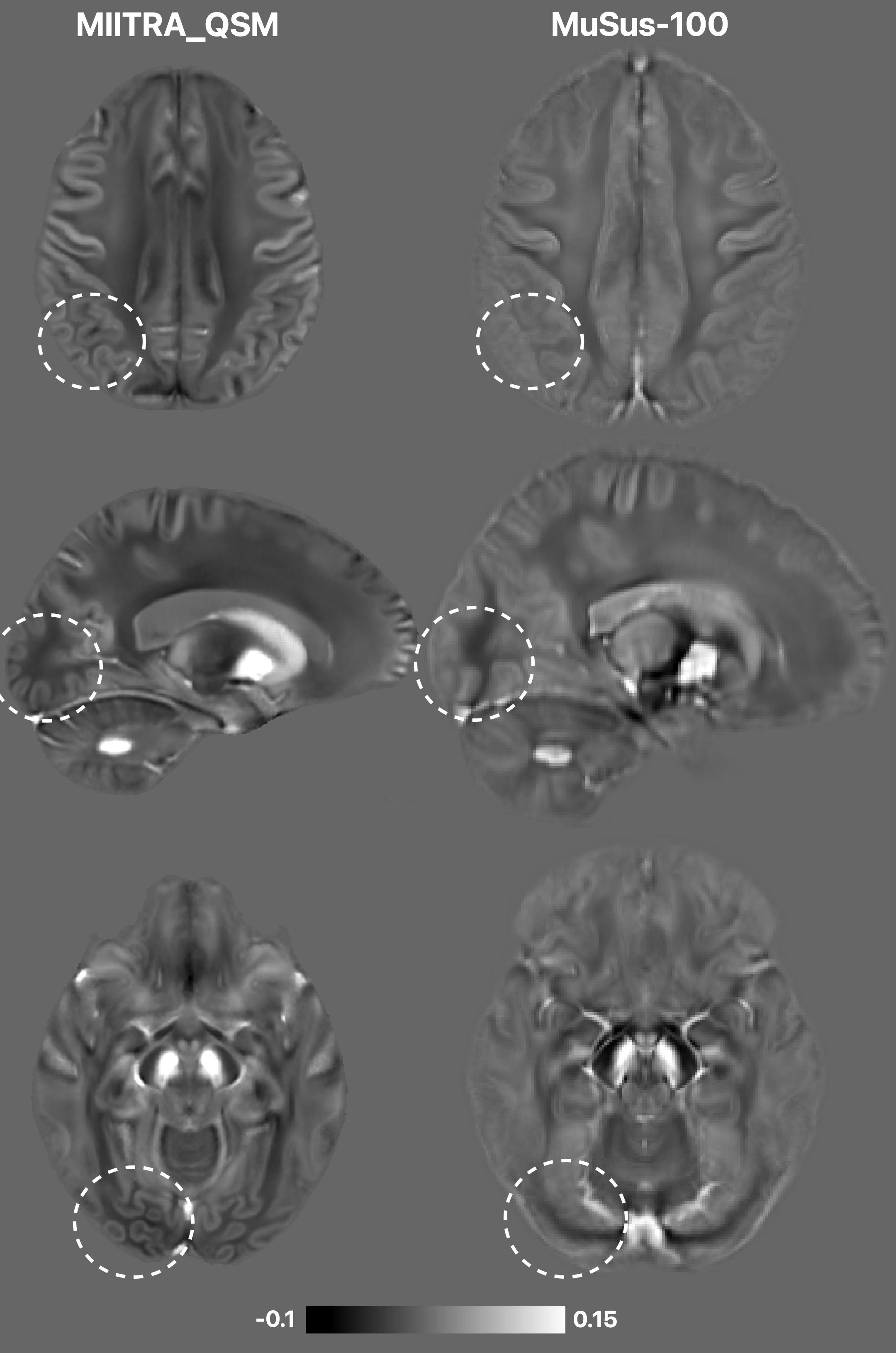
Since no other magnetic susceptibility template of the older adult brain is currently available, we compared the MIITRA_QSM template to a young adult template named MuSus-100 (1x1x1 mm3, 100 participants, mean age 24 years) [11] in terms of image quality and magnetic susceptibility properties.
Results
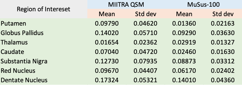
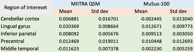
Visual inspection of MIITRA_QSM showed that the new template has the expected QSM contrast, highest values in subcortical, brainstem, and cerebellar structures known for their high magnetic susceptibility (especially in older adults), lower values in the white compared to gray matter, and higher values in white matter of the frontal lobe compared to the parietal and occipital lobes. In addition, MIITRA_QSM includes fine details, and is characterized by relatively low noise levels. When compared to MuSus-100, the MIITRA_QSM template showed better delineation of cortical structures and higher image sharpness in a number of brain areas (Fig.2). Magnetic susceptibility values in most subcortical brain structures were higher in MIITRA_QSM compared to MuSus-100 (Fig.3), while cortical structures from the two templates exhibited more similar magnetic susceptibility values (Fig.4).Discussion
This work constructed the first magnetic susceptibility template of the older adult brain, which we named MIITRA_QSM. The new template is of high quality and high resolution, it was constructed based on data from a large, diverse, community cohort of non-demented older adults, and is located in MIITRA space where other templates and resources are available. Magnetic susceptibility values in specific subcortical, brainstem and cerebellar structures are higher in the new template compared to a recently published young adult template, probably due to increased metal deposition in these structures with aging. Ongoing work includes an investigation of the improvement in spatial normalization precision achieved using the new template compared to other templates, an assessment of the enhancement in sensitivity achieved due to the more precise spatial normalization, as well as an assessment of the new template’s representativeness of the older adult population.Conclusion
In conclusion, the present work constructed a high quality, high resolution QSM template of the older adult brain in the space of the MITRA atlas based on data from a large, diverse, community cohort of non-demented older adults. The new template is expected to greatly enhance the accuracy of voxel-wise and atlas-based QSM studies on older adults.Acknowledgements
National Institute on Aging (NIA) R01AG052200
National Institute on Aging (NIA) P30AG010161
National Institute on Aging (NIA) P30AG072975
National Institute on Aging (NIA) R01AG017917
National Institute on Aging (NIA) RF1AG022018
National Institute on Aging (NIA) R01AG056405
National Institute on Aging (NIA) R01AG015819
National Institute on Aging (NIA) R01AG064233
National Institute of Neurological Disorders and Stroke (NINDS), UF1NS100599
References
[1] Betts, M.J., Acosta-Cabronero et al (2016): High-resolution characterisation of the aging brain using simultaneous quantitative susceptibility mapping (qsm) and r2* measurements at 7t. Neuroimage, 138, 43–63.
[2] Shmueli, K., de Zwart, J. A., van Gelderen, P., Li, T. Q., Dodd, S. J., & Duyn, J. H. (2009). Magnetic susceptibility mapping of brain tissue in vivo using MRI phase data. Magnetic Resonance in Medicine: An Official Journal of the International Society for Magnetic Resonance in Medicine, 62(6), 1510-1522.
[3] Wu Y, Ridwan AR, Niaz MR, Qi X, Zhang S, Alzheimer's Disease Neuroimaging Initiative, Bennett DA, Arfanakis K. Development of high quality T1-weighted and diffusion tensor templates of the older adult brain in a common space. Neuroimage. 2022 Oct 15;260:119417.
[4] Bennett DA, Buchman AS, Boyle PA, et al. Religious Orders Study and Rush Memory and Aging Project. J Alzheimers Dis. 2018;64(s1):S161-S189.
[5] Barnes LL, Shah RC, Aggarwal NT, et al. The Minority Aging Research Study: Ongoing Efforts to Obtain Brain Donation in African Americans without Dementia. Curr Alzheimer Res. 2012;9(6):734-745.
[6] Manjón JV, Coupé P, Buades A, et al. Non-local MRI upsampling. Med Image Analysis (2010) :784-92.
[7] Iglesias JE, Benjamin Billot, Yaël Balbastre, et al. Joint super-resolution and synthesis of 1 mm isotropic MP-RAGE volumes from clinical MRI exams with scans of different orientation, resolution and contrast. Neuroimage (2021): 118206.
[8] Avants et al. ANTS: Open-Source Tools for Normalization And Neuroanatomy.
[9] Wu Y., Niaz M.R., Ridwan A.R., Qi X., Bennett D.A., Arfanakis K. MIITRA atlas: Construction of high resolution T1w and DTI brain templates in a common space, based on 400 older adults. Proc. Int. Soc. for Magn. Reson. in Med. (ISMRM) 2021.
[10] Ridwan AR, Wu Y, Niaz MR, et al. Construction of an unbiased high resolution and detail-preserving structural T1-weighted template for use in studies on older adults. Proc. Intl. Soc. Mag. Reson. Med. 2021; 29.
[11] He, Chenyu, Xiaojun Guan, et al. Quantitative susceptibility atlas construction in Montreal Neurological Institute space: towards histological-consistent iron-rich deep brain nucleus subregion identification. Brain Structure and Function (2022): 1-23.
Figures