3501
Risk taking tendency correlate with imbalance functional link between brain networks following 36 hours total sleep deprivation
Jiyuan Li1, Yunlong Yue1, Yunlong Song2, and Yanfang Jin1
1Department of MRI, Beijing Shijitan Hospital, Capital Medical University, Beijing, China, 2Department of CT and MRI, The General Hospital of the Air Force People’s Liberation Army, Beijing, China
1Department of MRI, Beijing Shijitan Hospital, Capital Medical University, Beijing, China, 2Department of CT and MRI, The General Hospital of the Air Force People’s Liberation Army, Beijing, China
Synopsis
Keywords: Brain Connectivity, fMRI (resting state)
36 hours sleep deprivation produces a significant deficit in vmPFC functional connectivity and default mode networks (DMN), along with the enhanced functional link between vmPFC and executive control networks (ECN). Furthermore, the negative correlation between vmPFC-DMN and vmPFC-ECN coupling during RW were diminished after TSD. We also find that TSD induced the significantly negative correlation between vmPFC-ECN networks and risk-taking behavior. These results demonstrate that an absence of sleep substantially impaired the balance of large scale brain networks and which in turn predicts risk-taking behavior following 36 hours of TSD.Introduction
Evidences indicate that people intended to make more risk taking choices during total sleep deprivation (TSD) than rested wakefulness (RW) condition. Previous studies have revealed that the ventral medial prefrontal cortex (vmPFC) may have a crucial role in the psychophysiology of this impact. However, far less attention has been paid to investigating the intrinsic patterns of functional organization of vmPFC in the TSD brain. Based on the TSD influence on cognitive control, and vmPFC functional changes in clinic researches, we hypothesis that: 1) TSD disrupt the valuation process reflect by the functional communication between vmPFC and subcortical regions of valuation network; 2) SD enhanced the functional link with the top-down control areas among the valuation network.Methods
Thirty right-hand healthy male participants with mean age of 20.94 were recruited in this study. With the self controlled design, subjects were examined twice resting-state fMRI under the RW and 36 hours TSD conditions. And participants completed a risky gambles task after each MRI scanning. MRI scanning was performed using a GE 3.0T Discovery 750 scanner. Processing of the fMRI data was completed using Analysis of Functional NeuroImages (AFNI) software and FSL 5.0. The region-of-interest (ROI) of vmPFC was defined on the basis of a previous study1. Spherical ROIs with radius= 10 mm were created for left (center coordinates: x = -8, y = 40, z = -21) and right (center coordinates: x = 27, y = 18, z = -21) vmPFC in MNI space. We selected bilateral dorsolateral prefrontal cortex (DLPFC), bilateral inferior frontal gyrus (IFG), bilateral dorsal anterior cingulate gyrus (dACC), bilateral superior insula and bilateral parietal lobe as executive control networks (ECN) components, and selected bilateral dorsal medial prefrontal cortex (dMPFC), bilateral posterior cingulate cortex (PCC) and bilateral angular as the default mode networks (DMN) components. DMN and ECN components were extracted from Anatomical Automatic Labeling (AAL) template2. Mean time series of all voxels with the ROI , DMN, ECN and whole brain were extracted. Then, paired t-test was used to analyze the connection pattern change between bilateral vmPFC and whole brain before and after TSD. Finally, we compared the changes of correlation between DMN and ECN, vmPFC-ECN coupling and risky gambles task data before and after TSD.Results
Under RW and TSD conditions, the risky gambles task scores of the subjects were (3.12 ± 0.67) and (3.51 ± 0.81) respectively, with a statistically significant difference (t=2.16, P=0.034) (Fig. 1). 36 hours TSD produces a significant deficit in vmPFC functional connectivity and DMN, including middle frontal gyrus, posterior cingulate, angular and temporal lobe. Along with the enhanced functional link between vmPFC and ECN (Fig. 2, Table. 1). In RW state, there is a significant negative correlation between vmPFC-DMN coupling and vmPFC-ECN coupling (r=-0.55, P=0.0007). But there is no correlation between vmPFC-DMN coupling and vmPFC-ECN coupling after TSD (r=-0.066, P=0.7) (Fig. 3). It suggests that the negative correlation between vmPFC-DMN and vmPFC-ECN coupling during RW were diminished after TSD. Furthermore, We also find that TSD induced the significantly negative correlation between vmPFC-ECN networks and risk-taking behavior (Fig. 4).Conclusion
These results demonstrate that an absence of sleep substantially impaired the balance of large scale brain networks and which in turn predicts risk-taking behavior following 36 hours of TSD.Acknowledgements
No acknowledgement found.References
1. Liu, x, Hairston, J, Schrier M, et al. Common and distinct networks underlying reward valence and processing stages: A meta-analysis of functional neuroimaging studies. Neuroscience and Biobehavioral Reviews, 2011, 35(5): 1219-1236.
2. Zangemeister L,Grabenhorst F,Schultz W. Neural activity in human ventromedial prefrontal cortex reflecting the intention to save reward. Soc Cogn Affect Neurosci, 2019, 14(12): 1255-1261.
Figures
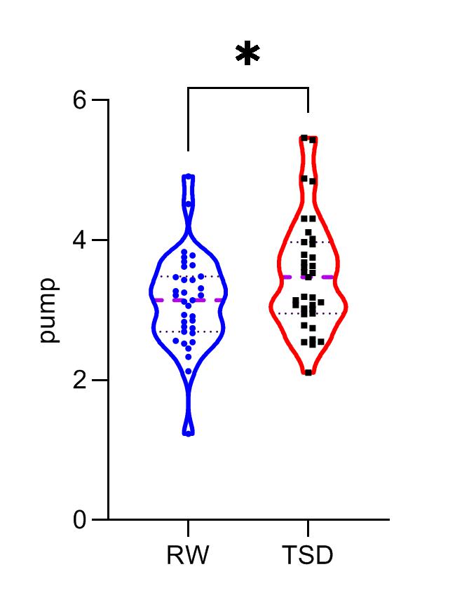
Figure 1 Comparison
of risky gambles task scores before and after 36 hours
sleep deprivation. RW, rested wakefulness;
TSD, total sleep deprivation
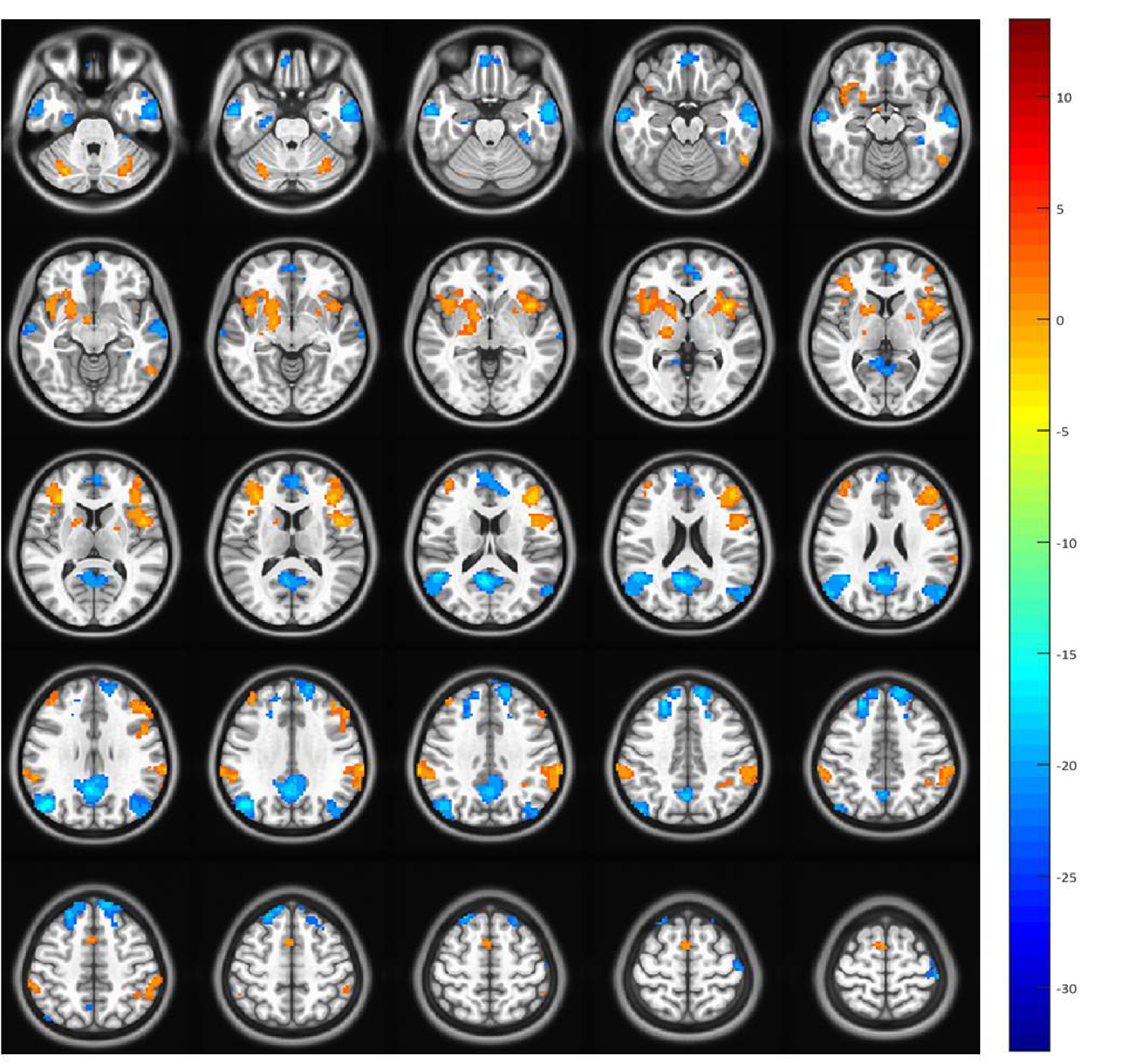
Figure 2 Changes of vmPFC functional
connectivity before and after sleep deprivation(RW-TSD). The warm color
represents the rise of functional connection, while the cold color represents
the decline of functional connection.
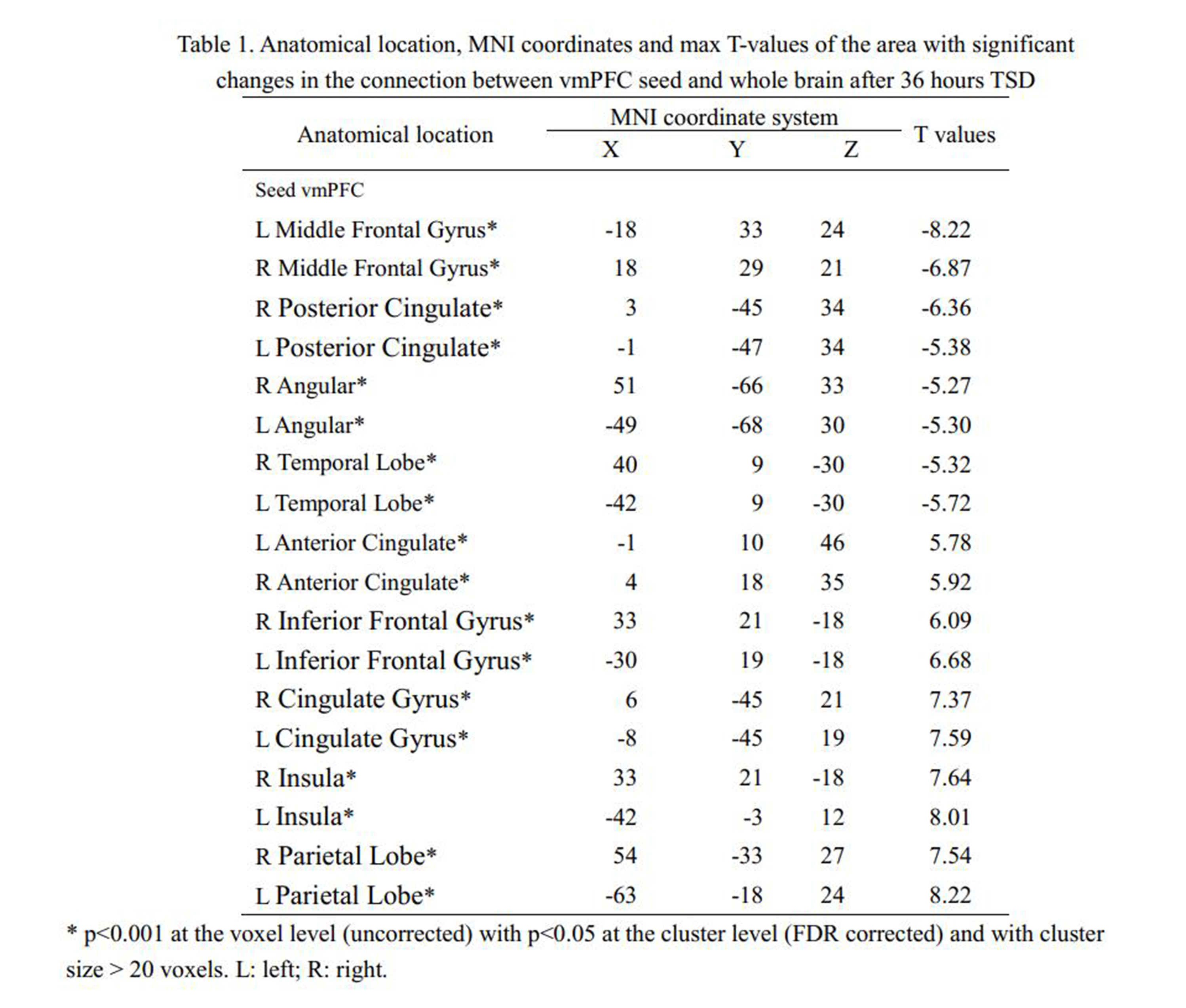
Table
1. Anatomical location, MNI coordinates and max T-values of the area with
significant changes in the connection between vmPFC seed and whole brain after
36 hours TSD
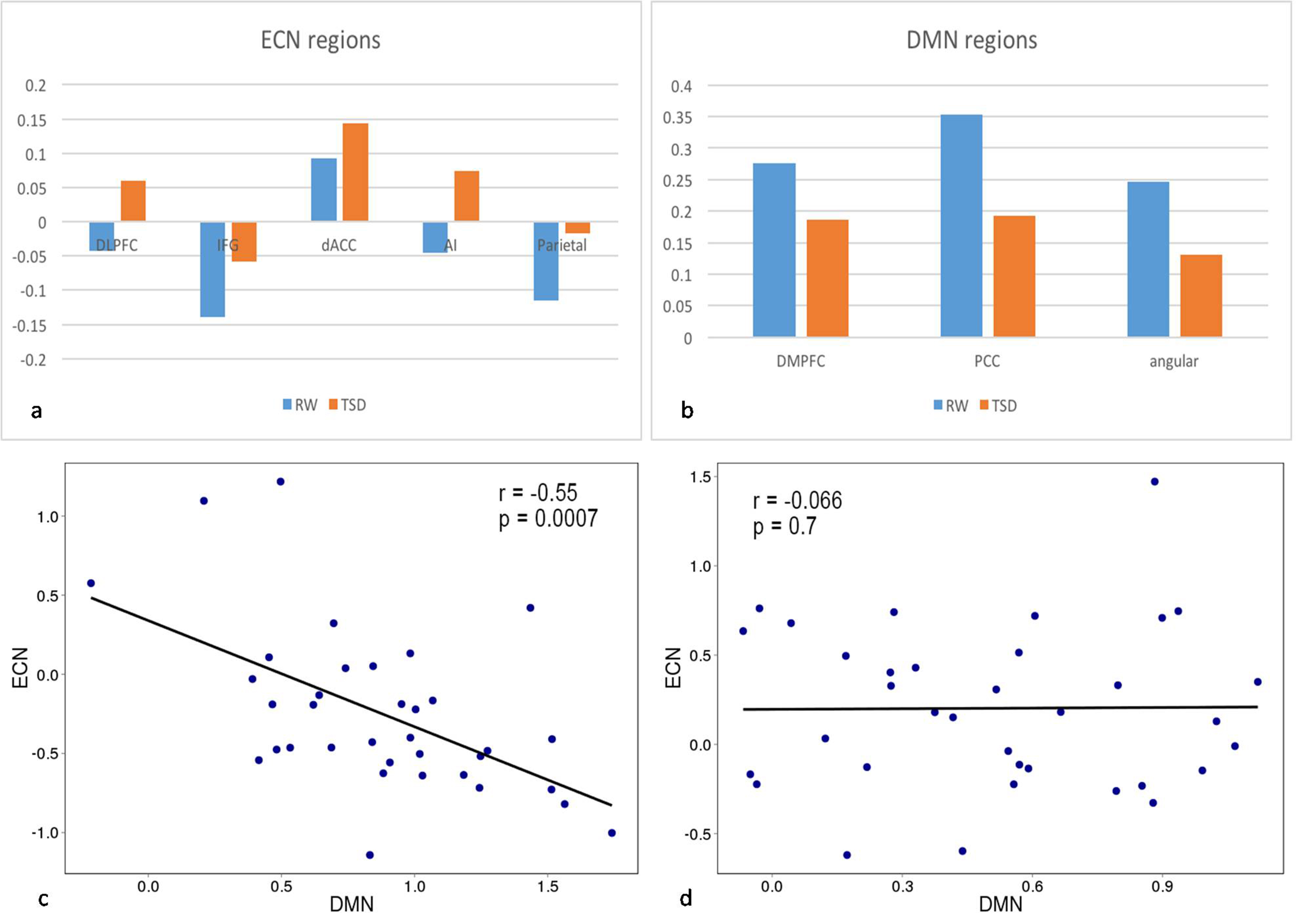
Figure 3 Quantification and correlation analysis of functional connectivity results of brain regions selected by ECN and DMN. a and b respectively quantify the functional connectivity results of brain regions selected by ECN and DMN. c shows a significant negative correlation between vmPFC-DMN coupling and vmPFC-ECN coupling in RW state. d shows that there is no correlation between vmPFC-DMN coupling and vmPFC-ECN coupling in TSD state.
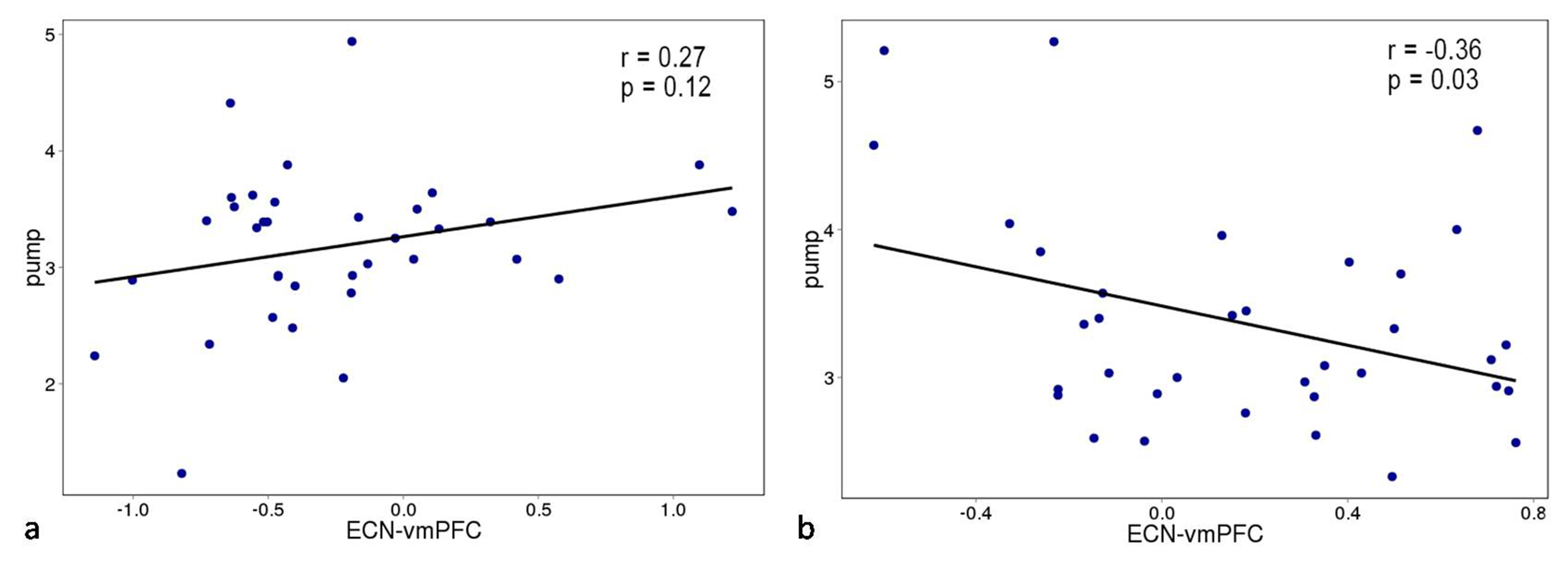
Figure 4 Correlation between vmPFC-ECN coupling
and the risky gambles task scores. There is a positive correlation between
vmPFC-ECN coupling and risk propensity in RW state, but there is a significant
negative correlation between vmPFC-ECN coupling and risk propensity in TSD
state. This shows that after sleep deprivation, the stronger the relationship
between ECN vmPFC, the smaller the individual's risk propensity.
DOI: https://doi.org/10.58530/2023/3501