3497
Abnormal neural activity in male chronic smokers revealed by resting-state functional MRI1Department of MRI, First Affiliated Hospital of Zhengzhou University, Zhengzhou, China
Synopsis
Keywords: Brain Connectivity, Brain, chronic smokers, amplitude of low frequency fluctuations, resting state functional connectivity, functional magnetic resonance imaging
We combined two methods of resting-state functional magnetic resonance imaging (rs-fMRI) to explore the abnormal neural activity in male chronic smokers. The amplitude of low frequency fluctuation (ALFF) was first calculated and brain regions with significant differences in ALFF between two groups were used as seeds for further resting-state functional connectivity (rs-FC) analysis. Our findings revealed increased spontaneous regional activity in the superior frontal gyrus (SFG) with reduced functional connectivity to visual attention areas and cerebellar subregions in smokers compared with controls, which may play an important role in the pathophysiology of smoking.Introduction:
Cigarette smoking is among the leading causes of preventable illness and death in the world, characterized by craving, withdrawal symptoms and relapsing1. Given the serious harms of smoking, understanding the neural mechanisms underlying it may contribute to guide brain-based smoking cessation treatments. Amplitude of low frequency fluctuation (ALFF) served as a common approach characterizes the spontaneous regional neural activity2, 3, which has shown to be significant for comprehending the neuropathologic and neurophysiological condition of disease4. It has reported that brain activity globally reduced in acute administration of nicotine in previous studies5. However, chronic smokers were reported to have abnormal spontaneous brain activity during resting state in only a few studies6-9. In particular, little was known about the relationship between the resting state abnormalities and smoking indicators. Besides, more than just the spontaneous regional activity within specific brain regions drives the addictive diseases, but also coordination between them10. Whether the functional connectivity between brain regions with abnormal spontaneous regional activity and other regions is altered in chronic smokers remains unexplored. Therefore, it is crucial to combine two different resting-state functional indicators including ALFF and resting-state functional connectivity (rs-FC) to explore the abnormal neural activity in chronic smokers.Methods:
A total of 142 participants (86 smokers and 56 nonsmokers) who met inclusion and exclusion criterion were recruited. MRI data were collected using a MAGNETOM Skyra 3T MR scanner (Siemens Healthcare, Erlangen, Germany) with 64 channel head coils. This functional imaging data set was preprocessed by using Data Processing Assistant for Resting-State fMRI Analysis Toolkit (DPARSF). ALFF was calculated based on Fast Fourier transform (FFT) and the time series of each voxel was converted to frequency domain without band-pass filtering. According to the results of ALFF, the brain regions with significant group differences in ALFF values between the smoking group and the control group were selected as region of interest (ROI) and the seed was defined as a 5-mm radius sphere that centered at the peak coordinate. Demographic and clinical characteristics were compared using two-sample t-tests between smokers and nonsmokers. Based on the MATLAB SPM12 toolkit, two tailed two-sample t-tests were conducted to explore the alternation of ALFF and ALFF-based rsFC between two groups with age and years of education as covariates (Gaussian random field theory [GRF] corrected, Pvoxel < 0.005, Pcluster < 0.05). In addition, we performed correlation analyses between the values of two functional indicators and smoking measurements (such as pack-years and FTND scores).Results:
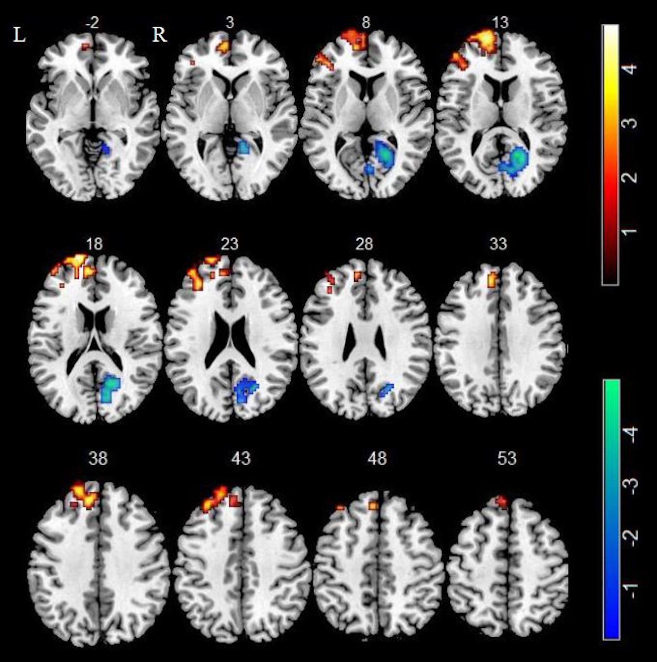
No significant differences were observed in age and education between the two groups (86 male smokers and 56 male nonsmokers) (all P-values > 0.05).In comparison of nonsmokers, smokers showed increased ALFF in left superior frontal gyrus (SFG), medial superior frontal gyrus (mSFG) and middle frontal gyrus (MFG) as well as decreased ALFF in right calcarine sulcus (GRF corrected P < 0.005) (Figure 1).
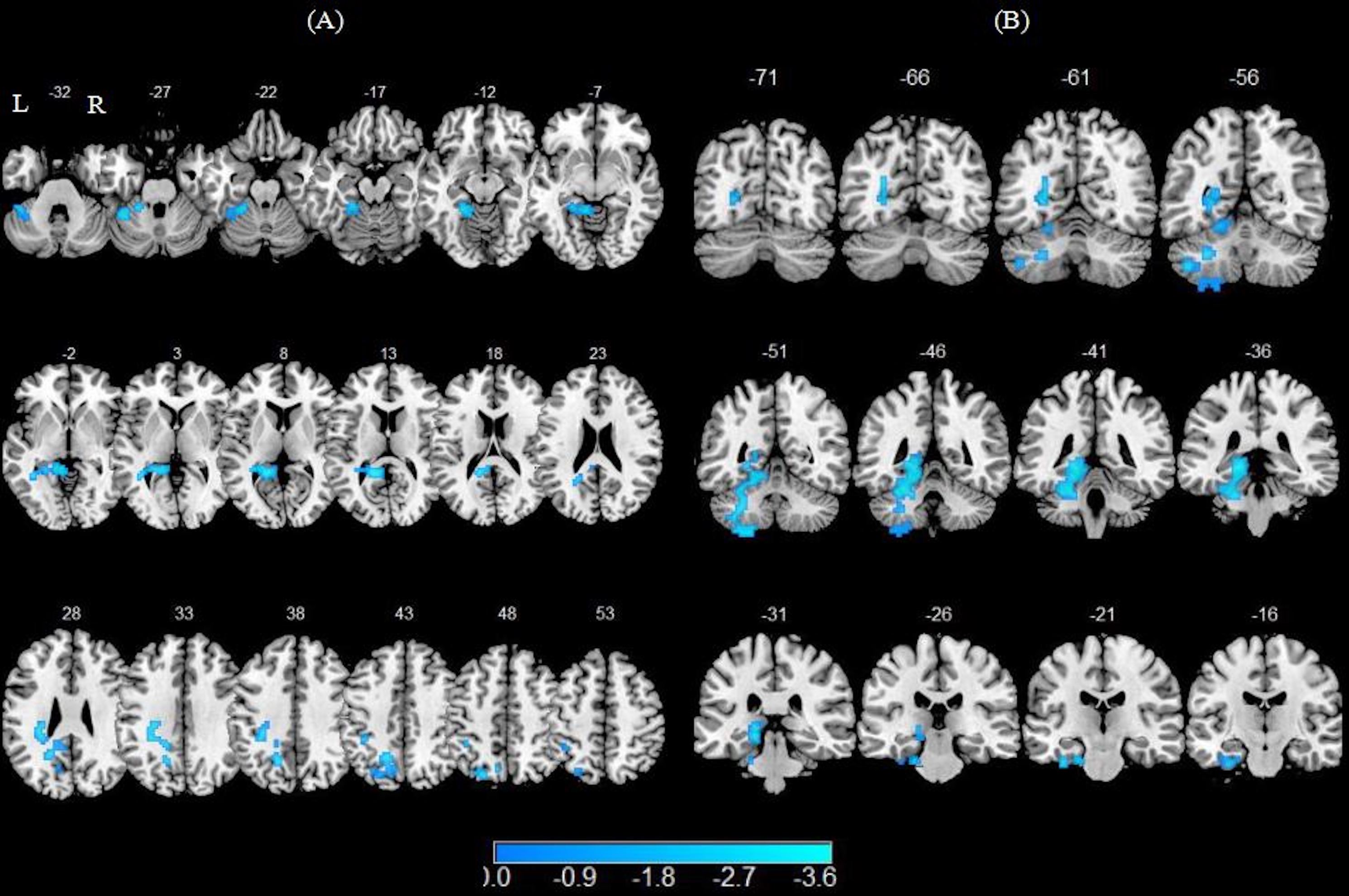
In comparison of nonsmokers, smokers showed attenuated functional connectivity with left SFG in left precuneus, fusiform, lingual gyrus, cerebellum 4 5 and cerebellum 6 as well as lower functional connectivity with left mSGF in left fusiform, lingual, parahippocampal gyrus (PHG), calcarine sulcus, cerebellum 4 5, cerebellum 6 and cerebellum 8 (GRF corrected P < 0.005) (Figure 2).
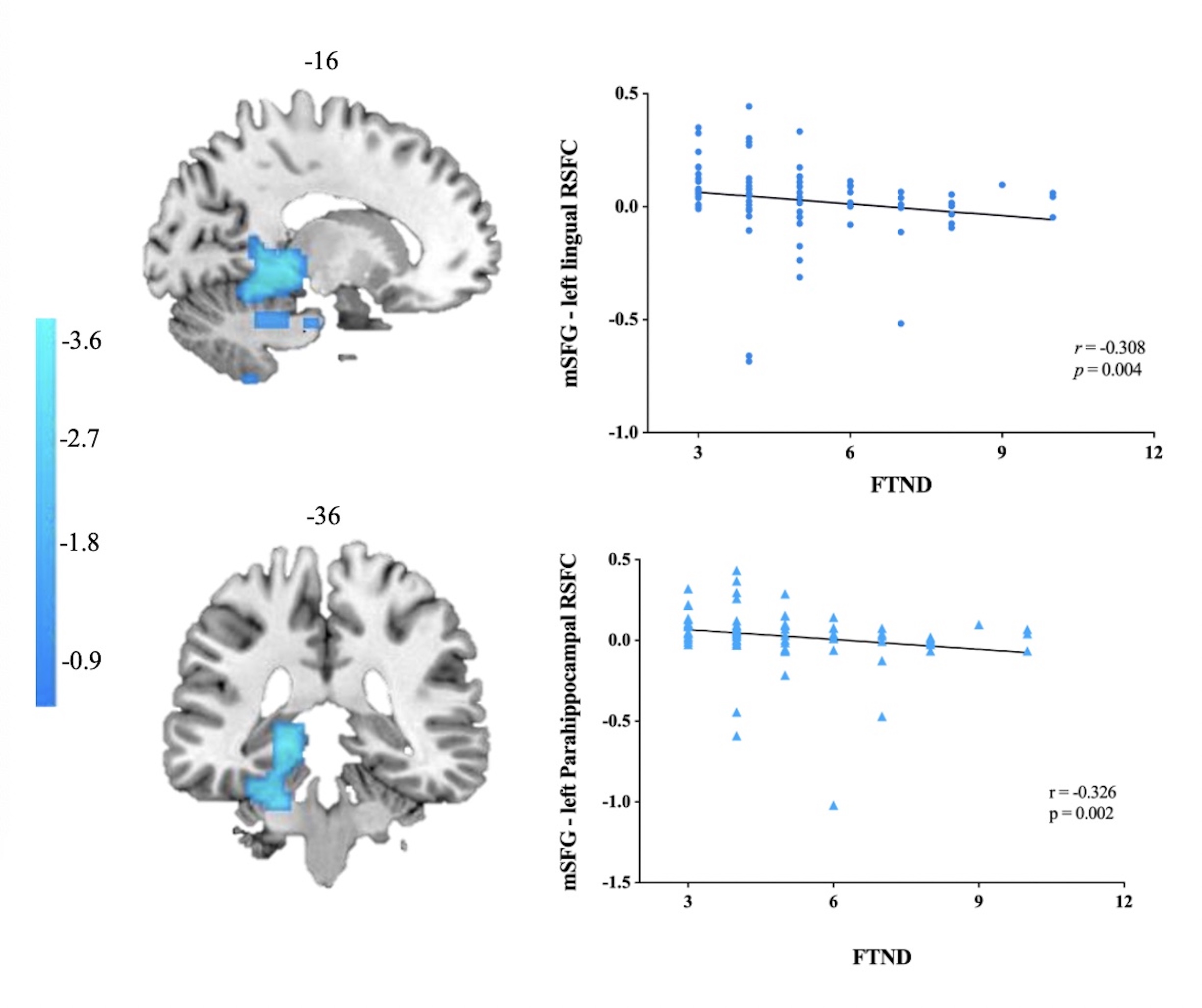
The results of correlation analysis showed that attenuated functional connectivity with left mSGF in left lingual gyrus and PHG was negatively correlated with FTND scores (r = -0.308, p = 0.004; r = -0.326, p = 0.002 Bonferroni corrected) (Figure 3).
Discussion:
Our study combined amplitude of low frequency fluctuation (ALFF) and resting-state functional connectivity (rs-FC) to identify the alterations of intrinsic brain activity and neural connectivity abnormalities in smokers. The results showed that smokers had increased spontaneous brain activity in the PFC including left SFG, mSFG and MFG. The PFC plays an important role in reward evaluation and is the basis of working memory, which enables rapid integration and updating of other information to guide target behaviors11. The effects of nicotine on the PFC can create pathological valuations and interfere with top-down behavioral control, making smokers unable to resist the urge to smoke even though they know the dangers of smoking. In addition, we also noted that smokers showed attenuated functional connectivity between the left mSFG with visual attention areas (such as the left calcarine sulcus, lingual gyrus and fusiform gyrus) and parahippocampal gyrus (PHG) involved in episodic memory, recollection12-14 and visuospatial processing15-17. The mSFG is functionally related to cognitive control 18, 19. In the current study, functional connectivity between mSFG and lingual gyrus as well as PHG was decreased, and it was negatively correlated with FTND scores. These reflected a disruption of the integrity of functional connectivity between cognitive control and visual episodic memory in smokers, and that the greater the nicotine dependence, the weaker the functional connectivity. The neural basis of cigarette smoking was thought to be mainly related to dopaminergic neural circuits composed of the prefrontal cortex, insula, striatum and so on20. However, our findings noted the role of the cerebellum in cigarette smoking extending previous findings that have primarily focused on changes in cerebrum. We hope our findings may complement the current understanding of the neural mechanisms of smoking.Acknowledgements
This study was supported by the Natural Science Foundation of China (81601467, 81871327) and Medical Science and Technology Research Project of Henan Province (201701011).References
1. Asma S, Song Y, Cohen J, et al. CDC Grand Rounds: global tobacco control. MMWR Morb Mortal Wkly Rep. 2014;63(13):277-280.
2. Zang Y-F, He Y, Zhu C-Z, et al. Altered baseline brain activity in children with ADHD revealed by resting-state functional MRI. Brain Dev. 2007;29(2):83-91.
3. Wang S, Rao B, Chen L, et al. Using Fractional Amplitude of Low-Frequency Fluctuations and Functional Connectivity in Patients With Post-stroke Cognitive Impairment for a Simulated Stimulation Program. Front Aging Neurosci. 2021;13:724267.
4. Raichle ME. Neuroscience. The brain's dark energy. Science. 2006;314(5803):1249-1250.
5. Brody AL. Functional brain imaging of tobacco use and dependence. J Psychiatr Res. 2006;40(5):404-418.
6. Wu G, Yang S, Zhu L, Lin F. Altered spontaneous brain activity in heavy smokers revealed by regional homogeneity. Psychopharmacology (Berl). 2015;232(14):2481-2489.
7. Wen M, Yang Z, Wei Y, et al. More than just statics: Temporal dynamic changes of intrinsic brain activity in cigarette smoking. Addict Biol. 2021;26(6):e13050.
8. Yu R, Zhao L, Tian J, et al. Regional homogeneity changes in heavy male smokers: a resting-state functional magnetic resonance imaging study. Addict Biol. 2013;18(4):729-731.
9. Chu S, Xiao D, Wang S, et al. Spontaneous brain activity in chronic smokers revealed by fractional amplitude of low frequency fluctuation analysis: a resting state functional magnetic resonance imaging study. Chin Med J (Engl). 2014;127(8):1504-1509.
10. Wang L, Zhang Y, Lin X, Zhou H, Du X, Dong G. Group independent component analysis reveals alternation of right executive control network in Internet gaming disorder. CNS Spectr. 2018;23(5):300-310.
11. Hyman SE, Malenka RC, Nestler EJ. Neural mechanisms of addiction: the role of reward-related learning and memory. Annu Rev Neurosci. 2006;29:565-598.
12. Zola-Morgan S, Squire LR, Amaral DG, Suzuki WA. Lesions of perirhinal and parahippocampal cortex that spare the amygdala and hippocampal formation produce severe memory impairment. J Neurosci. 1989;9(12):4355-4370.
13. Davachi L, Mitchell JP, Wagner AD. Multiple routes to memory: distinct medial temporal lobe processes build item and source memories. Proc Natl Acad Sci U S A. 2003;100(4):2157-2162.
14. Diana RA, Yonelinas AP, Ranganath C. Medial temporal lobe activity during source retrieval reflects information type, not memory strength. J Cogn Neurosci. 2010;22(8):1808-1818.
15. Ekstrom AD, Kahana MJ, Caplan JB, et al. Cellular networks underlying human spatial navigation. Nature. 2003;425(6954):184-188.
16. Epstein R, Kanwisher N. A cortical representation of the local visual environment. Nature. 1998;392(6676):598-601.
17. Stevens WD, Kahn I, Wig GS, Schacter DL. Hemispheric asymmetry of visual scene processing in the human brain: evidence from repetition priming and intrinsic activity. Cereb Cortex. 2012;22(8):1935-1949.
18. Sohn M-H, Albert MV, Jung K, Carter CS, Anderson JR. Anticipation of conflict monitoring in the anterior cingulate cortex and the prefrontal cortex. Proc Natl Acad Sci U S A. 2007;104(25):10330-10334.
19. Ursu S, Clark KA, Aizenstein HJ, Stenger VA, Carter CS. Conflict-related activity in the caudal anterior cingulate cortex in the absence of awareness. Biol Psychol. 2009;80(3):279-286.
20. Yang Z, Zhang Y, Cheng J, Zheng R. Meta-analysis of brain gray matter changes in chronic smokers. European journal of radiology. 2020;132:109300.
Figures