3485
Altered neurovascular coupling in patients with MELAS evaluated by combining cerebral blood flow and regional homogeneity
Rong Wang1, Yuxin Li1, Jie Lin1, Yong Zhang2, and Jiankun Dai3
1Huashan Hospital, Shanghai, China, 2GE Healthcare, Shanghai, China, Shanghai, China, 3MR Research, GE Healthcare, Beijing, China, Shanghai, China
1Huashan Hospital, Shanghai, China, 2GE Healthcare, Shanghai, China, Shanghai, China, 3MR Research, GE Healthcare, Beijing, China, Shanghai, China
Synopsis
Keywords: Stroke, fMRI (resting state)
This is the first study that used a combined arterial spin labeling (ASL) and resting-state fMRI approach to assess the neurovascular coupling in patients with mitochondrial myopathy, encephalopathy, lactic acidosis and stroke-like episodes (MELAS), which may provide a new mechanistic perspective into understanding numerous brain diseases.Background and purpose
Mitochondrial myopathy, encephalopathy, lactic acidosis and stroke-like episodes (MELAS) is the most common type of mitochondrial disease [1]. It is a rare maternally inherited genetic disease mostly affecting children and young adults [2]. Coupling between neuronal activity and blood perfusion is termed neurovascular coupling [3]. Although abnormal brain activity and blood supply have been separately reported in MELAS [4, 5], whether anomalous neurovascular coupling would be presented in such disease is still unknown. Thus, in this study, the neuronal activity and blood supply were measured using the regional homogeneity (ReHo) and cerebral blood flow (CBF). The voxel-wise CBF–ReHo correlations and CBF/ReHo ratio were separately used to assess global and local neurovascular coupling in participants.Methods
A total of 24 patients with MELAS at acute stage of stroke-like episodes and 24 healthy controls (HC) were recruited in this study, respectively. The MRI examination was performed on a 3.0 Tesla scanner (Discovery MR750, General Electric, Boston, MA, USA) equipped with an 8-channel head coil. Main scan parameters were as follows: Pseudo-continuous 3D ASL (TR/TE = 5327/10.5 ms, post-labeling delay = 1.5 s, slice thickness = 4.0 mm, slices = 35, FOV = 240 mm × 240 mm). The rs-fMRI images were obtained using a single-shot gradient-recalled echo planar imaging sequence with the following parameters: TR = 2000 ms, TE = 30 ms, slice thickness = 4 mm, flip angle = 90◦, slices = 35, field of view (FOV) = 240 mm × 240 mm, matrix size = 64 × 64, No. of volumes = 210. On the basis of MATLAB 2016a (MathWorks, Natick, MA, USA), rs-fMRI and CBF images were preprocessed using DPABI (http://rfmri.org/dpabi) and SPM 12 (SPM, www.fil.ion.ucl.ac.uk/spm).Results
The spatial distributions of CBF, ReHo and the CBF/ReHo ratio are similar between MLEAS and HC groups (Figure 1a). Both groups showed higher values of CBF, ReHo and the CBF/ReHo ratios in the visual and auditory cortices, the default mode network (including the posterior cingulate cortex, precuneus and medial prefrontal cortex), the sensorimotor network (including the paracentral gyrus, comprising the precentral and postcentral gyri). In addition, compared with the HC, the MELAS patients showed lower global CBF (MELAS: 62.37 ± 1.82 ml/ 100 g/min; HC: 76.35 ± 2.44 ml/100 g/min, P < 0.001) and global ReHo (MELAS: 1.31 ± 0.049; HC: 1.62 ± 0.063, P < 0.001) in whole gray matter. Significant correlations between CBF and FCS were identified in all participants. Two representative correlation maps from one MELAS patient and one HC are displayed in Figure 1(b); the MELAS patients showed reduced CBF-ReHo coupling compared with the HC in whole gray matter (P = 0.0002) and stroke-like lesion (P < 0.0001).Conclusion
The combination of CBF and ReHo was exploited for mapping abnormal neurovascular coupling in patients with MELAS in the current study. Specifically, patients with MELAS exhibited reduced global and focal neurovascular coupling, which was reflected by decreased voxel-wise CBF–ReHo correlation. It might may provide a new perspective in understanding the neuropathological underpinning of such disease.Acknowledgements
No acknowledgement found.References
[1]. El-Hattab AW, Adesina AM, Jones J, Scaglia F. MELAS syndrome: Clinical manifestations, pathogenesis, and treatment options. Mol Genet Metab. 2015 116: 4-12. [2]. Pavlakis SG, Phillips PC, DiMauro S, De Vivo DC, Rowland LP. Mitochondrial myopathy, encephalopathy, lactic acidosis, and strokelike episodes: a distinctive clinical syndrome. Ann Neurol. 1984 16: 481-488. [3]. Iadecola, C., 2017. The neurovascular unit coming of age: a journey through neurovascular coupling in health and disease. Neuron. 96, 17–42. [4]. Wang R, Li Y, Lin J, Sun C, Chen N, Xu W, Hu B, Liu X, Geng D, Yang L. Altered spontaneous brain activity at attack and remission stages in patients with mitochondrial encephalomyopathy, lactic acidosis and stroke-like episodes (MELAS): Beyond stroke-like lesions. Mitochondrion 2020, 54: 49-56. [5]. Li Y, Xu W, Sun C, Lin J, Qu J, Cao J, Li H, Yang L. Reversible Dilation of Cerebral Macrovascular Changes in MELAS Episodes. Clinical neuroradiology 2019, 29(2): 321-329.Figures
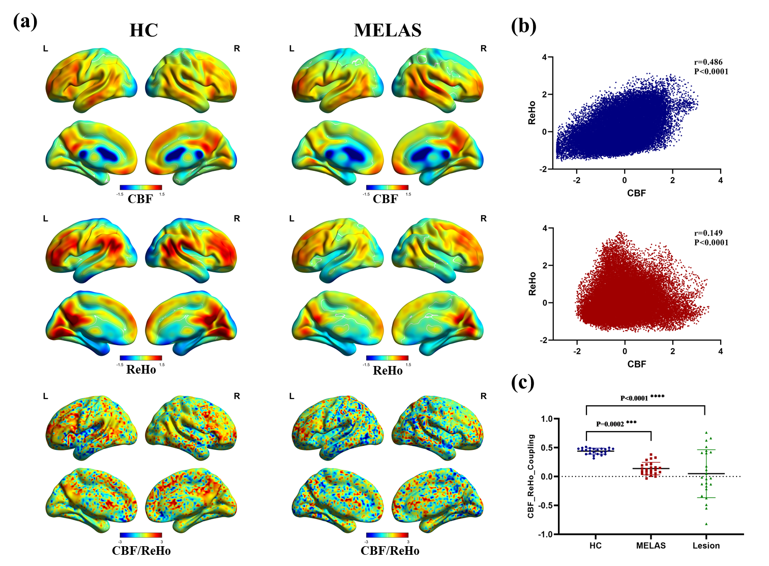
Figure 1: Spatial distribution maps and reduced neurovascular coupling in patients
with MELAS. (a) Examples of the mapped spatial distributions of CBF, ReHo and
the CBF/ReHo ratio in a MELAS patient and a HC. The two subjects exhibited
similar spatial distributions in these measures. (b) Scatter plots
demonstrating the spatial correlations across voxels between CBF and ReHo in
the same subjects (blue, patient; red, HC) represented in Figure 1(a). (c)
Compared with HC, MELAS patients showed reduced CBF-ReHo coupling within gray
matter (P = 0.0002) and stroke-like lesion (P < 0.0001).
DOI: https://doi.org/10.58530/2023/3485