3465
Feasibility of ultrafast DCE-MRI in differentiating benign and malignant breast tumors1Paul C. Lauterbur Research Center for Biomedical Imaging, Shenzhen Institute of Advanced Technology, Chinese Academy of Sciences, ShenZhen, China, 2Faculty of Information Engineering and Automation, Kunming University of Science and Technology, Kunming, China, 3Yibin Second People's Hospital, Yibin, China, 4Chengdu Pidu District People's Hospital, Chengdu, China, 5Paul C. Lauterbur Research Center for Biomedical Imaging, Shenzhen Institute of Advanced Technology, Chinese Academy of Sciences, Shenzhen, China
Synopsis
Keywords: Breast, Data Analysis, Conventional DCE-MRI, ultrafast DCE-MRI, quantitative parameters, semiquantitative parameters
The conventional dynamic contrast-enhanced magnetic resonance imaging (DCE-MRI) technique has been shown to be very sensitive in detecting benign and malignant breast cancer lesions. However, it requires a relatively long acquisition time and hence a semiquantitative imaging technique. To reduce the acquisition time, ultrafast DCE-MRI techniques have been proposed for breast tumor imaging. Our study aims to investigate the feasibility of the ultrafast DCE-MRI technique in differentiating benign and malignant breast tumors. The results show that the semiquantitative and quantitative parameters obtained with ultrafast DCE-MRI are superior to those obtained with conventional DCE-MRI in differentiating benign and malignant breast tumors.INTRODUCTION
Breast cancer is the most common malignant tumor affecting women's lives, and it is therefore important to accurately diagnose benign and malignant lesions of breast cancer 1. Dynamic contrast-enhanced magnetic resonance imaging (DCE-MRI) is currently the most sensitive method for detecting breast cancer 2. However, conventional DCE-MRI takes too long to acquire images and makes it difficult to accurately capture signal changes in the tumor area, which in turn affects the diagnosis of benign and malignant lesions. To improve the accuracy of sampling, researchers have proposed the use of an ultrafast DCE-MRI protocol to acquire information on breast tumors in the early stages of contrast injection and compared it with conventional DCE in terms of accuracy in semiquantitative analysis of breast tumors 3-7. To evaluate ultrafast DCE-MRI more comprehensively, we not only compared the performance of semiquantitative parameters of ultrafast DCE-MRI and conventional DCE-MRI in identifying benign and malignant breast tumors but also used quantitative parameters of ultrafast DCE-MRI to identify benign and malignant breast tumors. The aim of this study was to investigate the feasibility of replacing conventional DCE-MRI with ultrafast DCE-MRI in identifying benign and malignant breast tumors.METHODS
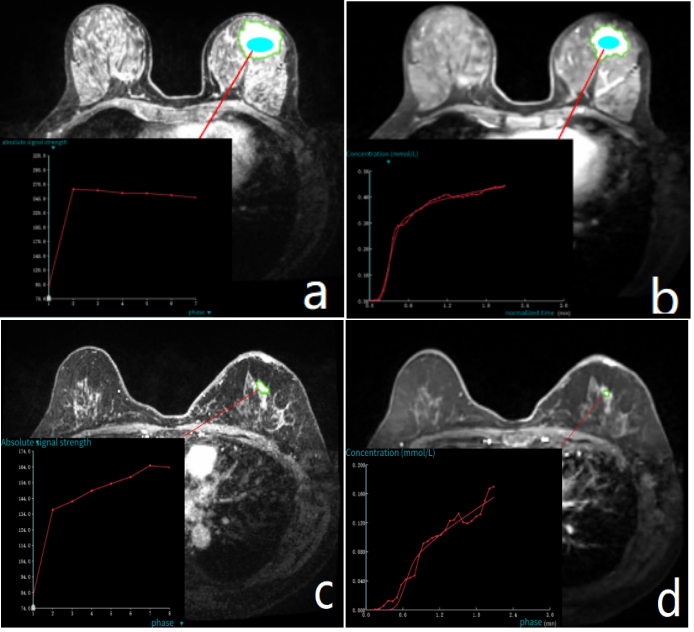
A total of 89 patients were enrolled in this prospective study. In this study, the 3.0T MRI system (uMR 790, United Imaging Healthcare, Shanghai, China) was used to acquire 30-phase ultrafast DCE-MRI images and 7-phase conventional DCE-MRI images. The imaging parameters of ultrafast DCE-MRI were as follows: spatial resolution=1.69×1.69×3 mm3, temporal resolution=4.4 s, field of view=340 mm, TR/TE=2.98/1.37 ms, FA=10, number of slices=96, and number of dynamics=30. The imaging parameters of conventional DCE-MRI were as follows: spatial resolution=1.06×1.06×1.1 mm3, temporal resolution=57.8 s, field of view=340 mm, TR/TE=4.66/1.97 ms, FA=10, number of slices=138, and number of dynamics=7. The Mann‒Whitney U test was used to analyze whether there were significant differences in semiquantitative parameters obtained by conventional DCE-MRI between benign and malignant breast tumors (p<0.05) and whether there were significant differences in semiquantitative and quantitative parameters obtained by ultrafast DCE-MRI between benign and malignant breast tumors (p<0.05). The area under the curve (AUC) of the ROC curve was used to evaluate the diagnostic performance of the parameters, and support vector machines (SVMs) were also used to determine the benign and malignant classification effects of quantitative and semiquantitative parameters of ultrafast DCE-MRI versus those of semiquantitative parameters of conventional DCE-MRI.RESULTS
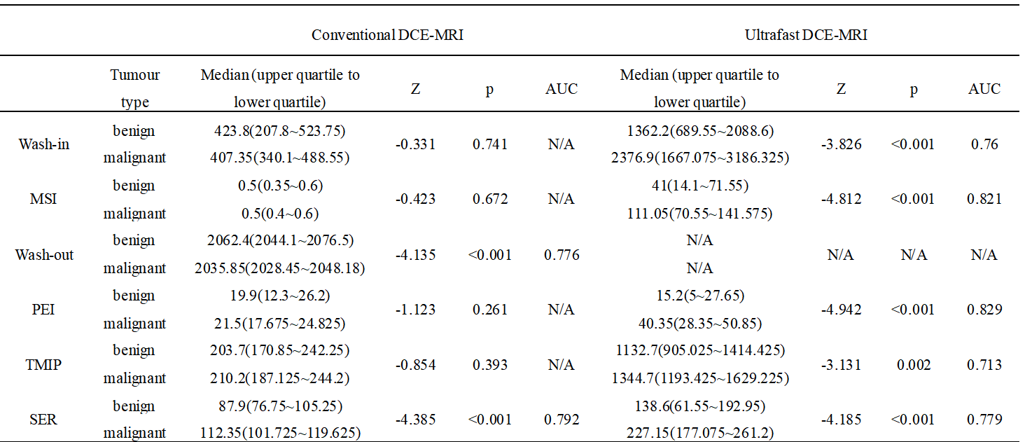
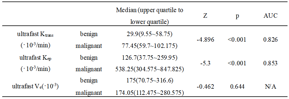
There were 103 lesions in 89 patients, among which 78 were malignant and 25 were benign. Table 1 compares the performance of conventional DCE-MRI and ultrafast DCE-MRI in identifying benign and malignant breast tumors in terms of semiquantitative parameters, including wash-in rate, wash-out rate, maximum slope of increase (MSI), positive enhancement integral (PEI), time-maximum intensity projection (TMIP) and signal enhancement ratio (SER). For conventional DCE-MRI, except for wash-out and SER, there were no significant differences in other parameters between benign and malignant breast tumors (p>0.05). The AUC of wash-out and SER to determine benign and malignant lesions was 0.776 and 0.792, respectively. For ultrafast DCE-MRI, all semiquantitative parameters were significantly different in identifying benign and malignant breast tumors (p<0.05), except wash-out, and the AUCs of these parameters were 0.76 (wash-in), 0.821 (MSI), 0.829 (PEI), 0.713 (TMIP) and 0.779 (SER).Table 2 summarizes the performance of the quantitative parameters of ultrafast DCE-MRI in identifying benign and malignant breast tumors. There were no significant differences in the values of the extracellular volume fraction of the imaged tissue (Ve) between benign and malignant breast tumors. However, the volume transfer constant (Ktrans) and rate constant (Kep) were significant in discriminating benign from malignant lesions (P<0.05). The AUCs for Ktrans and Kep to identify benign and malignant tumors were 0.826 and 0.853, respectively.
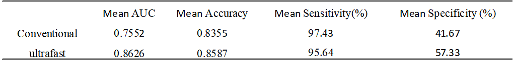
In Table 3, the semiquantitative and quantitative parameters of ultrafast DCE-MRI had a mean AUC of 0.8626, a mean accuracy of 0.8587 and a mean specificity of 57.33% for the identification of benign and malignant tumors. The semiquantitative parameters of conventional DCE-MRI had a mean AUC of 0.7552, a mean accuracy of 0.8355 and a mean specificity of 41.67% in identifying benign and malignant tumors.
DISCUSSION
The semiquantitative parameters washout, MSI, PEI and TMIP of ultrafast DCE-MRI were superior to those of conventional DCE-MRI in identifying breast tumors. The reason is that ultrafast DCE-MRI has a higher temporal resolution and a higher sampling rate. The SER of ultrafast DCE-MRI is lower than that of conventional DCE-MRI in identifying breast tumors due to the short overall ultrafast sampling time; therefore, a peak signal may not be obtained.The quantitative parameters of ultrafast DCE-MRI not only identified breast tumors, but the quantitative parameters also had the best performance in identifying tumors. The reason is that quantitative parameters are less affected by MRI scanning sequence, time resolution and other factors.
Ultrafast DCE-MRI is superior to conventional DCE-MRI in differentiating benign and malignant breast tumors, indicating that most of the information that is meaningful for clinical interpretation is obtained early. Therefore, ultrafast DCE-MRI can be used to replace conventional DCE-MRI at the early stage of contrast agent injection.
CONCLUSION
Ultrafast DCE-MRI can effectively identify benign and malignant breast tumors and is more accurate than conventional DCE-MRI.Acknowledgements
No acknowledgement found.References
[1] Lin Y, Wang C, Zhong Y, et al. Striking life events associated with primary breast cancer susceptibility in women: a meta-analysis study. Journal of Experimental & Clinical Cancer Research, 2013, 32(1): 1-8.
[2] Morris E A, Comstock C E, Lee C H, et al. ACR BI-RADS® magnetic resonance imaging. ACR BI-RADS® atlas, breast imaging reporting and data system, 2013, 5.
[3] Ohashi A, Kataoka M, Kanao S, et al. Diagnostic performance of maximum slope: A kinetic parameter obtained from ultrafast dynamic contrast-enhanced magnetic resonance imaging of the breast using k-space weighted image contrast (KWIC). European Journal of Radiology, 2019, 118: 285-292.
[4] Ohashi A, Kataoka M, Iima M, et al. A multiparametric approach to diagnosing breast lesions using diffusion-weighted imaging and ultrafast dynamic contrast-enhanced MRI. Magnetic Resonance Imaging, 2020, 71: 154-160.
[5] Milon A, Perre S V, Poujol J, et al. Abbreviated breast MRI combining FAST protocol and high temporal resolution (HTR) dynamic contrast enhanced (DCE) sequence. European Journal of Radiology, 2019, 117: 199-208.
[6] Lee S J, Ko K H, Jung H K, et al. The additional utility of ultrafast MRI on conventional DCE-MRI in evaluating preoperative MRI of breast cancer patients. European Journal of Radiology, 2020, 124: 108841.
[7] Honda M, Kataoka M, Onishi N, et al. New parameters of ultrafast dynamic contrast‐enhanced breast MRI using compressed sensing. Journal of Magnetic Resonance Imaging, 2020, 51(1): 164-174.
Figures