3462
Feasibility of 3D Quantitative Synthetic MRI for Discriminating Immunohistochemical Status in Invasive Ductal Carcinoma of the Breast
Maki Amano1,2, Shohei Fujita1,3, Naoyuki Takei4, Katsuhiro Sano1, Akihiko Wada1, Kanako Sato1, Junko Kikuta1, Yoshiki Kuwatsuru1, Rina Tachibana1,5, Towa Sekine1,5, Yoshiya Horimoto6, and Shigeki Aoki1
1Radiology, Juntendo University, Tokyo, Japan, 2Radiology, Nihon University Hospital, Tokyo, Japan, 3Radiology, The University of Tokyo Graduate School of Medicine, Tokyo, Japan, 4MR Applications and Workflow, GE Healthcare Japan, Tokyo, Japan, 5Radiological Sciences, Graduate School of Human Health Sciences, Tokyo Metropolitan University, Tokyo, Japan, 6Breast Oncology, Juntendo University, Tokyo, Japan
1Radiology, Juntendo University, Tokyo, Japan, 2Radiology, Nihon University Hospital, Tokyo, Japan, 3Radiology, The University of Tokyo Graduate School of Medicine, Tokyo, Japan, 4MR Applications and Workflow, GE Healthcare Japan, Tokyo, Japan, 5Radiological Sciences, Graduate School of Human Health Sciences, Tokyo Metropolitan University, Tokyo, Japan, 6Breast Oncology, Juntendo University, Tokyo, Japan
Synopsis
Keywords: Breast, Cancer
The present study aimed to evaluate the feasibility of 3D quantitative synthetic MRI for discriminating the immunohistochemical (IHC) status of invasive ductal carcinoma (IDC) of the breast. We found that volumetric T1 and T2 values of IDC varied according to IHC status, including those for hormone receptors and Ki-67, indicating the feasibility of this method for discriminating the IHC status of IDC of the breast.INTRODUCTION
Breast cancer is classified by molecular subtypes based on the levels of expression of hormone receptors (HR), HER2, and Ki-67, as usually determined by immunohistochemical (IHC) staining1. Molecular subtyping is used to evaluate tumor heterogeneity. Tumor heterogeneity can even be observed within an individual tumor. Core needle biopsy (CNB) is widely applied for molecular subtyping in a preoperative setting. Major limitations of CNB include its invasiveness and sampling error, which may be overcome using imaging markers by assessing the IHC status of an entire tumor. MRI is widely used for diagnosing breast cancer. Several studies have investigated the relationships between quantitative parameters, including T1, T2, pharmacokinetic parameters, and apparent diffusion coefficient (ADC), and molecular subtypes of breast cancer2,3. Synthetic MRI using multiecho and multidelay sequences provides simultaneous measurement of T1, T2, and proton density of the tissues. To date, 2D sequences have been used for quantitative synthetic MRI of the breast4-6, but they have a limited spatial resolution in the slice direction compared with dynamic contrast-enhanced (DCE) imaging. 3D Quantification using an interleaved Look–Locker acquisition sequence with a T2 preparation pulse (QALAS) has been developed for simultaneous and volumetric quantification of T1 and T2 in cardiac and brain MRI. Here, we have modified the sequence to apply it to breast MRI. We hypothesized that 3D synthetic MRI using QALAS would be useful in identifying the IHC status of breast cancer with high accuracy. Thus, the present study aimed to evaluate the feasibility of 3D synthetic MRI for discriminating the IHC status of invasive ductal carcinoma (IDC) of the breast.METHODS
Patients. We recruited 33 female patients with IDC (mean age, 52.3 years).MRI Acquisition. Breast MRI was performed using a 3.0 T system (Signa Architect, GE Healthcare, Milwaukee, WI, USA). The 3D synthetic MRI using 3D QALAS and DCE MRI was performed with the imaging parameters shown in Table 1. To adapt 3D-QALAS to breast MRI, a B1-insensitive rotation T2 preparation sequence (BIR4) 7 was used to achieve uniform T2 preparation across the imaging volume. The tan/tanh-modulated AHP of duration with 10 ms was used in the BIR-4 pulse with a T2Prep time of 90ms.
Data Processing. Raw image data of QALAS were processed, and quantitative parametric maps (i.e., T1 and T2 maps) were generated using SyMRI version 0.45.32 software (Synthetic MRI, Linkoping, Sweden) (Fig. 1). We first performed a rigid registration between the first phase images of DCE MRI and synthetic T1-weighted images using open-source software, Ants (https://sourceforge.net/projects/advants/files/ANTS/), automatically. Then, a 3D ROI of the whole tumor was semiautomatically delineated on the registered DCE MRI images using LIFEx software (https://www.lifexsoft.org/). Two radiologists with 12 years of experience in breast imaging and 3 years of experience in general radiology individually delineated the tumor contour on the DCE MRI. The 3D ROI was eroded by a voxel in any direction to minimize the partial volume effects between cancer and surrounding tissues, and then applied to the T1 and T2 maps to measure T1 and T2 values.
Statistical Analysis. The means of T1 and T2 values of each tumor were calculated automatically by LIFEx. For univariate analysis, the relationship between the quantitative values, including the mean T1 and mean T2, and the HR, HER2, Ki-67, or triple-negative (TN) status was assessed using a Mann–Whitney U test. In addition, ROC analysis and multivariate analyses using multivariate logistic regression analysis were performed. All statistical analyses were performed using SPSS software (IBM Corp, Armonk, NY, USA), and p < 0.05 was defined as statistical significance for all analyses.
RESULTS
The interclass correlation coefficients between the two radiologists for the mean of the T1 and T2 values were excellent (0.934 and 0.828, respectively). Univariate analysis showed that the mean T1 value differed between HR-positive and -negative (p = 0.024, AUC 0.78), while the mean T2 value differed between HR-positive and -negative (p = 0.012, AUC 0.81), the Ki-67 level >14% and ≥14% (p = 0.028, AUC 0.82), and TN and non-TN (p = 0.014, AUC 0.84). Multivariate analysis showed that the mean T2 of the HR-negative was significantly higher than that of the HR-positive (p = 0.017) (Tables 2 and 3, Fig. 2).DISCUSSION
3D Synthetic MRI using QALAS provided volumetric T1 and T2 values of the IDC, which were significantly correlated with the IHC status, with an excellent correlation between the observers. MRI may compensate for CNB limitations by estimating the IHC status of the entire tumor. Although the mean T1 and T2 values were not useful for discriminating HER2 status, this has been distinguished previously using ADC values. As a future direction, combining 3D-synthetic MRI and diffusion-weighted imaging may improve IDC subtyping. In addition, because volumetric ROI can measure tumor volume, it may be useful to evaluate the effect of neoadjuvant chemotherapy when it is calculated together with tissue relaxation time by 3D synthetic MRI.CONCLUSION
3D quantitative synthetic MRI using QALAS may be feasible for discriminating the IHC statuses of IDC of the breast.Acknowledgements
This study was supported by JSPS KAKENHI Grant Number JP20K08035.
References
- Januškevičienė I, Petrikaitė V. Heterogeneity of breast cancer: The importance of interaction between different tumor cell populations. Life Sci. 2019;239:117009.
- Montemezzi S, Camera L, Giri MG, et al. Is there a correlation between 3T multiparametric MRI and molecular subtypes of breast cancer? Eur J Radiol. 2018;108:120-127.
- Nagasaka K, Satake H, Ishigaki S, et al. Histogram analysis of quantitative pharmacokinetic parameters on DCE-MRI: correlations with prognostic factors and molecular subtypes in breast cancer. Breast Cancer. 2019; 26:113-124.
- Du S, Gao S, Zhang L, et al. Improved discrimination of molecular subtypes in invasive breast cancer: Comparison of multiple quantitative parameters from breast MRI. Magn Reson Imaging. 2021 ;77:148-158.
- Li Q, Xiao Q, Yang M, et al. Histogram analysis of quantitative parameters from synthetic MRI: Correlations with prognostic factors and molecular subtypes in invasive ductal breast cancer. Eur J Radiol. 2021;139:109697.
- Matsuda M, Tsuda T, Ebihara R, et al. Triple-negative breast cancer on contrast-enhanced MRI and synthetic MRI: A comparison with non-triple-negative breast carcinoma. Eur J Radiol. 2021;142:109838.
- Garwood, M. and Ke, Y. Symmetric pulses to induce arbitrary flip angles with compensation for RF inhomogeneity and resonance offsets. J. Magn. Reson. 1991; 94: 511–525.
Figures
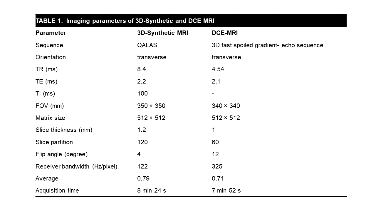
Table 1. Imaging parameters for 3D synthetic and dynamic
contrast-enhanced imaging.
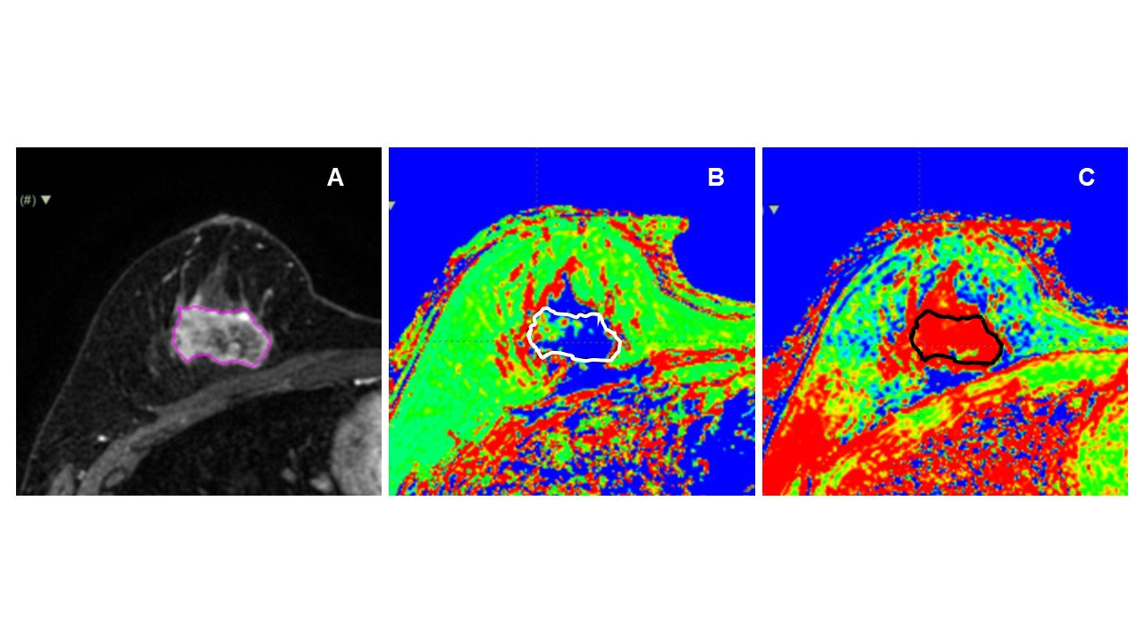
Figure 1. A. The 3D ROI is placed semiautomatically on the
dynamic contrast-enhanced images registered with synthetic T1-weighted images
(not shown). B. T1 map, C. T2 map.
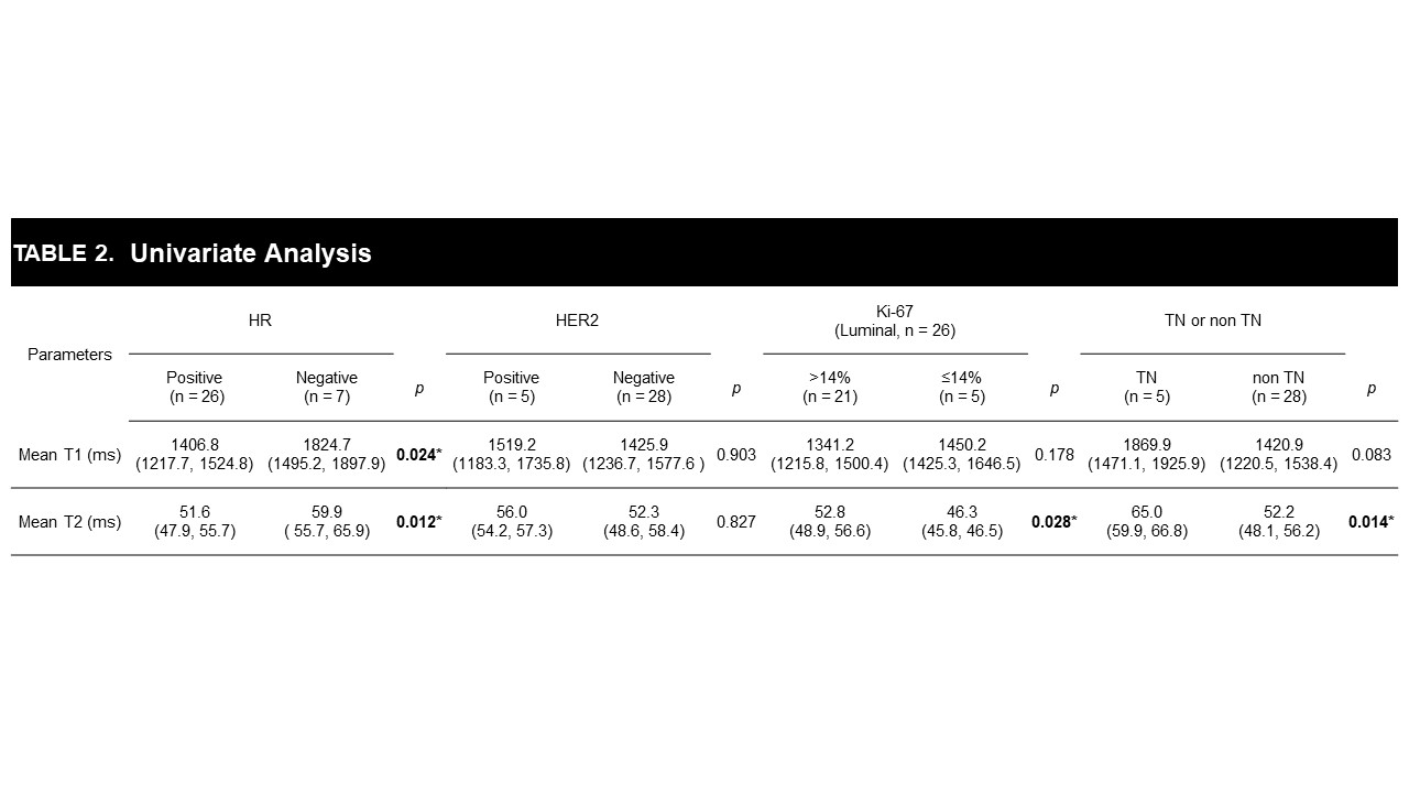
Table 2. Relationship between T1 or T2 values of invasive
breast carcinoma (IDC) and immunohistochemical (IHC) findings. There were
significant differences in T1 and T2 according to the IHC status of IDC (p
< 0.05).
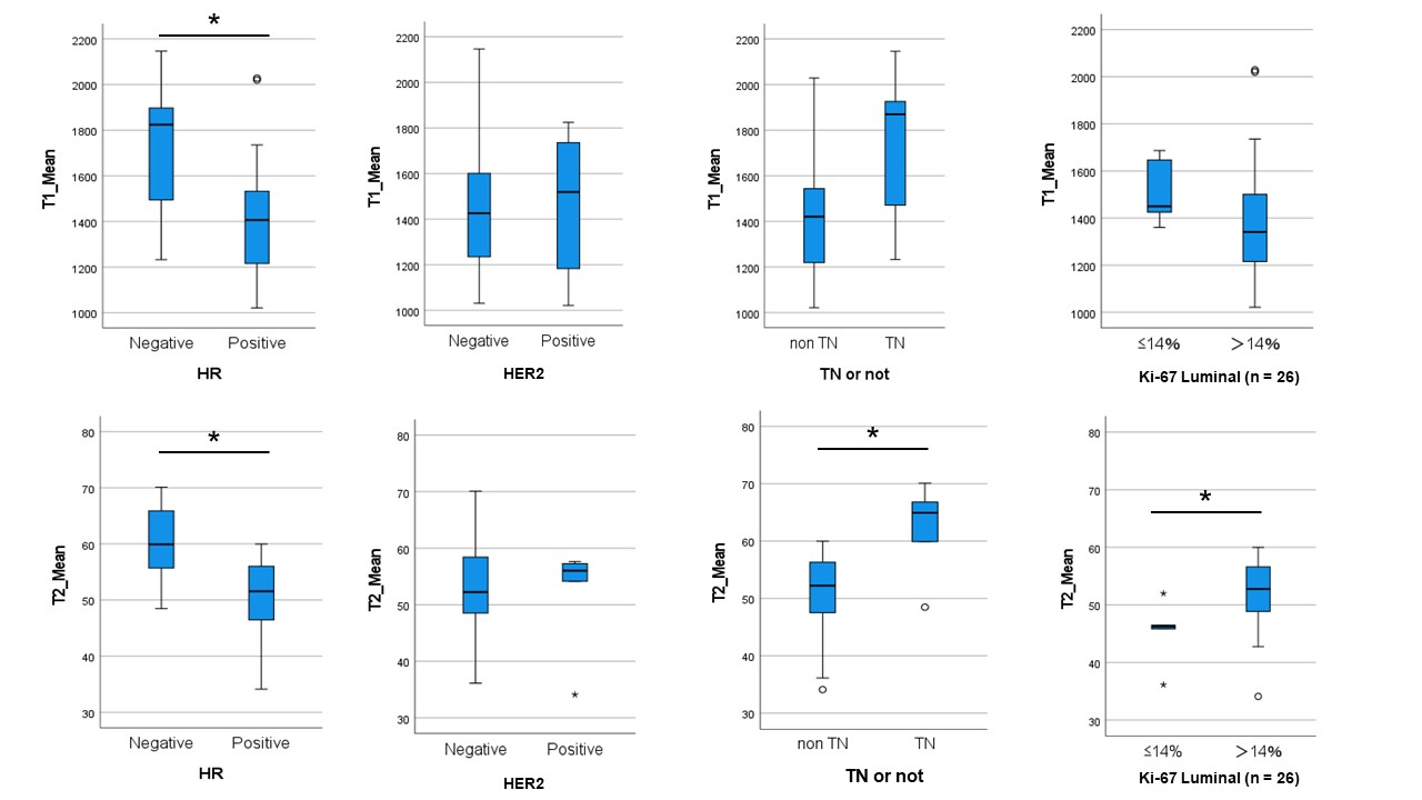
Figure 2. The mean T1 value differs only between hormone
receptor (HR)-positive and -negative status (*p = 0.024), while the mean
T2 value differs between HR-positive and -negative status (*p = 0.012),
triple-negative status and HR-positive or HER2-positive status (*p =
0.014), and the Ki-67 level >14% and £14% (*p = 0.028).
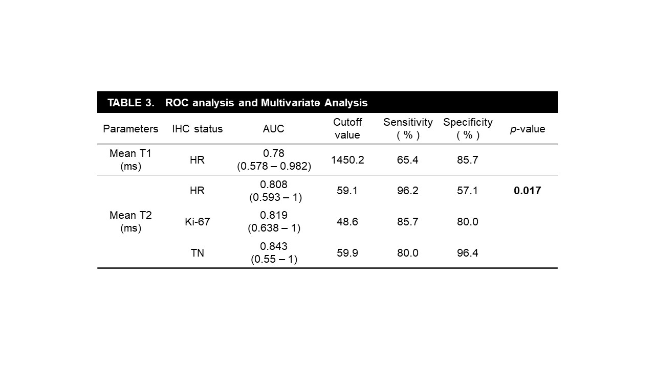
Table 3. ROC and multivariate analyses of the relationship
between T1 or T2 values of the invasive breast carcinoma (IDC) and immunohistochemical (IHC) findings. The mean T1
and T2 values acquired by synthetic quantitative 3D MRI discriminated the IHC
status of IDC.
DOI: https://doi.org/10.58530/2023/3462