3419
Investigation of Susceptibility Gradient Artifacts in EPI at 7T
Chan Hong Moon1, Hoby Hetherington2,3, and Jullie W. Pan2,4
1MRRC, Department of Radiology, University of Pittsburgh, Pittsburgh, PA, United States, 2NEXTGEN, Department of Radiology, University of Missouri, Columbia, MO, United States, 3Resonance Research, Inc., Billerica, MA, United States, 4University of Missouri, Columbia, MO, United States
1MRRC, Department of Radiology, University of Pittsburgh, Pittsburgh, PA, United States, 2NEXTGEN, Department of Radiology, University of Missouri, Columbia, MO, United States, 3Resonance Research, Inc., Billerica, MA, United States, 4University of Missouri, Columbia, MO, United States
Synopsis
Keywords: Artifacts, High-Field MRI, EPI, 7T, Susceptibility, GRAPPA, Aliasing
With the development of high-field 7T scanners, interest in high resolution neuroimaging for EPI-based functional and diffusion MRI has increased. However, EPI artifacts at 7T (spatial distortion and signal loss) increase compared to that seen at 3T due to increased B0 inhomogeneity. Furthermore, EPI artifacts become more significant and complicated when parallel imaging is used. As a result, 7T fMRI and DWI suffers from B0 inhomogeneity-induced aliasing as well as the more common EPI artifacts. In this study, the source of B0 inhomogeneity artifact in parallel imaging was investigated and methods to reduce these artifacts are proposed/tested at 7T.Introduction
EPI is known to suffer from spatial distortion and signal dropout in high B0 inhomogeneity (dB0) brain regions (eyeball, pre-frontal cortex and nasal cavities). Such artifacts are commonly worse at higher field. Parallel imaging (PI) such as GRAPPA achieves faster phase/slice encoding through k-space subsampling. This decreases artifacts but can cause additional artifacts - aliasing/ghosting. This is particularly problematic at 7T because of the different spectral characteristics between calibration and imaging data. dB0 consists of an offset as well as linear/nonlinear gradients. At 7T, EPI artifacts in those regions need to be minimized or compensated for in fMRI and DWI scans. In this study, we investigated the susceptibility gradient effects on GRAPPA at 7T in GE- and SE-EPI, and proposed higher order B0 shimming and apodization to reduce the artifacts.Theory
It is well known that EPI phase encoding (PE) is slow such that the sampling bandwidth is narrow, which causes spatial distortion in PE of the image. In addition to spatial encoding gradient, B0 susceptibility gradient (dB0/dy) produces an effective third encoding. This incidental encoding can be identified (from eyeball and nasal cavities) even if no PE is performed (Fig. 2, middle). This effect can be better identified in SE-EPI where all signals (both imaging and susceptibility gradients) are recorded at TE, but the shape of the object is distorted resulting in compression or expansion (Fig. 2A). This effect in a GE-EPI results in the echo being shifted away from TE where the imaging echo is generated (Fig. 1B). If dB0/dy is high compared to the spatial encoding gradient, dB0/dy echo could be truncated or not sampled in acquisition, i.e., outside temporal acquisition window of EPI signal during PE. This type of signal loss occurs frequently in 7T EPI. In GRAPPA, the loss due to the susceptibility echo can cause GRAPPA kernel mismatch between calibration data (ACS) and subsampled imaging data, generating susceptibility-based aliasing artifacts (Fig. 2, right).Methods
In silico, the susceptibility generated echo shift and distortion due to dB0/dy was simulated using Bloch equation. All data were acquired on an 8ch pTx 7T Magnetom (Siemens) with vendor 1st - 2nd shims and a high degree head shim insert coil (Resonance Research Inc.) with 3rd and 4th degree shims1. Dynamic shim updating (DSU) of all shims over each imaging slice was achieved using an RRI power and console dynamically controlled via a trigger signal synched with sequence1-2. The shim optimization was performed over the entire 3D volume for ‘static’ and ‘dynamic’ multi-slice shimming. EPI data were acquired at 2-mm, 96x96 matrix and acceleration factor 3 under static 1st-2nd, static1st- 4th+, and DSU 1st-4th+ order shims. To visualize imaging artifacts due to intrinsic susceptibility gradients, the conditions of a) A>P (“expanded” eyeball), b) P>A (“compressed” eyeball) and c) no PE was applied in EPI sequence. All data were acquired using a 16ch transceiver.Results
Simulated GE-EPI with dB0/dy shows that susceptibility echo separates from imaging echo, occurring either prior to or after TE. Further, the shape of that echo is distorted (Fig. 1B). Extending PE window can sample dB0/dy signal, but this also produces ringing artifacts. dB0/dy can produce spatial information of susceptibility gradient objects such as eyeball and the region close to nasal cavity (Fig. 2, middle). dB0/dy distorts the susceptibility object, e.g., eyeball with A>P encoding (“expanded”) and P>A (“compressed”) (Fig. 2, left). No artifact is found in expanded object image except echo shrinks in PE (Fig. 2, top-left), while ringing artifacts appear in compressed object image (echo expansion in k-space PE) because the susceptibility echo is truncated given low bandwidth sampling of ACS data (Fig. 2, bottom-left). Similar distortion artifacts are shown in GRAPPA images but with additional aliasing from the eyeball (Fig. 2, bottom-right). These artifacts can be reduced with higher order shimming. Fig. 3 shows that use of the high order shim and dynamic shimming can remove the aliasing (green circles) as well as reduce the extent of distortion (yellow contours) and signal loss (red contours). However, not all such aliasing and “zebra striping” effects in the SE- and GE-EPI respectively, can be removed (Fig. 4). The residual artifacts can be further improved by selecting the preferred A>P or P>A direction, dependent on shim performance and slice within TR, and k-space apodizing in PE.Discussion/Conclusions
Susceptibility gradient artifacts in EPI were demonstrated at 7T. As shown, the severity can be significantly decreased by using advanced shim and dynamic updating of the shims. Given the nature of susceptibility gradient signal in SE- and GE-EPI, changing basic aspects of the sequence and parameters (e.g., longer ACS time window, PE direction, extended PE acquisition, etc.) can also reduce the artifacts. However, some of the necessary parameter changes can be difficult for EPI scans, given their relatively short TE, 20ms and high resolution, <1.5-mm isotropic. There are new GRAPPA strategies which produce fewer susceptibility artifacts3. However, the methods proposed in this work can improve EPI quality (less artifact and higher signal in susceptibility regions) while preserving standard EPI acquisition. These methods are important given the clinical interest in 7T BOLD and tractography DWI.Acknowledgements
This work was supported by NIH EB011639, EB009871, NS090417, and NS081772.References
1. Moon CH, Schwerter M, Pan JW , Shah NJ, and Hetherington H, Constrained optimization for static and dynamic B0 shimming, Proc. Intl. Soc. Mag. Reson. Med., 26, 2018.
2. Hetherington HP, Moon CH, Schwerter M, Shah NJ, Pan JW. Dynamic B0 shimming for multiband imaging using high order spherical harmonic shims. Magnet Reson Med. 2020; doi: 10.1002/mrm.28438.
3. Hoge SW, Polimeni JR. Dual-polarity GRAPPA for simultaneous reconstruction and ghost correction of echo planar imaging data. Magnetic Resonance in Medicine. 2016;76(1):32–44. doi: 10.1002/mrm.25839.
Figures
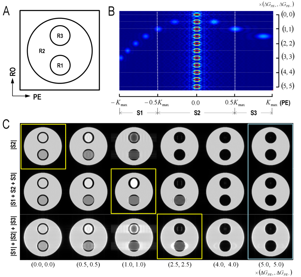
Figure 1. EPI simulation of GE-EPI under different strength and polarity of susceptibility gradient (dB0/dy). (A) Phantom; R2, R1 & R3 w/ zero, positive & negative dB0/dy, respectively, (B) Echoes generated at different dB0/dy, and (C) Reconstructed images. S1, S2, and S3 is different acquisition time window. The time window of echo acquisition was adjusted to see if the extended PE acquisition could compensate loss of dB0/dy signal.
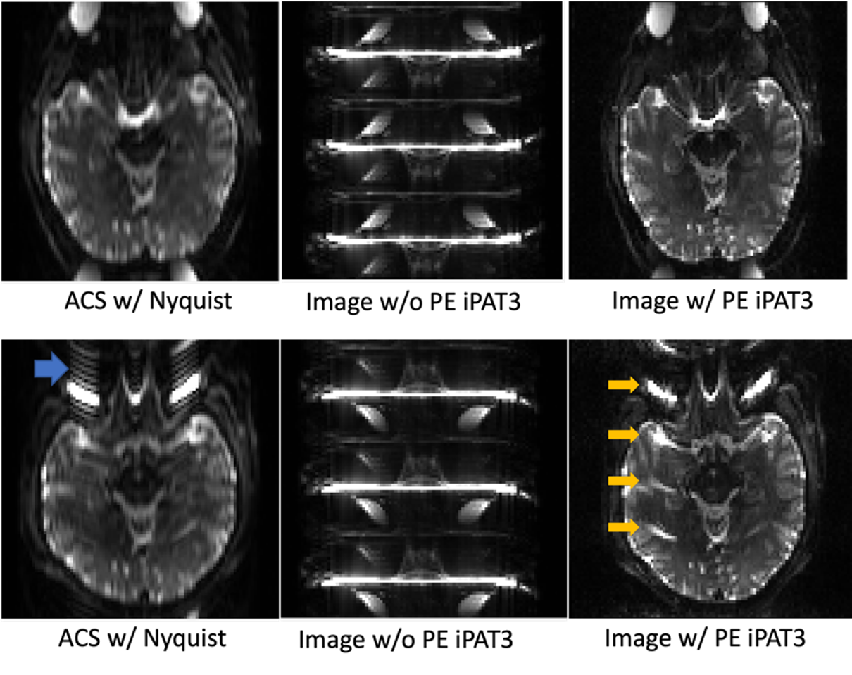
Figure 2. SE-EPI at 7T to demonstrate the artifacts from susceptibility gradient. (Top, Bottom panels) EPI with A>P and P>A PE direction. (Left panel) ACS images w/ Nyquist sampling, (middle) GRAPPA images w/o PE, and (right) GRAPPA images w/ PE. Cyan arrow indicates truncation ringing artifacts, and yellow does aliasing in GRAPPA due to susceptibility gradient.
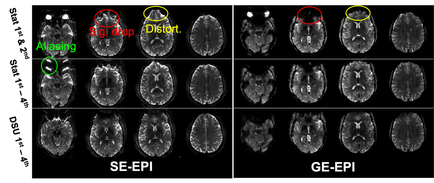
Figure 3. Compensation of susceptibility gradient artifacts in SE-EPI (upper) and GE-EPI (lower) by high order B0 shimming. In each EPI, static 2nd(top), 4th+(middle) and DSU 4th+(lower) was applied. PE is applied in A>P direction.
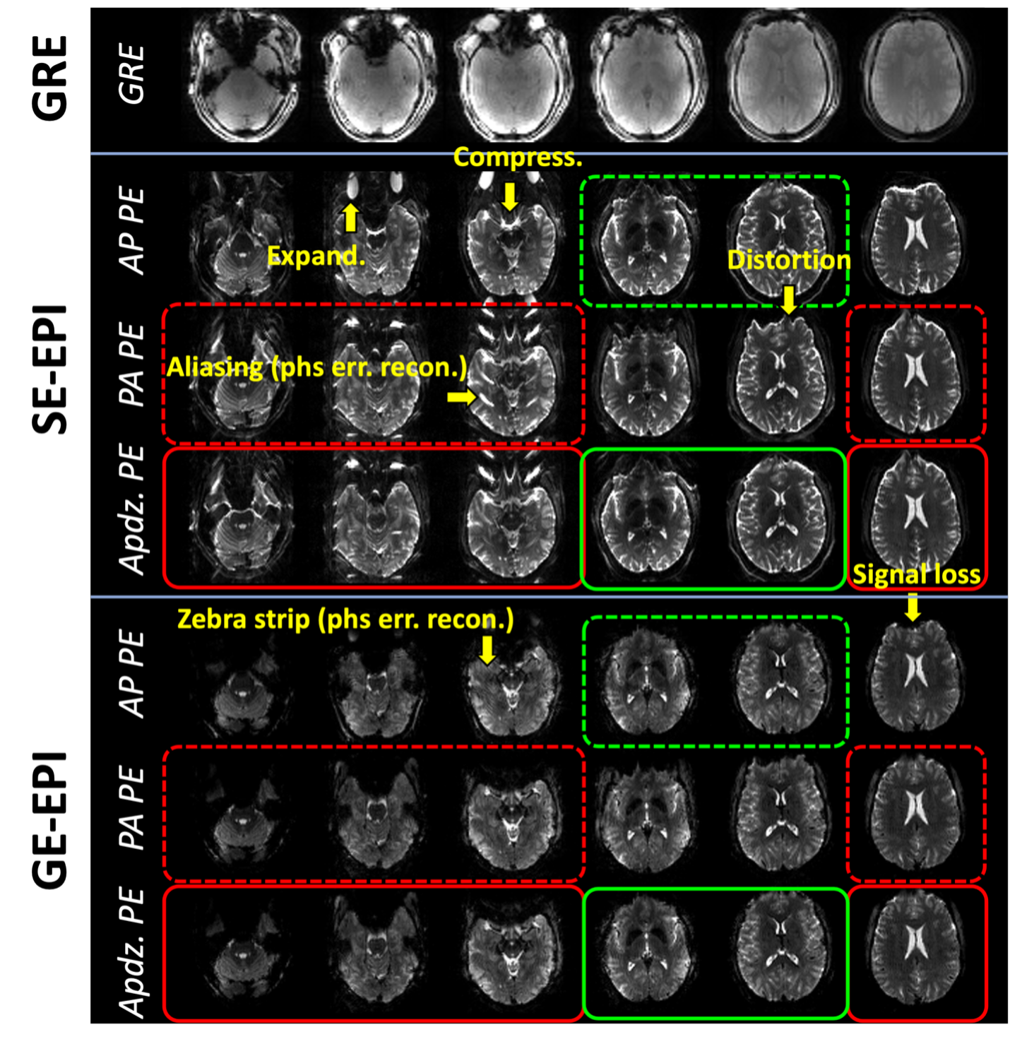
Figure 4. Further reduction of residual artifacts due to susceptibility gradient in EPI by using 1) DSU B0 high order shimming, 2) flexible PE direction (A>P – green box or P>A – red box) in an imaging volume of EPI, TR, and 3) one-side apodizing filter in PE k-space was applied. In GE-EPI, the aliasing artifact in SE-EPI changes to zerbra stripping artifact.
DOI: https://doi.org/10.58530/2023/3419