3415
Denoising of 7T MP2RAGE MRI with Preserving the Brain Signal
Kwan-Jin Jung1
1Biomedical Imaging Center, Beckman Institute, University of Illinois at Urbana-Champaign, Urbana, IL, United States
1Biomedical Imaging Center, Beckman Institute, University of Illinois at Urbana-Champaign, Urbana, IL, United States
Synopsis
Keywords: Artifacts, High-Field MRI, MP2RAGE, 7T
MP2RAGE requires denoising of the synthesized T1-weighted image. The conventional denoising method is to add a constant in the synthesis that alters the brain signal. This affects brain segmentation. A new denoising method has been developed to denoise it with preserving the brain signal. The new method estimates the bias field from two inversion images and sets the noise floors at the low and high noise levels in proportion to the bias field. The denoise weight is calculated at each voxel using the two noise floors. The proposed denoising method worked reliably with preserving the brain signal.Introduction
The MP2RAGE obtains two images, i.e., INV1 and INV2, at two different inversion times, and the two images are synthesized to a T1-weighted image, i.e., UNI. This synthesized image suffers from noise in the background and low signal in some brain regions. The amplified noise of the UNI image can be denoised by using a simple approach of modifying the synthesis equation with the addition of a constant [1]. However, this method results in changing the brain signal of the UNI image, which affects the brain segmentation and other analyses [2]. The purpose of this study is to develop a new denoising method of the UNI image while preserving the brain signal.Methods
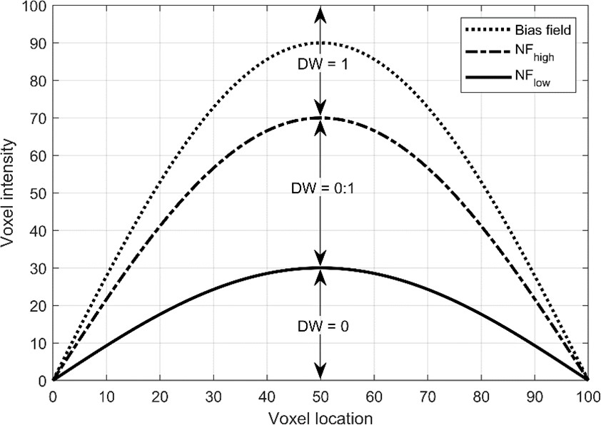
The noise of the UNI image was denoised by introducing a constant term b as in the following equation (1) [1]: UNI = [real(INV1* x INV2) - b x INV2mean] / [|INV1|2 + |INV2|2 + 2b x INV2mean] , (1)where INV2mean denotes a mean of the INV2 image and is introduced here to make the added constant proportional to the image intensity. This conventional denoising method is named ‘KO’. The effect of b on the UNI image is amplified when the image intensity of INV1 and INV2 is low. The noise in the UNI image can be directly removed by thresholding the UNI image without changing the synthesis equation. However, the noise is superimposed on the bias field that stems from the nonuniformity of the RF transmit and receive sensitivity. Therefore, the noise threshold needs to be spatially dependent. The bias field is used to construct two noise floors of low and high thresholds (Figure 1). The two noise floors are obtained by multiplying the bias field with the user-provided noise range. The denoise weight, i.e., DW, in each voxel is determined by the noise floors using the following equation: DW = 0 when a ≤ 0; DW = α when 0 < α < 1; DW = 1 when α ≥1, where a =(SoS-NFlow) / (NFhigh - NFlow). (2)
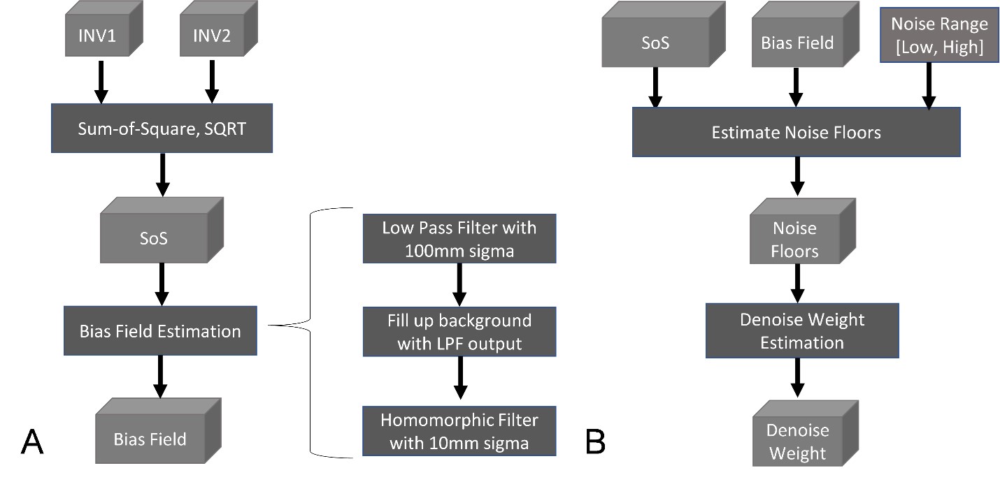
The proposed method affects only the voxel in which the intensity is within the noise window that is spatially dependent. This proposed method is named ‘KJ’ for convenience. The bias field is estimated from the square-root of the sum-of-square, SoS, of INV1 and INV2 to increase the signal using a background-filled homomorphic filter, as illustrated in Figure 2A. SoS was filtered with a Gaussian kernel of sigma = 100 mm and this lowpass-filtered image is filled into the background of the SoS image for the next step of the homomorphic filter with sigma = 10 mm. The lowpass-filtered image from the homomorphic filter is used as an estimated bias field. The bias field is multiplied with the input parameters for the low and high noise floors, as illustrated in Figure 2B. From the two noise floors, a 3-dimensional volume of the denoise weight is constructed. The proposed KJ method was compared with the conventional KO method using 7T MP2RAGE images of 53 subjects [3]. All 53 subjects were processed with equal processing conditions and compared for brain segmentation using FreeSurfer v.7 [4].
Results
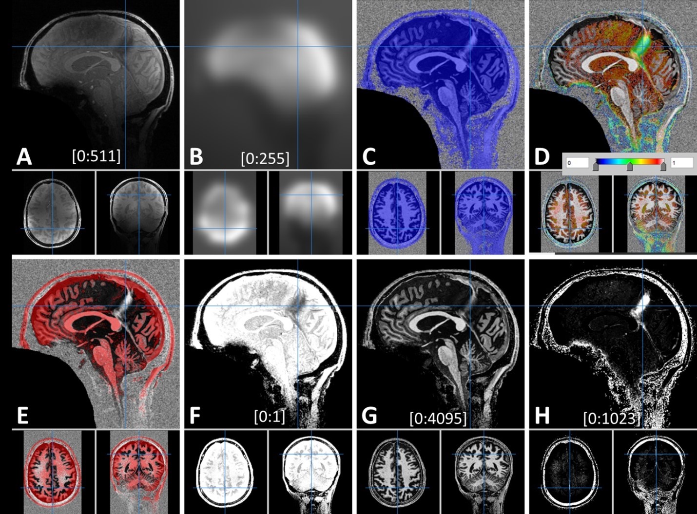
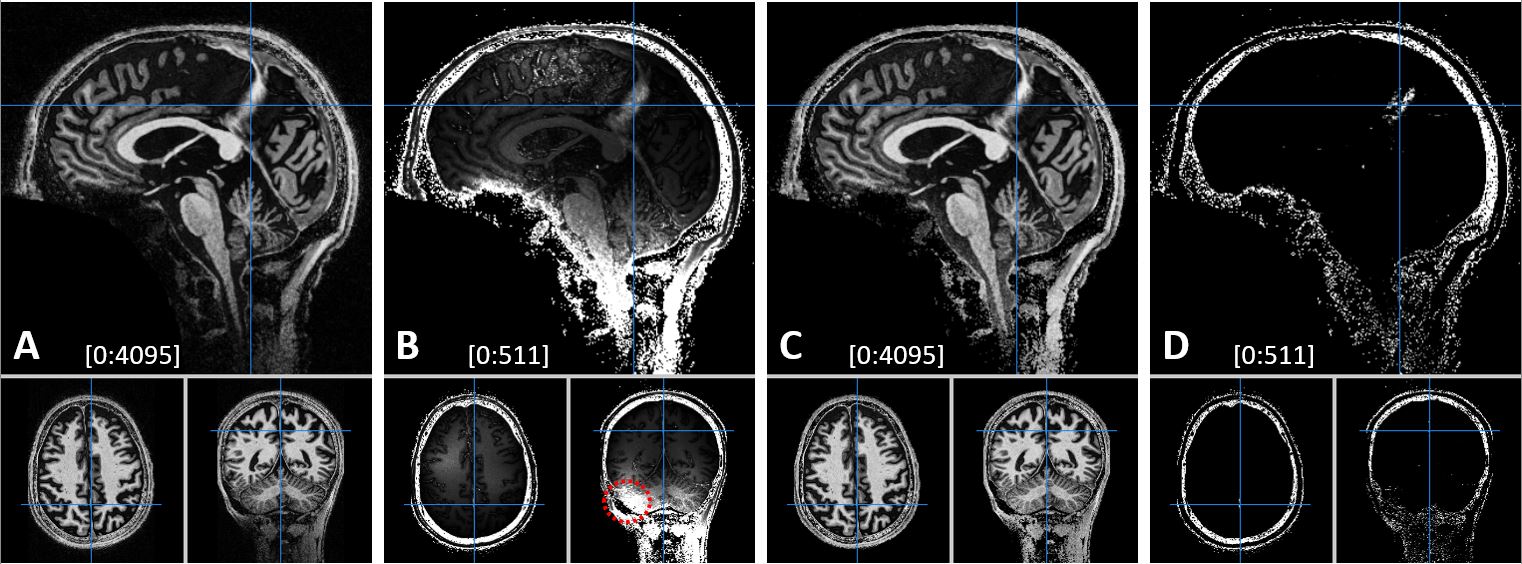
The bias field estimated from the SoS image (Figure 3A) is shown in Figure 3B. The noise floors were overlaid on the UNI image (Figures 3C, 3D, and 3E). The resulting denoise weight cleaned the noise not only in the background but also in the brain region of a low signal as shown in Figure 3E. The denoised UNI image and the difference between the UNI and the denoise image are shown in Figure 3G and 3H, respectively. The change of brain signal achieved by the denoising method was compared between the conventional KO and the proposed KJ methods in Figure 4. The effect of denoising on brain segmentation is shown in Figure 5.Discussion
It was confirmed that the estimated bias field in this proposed method was partially like the measured B1 transmit field using the LAYNII sample data [5]. It was noted that the acceptable b in equation (1), varies significantly depending on the data set, even with using the mean value of the INV2 image. For example, it was necessary to increase b to 20 to suppress the background noise and head cushions in the LAYNII sample data. It was noticed that the bias field was significantly asymmetrical laterally at the cerebellar level, suggesting that the B1 field at 7-Tesla was compromised at this level. The artifactually pronounced signal in some brain regions, such as in the longitudinal fissure, might need extra precautions during the interpretation of the MP2RAGE image for diagnosis.Conclusion
The conventional denoising method of the MP2RAGE decreases the image intensity of the synthesized T1-weighted UNI image, which affects brain segmentation. The proposed method can denoise the UNI image while preserving the brain signal at a reliable range of the processing parameters. Furthermore, it can suppress artifactual image intensities in the longitudinal fissure at a minimum effect on other brain signals.Acknowledgements
No acknowledgement found.References
1. O'Brien, K.R., et al., Robust T1-weighted structural brain imaging and morphometry at 7T using MP2RAGE. PLoS One, 2014. 9(6): p. e99676.
2. Haast, R.A.M., D. Ivanov, and K. Uludag, The impact of B1+ correction on MP2RAGE cortical T1 and apparent cortical thickness at 7T. Hum Brain Mapp, 2018. 39(6): p. 2412-2425.
3. Forstmann, B.U., et al., Multi-modal ultra-high resolution structural 7-Tesla MRI data repository. Sci Data, 2014. 1: p. 140050.
4. Dale, A.M., B. Fischl, and M.I. Sereno, Cortical surface-based analysis. I. Segmentation and surface reconstruction. Neuroimage, 1999. 9(2): p. 179-94.
5. Huber, L.R., et al., LayNii: A software suite for layer-fMRI. Neuroimage, 2021. 237: p. 118091.
Figures
Figure
1. The basic concept of
the denoise weighting of UNI image. DW stands for denoise weight. NFlow
and NFhigh denote the noise floor for the low and high fraction of
the estimated bias field, respectively.
Figure
2. Functional block
diagrams of the proposed denoising method. (A) Estimation of the bias field
from INV1 and INV2 images. (B) Construction of the denoise weight from the
previously estimated bias field and the selected noise range.
Figure
3. A set of images of the
proposed KJ method. (A) Sum-of-Square-Squared (SoS), (B) Bias field, (C) Low
noise floor at 0.3 fractions of the bias field, (D) Transition noise floor, (E)
High noise floor at a 1.2 fraction of the bias field, (F) Denoise weight, (G)
The denoised UNI image, and (H) A difference from the UNI image within the
brain. The noise floors are overlaid on the UNI image. The numbers in the
bracket in each image denote the display window.
Figure
4. A comparison of the
brain signal change between the conventional KO method with b = 3 (A and B) and the proposed KJ method with
the noise floors at 0.3 and 0.7 (C and D). (B) and (D) are the difference from the UNI
image within the brain. The numbers in the bracket in each image denote the
display window. Note the large difference on the cerebellum marked with a
dotted circle in B.
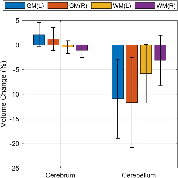
Figure
5. An effect of the
denoising method KO on brain segmentation. The volume change was in reference
to the proposed KJ method. The segmented volumes were noticeably affected by
the denoising method, particularly at the cerebellum. The black line in each
bar denotes the standard deviation.
DOI: https://doi.org/10.58530/2023/3415