3414
RF Interference Manifested Zipper Detection And Removal Using Channel Compression
Megha Goel1, Preetham Shankpal1, Suresh Emmanuel Joel1, Rajagopalan Sundareshan1, and Harsh Agarwal1
1GE Healthcare, Bengaluru, India
1GE Healthcare, Bengaluru, India
Synopsis
Keywords: Artifacts, Image Reconstruction, Zipper-removal
Zipper artifacts are commonly seen in MR images due to spurious radio-frequency signals or improper RF-shielding. Zipper-riddled images lose diagnostic value and are usually sent for rescan. Here, we attempt to mitigate zipper artifacts in the post-processing pipeline after scan has been acquired. We do this after channel combination technique has limited zipper appearance to 1-2 pseudo-channels, which we detect and remove from channel-combination process. We evaluated this on various brain contrasts and confirm reduction of zipper presence visually in the images. Given that zippers manifest as bright/dark discontinuous lines irrespective of the anatomy/contrast, the method should be generalizable.INTRODUCTION
Zipper artifacts1,2 arise due to narrow band unwarranted RF received by the receiver coil/s. The common sources of these narrow band RF include improper closure of RF shield door, untested power supply of the device kept in the shield room and improper routing of cable inside the shield room. Zipper riddled images lose diagnostic value and significant amount of time is spent by the MR technologist to detect and remove the source of RF interference. If the source of RF interference is not detected, then scanning session is suspended awaiting vendor support. In this abstract, we have proposed method to mitigate zipper artifacts during MR image reconstruction after the scan has been acquired. The proposed method can be potentially used in shield-less MR and high RF interference environments.METHODS
Diagnostic MR images are typically acquired with multi-channel receive coils. In order to minimize the computational need of MR image reconstruction, channel compression3 is done wherein information from different channels is re-distributed using singular-value-decomposition to generate virtual channel. Selected few virtual-channels were used in MR image reconstruction resulting in channel compression or reduction. We have observed that the zipper artifacts which were present in each of the native receive coil image was present in only one or two of the virtual channels which could be due to the fact that the RF interference received by multi-channel receive coil is highly correlated. Here we propose a two-step method,Zipper manifested virtual channel detection: Zipper dominated virtual channels were detected in this step. As shown in Figure 1, a custom zipper amplitude detection algorithm has been implemented, which analyzes the mean intensity profiles of each virtual-channel image. The mean intensity has been taken along the phase-encode direction, since zippers will manifest as sharp discontinuities here.
Zipper manifested virtual-channel removal: Zipper corrupted virtual-channels were dropped during channel compression step of MR image reconstruction
RESULTS AND DISCUSSION
Institution’s IRB board approved study was conducted on a volunteer being scanned at the research 0.5T MRI scanner. The RF interference was injected into the system through a metallic pipe which is routed in the MR shield room without the waveguide. Common brain MR contrasts (T2w, T2-FLAIR, BRAVO) were acquired using 6 channels of the 14-channel research HNU coil surrounding brain anatomy.Figure 2 shows the virtual-coil channel images of one slice and highlight two of those virtual channels, in one of which zippers are present. The projection image collapsed along the readout direction shows spike in underlying anatomy depicting the pixel/frequency of the RF interference which is causing zipper artifact.
Zipper artifact removal is demonstrated in the T2 FLAIR MRI in Figure 3. This method should work well for any anatomy in general, given that zippers manifest as bright or dark discontinuous lines in any MR image, irrespective of the anatomy or contrast.
A point worth mentioning here would be that this method promises to be most impactful in acquisitions where there are fewer coils/channels (2-8) left after channel compression, since with fewer channels to derive information from, zipper effects are more distinguishably noticed.
RF interference is typically mitigated by better RF shielding1,2, using hardware methods which are restrictive and expensive. This method uses legacy recon methods to condense zipper artefacts to 1 or 2 pseudo-channels of the acquisition, and by removing these corrupted pseudo-channels, reduces zipper artefacts with minimal loss in signal.
The proposed method can lead to potential loss of anatomy if significant imaging signal is present in the zipper riddled virtual channel. Also the proposed method can be employed only with multi-channel receive system.
CONCLUSION
The proposed strategy for zipper detection and removal proves to be a viable solution for post MR acquisition image quality enhancement. It can help with patient recall/rescan, since zipper affected scans are usually outright rejected by radiologists. Usually zipper removal is done through hardware modifications, checks for spurious data1,2, etc. Using channel compression in combination with line detection for zipper removal has not been attempted before as per our knowledge.Acknowledgements
No acknowledgement found.References
[1] Stadler, Alfred, et al. "Artifacts in body MR imaging: their appearance and how to eliminate them." European radiology 17.5 (2007): 1242-1255.
[2] Yanasak, Nathan E., and Michael J. Kelly. "MR imaging artifacts and parallel imaging techniques with calibration scanning: a new twist on old problems." Radiographics 34.2 (2014): 532-548.
[3] Huang, Feng, Sathya Vijayakumar, and James Akao. "Software compression for partially parallel imaging with multi-channels." 2005 IEEE Engineering in Medicine and Biology 27th Annual Conference. IEEE, 2006.
[4] Oliphant, Travis E. "Python for scientific computing." Computing in science & engineering 9.3 (2007): 10-20.
Figures
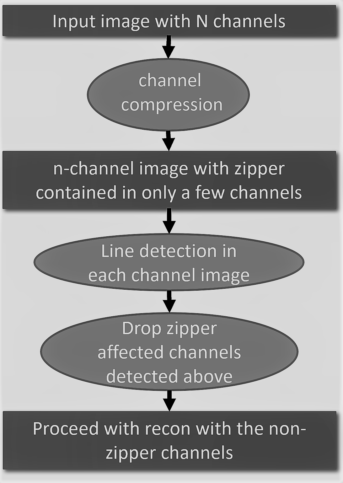
Fig. 1: Algorithm for RF Interference detection
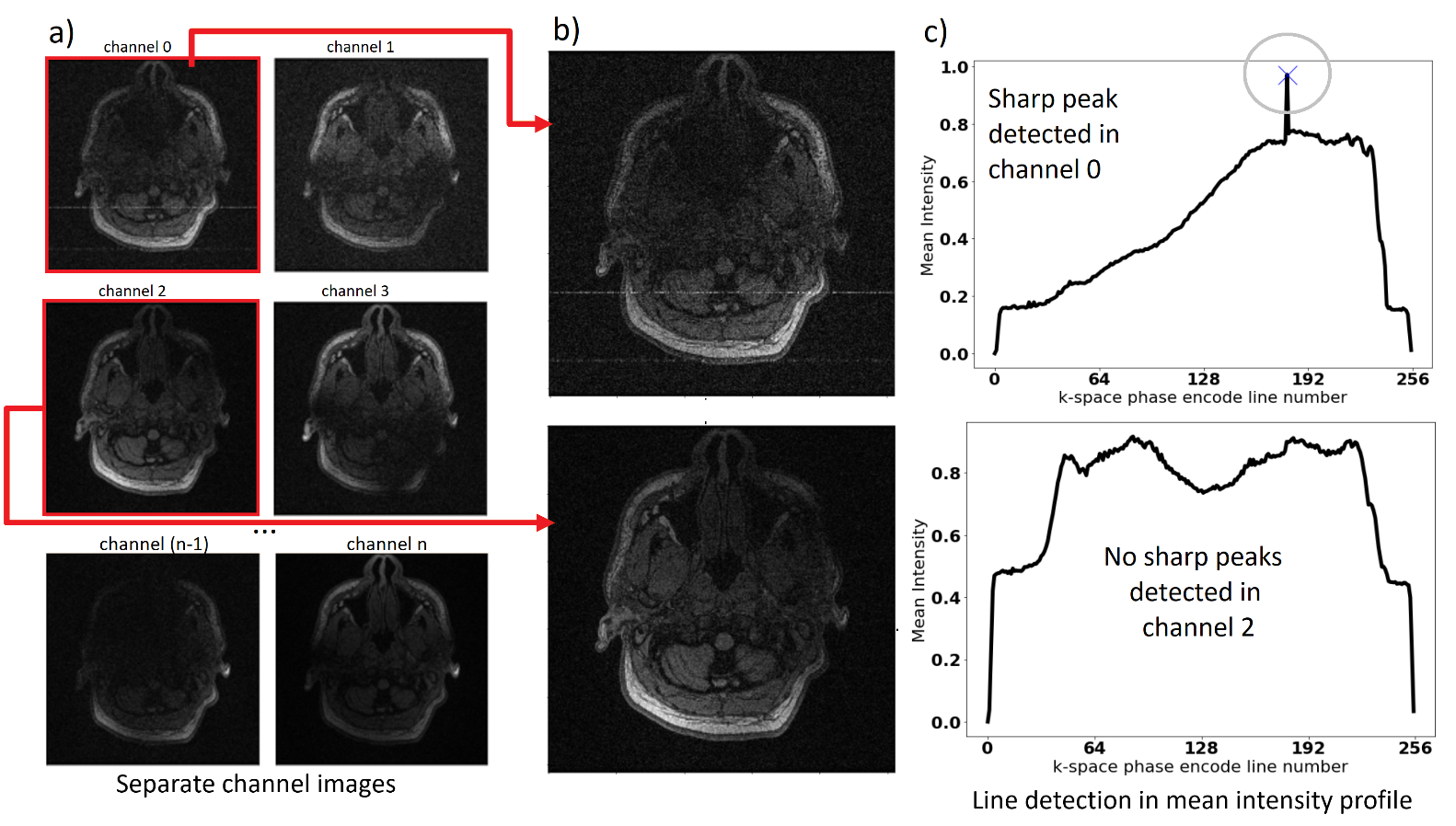
Fig. 2: The figure explains the working of the algorithm: The six images shown in (a) are different coil images acquired. (b) Each image is run
through the algorithm separately to determine the presence/absence of zipper
artifact. (c) Shows how the algorithm uses the mean intensity profile along the phase encode direction to determine presence of zipper by highlighting sharp
peaks in the profile. Here, channel 0 would be dropped and not considered for
further processing.
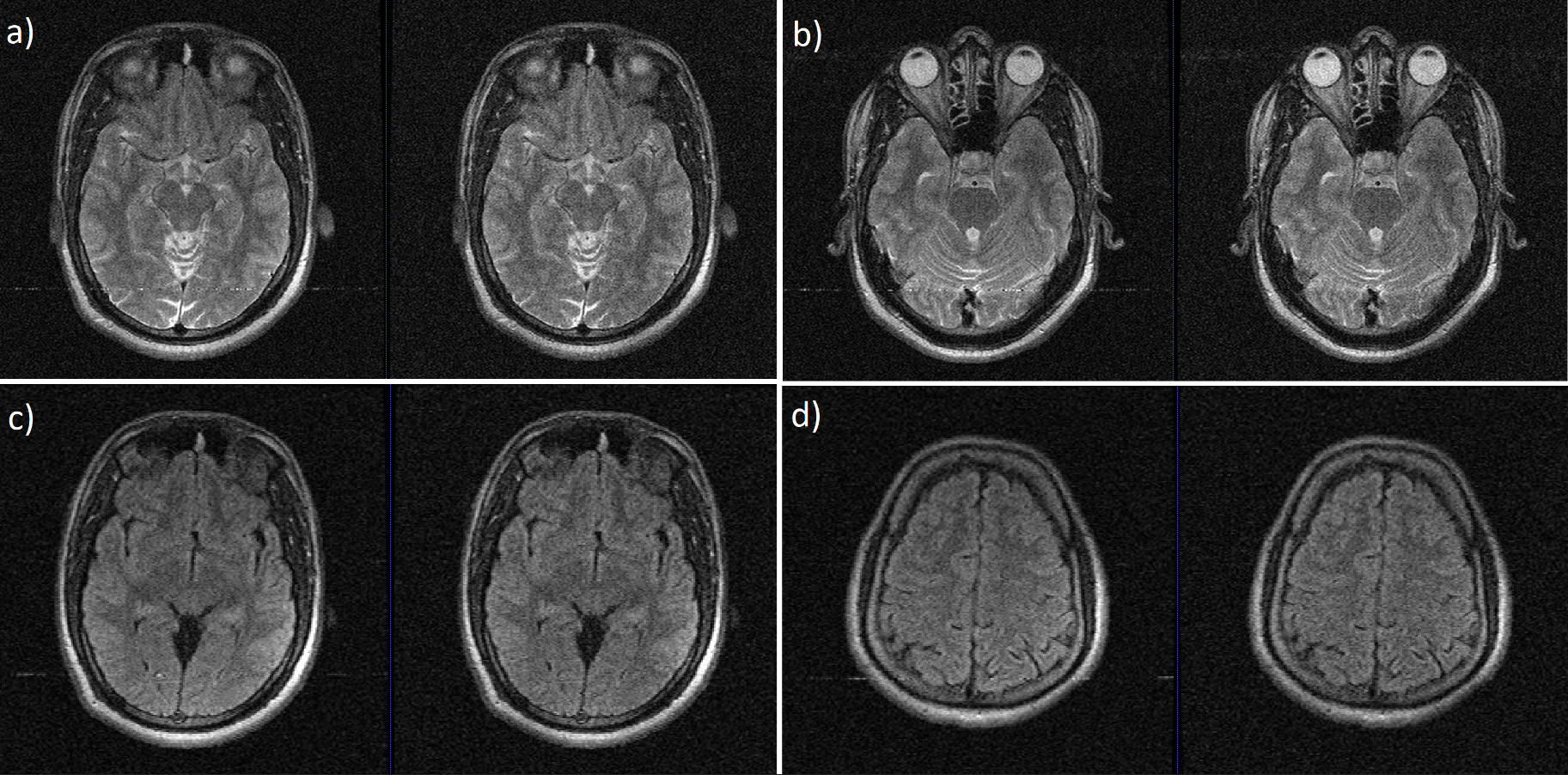
Fig. 3: The figure above shows the pre and post zipper correction results using our algorithm on two scans acquired with RF interference. The left side images are those reconstructed using all the coils, while the right-side images are reconstructed after zipper riddled virtual channel images have been found and removed by the algorithm. (a) and (b) show slices belonging to a T2-FSE scan, while (c) and (d) belong to a T2-FLAIR scan.
DOI: https://doi.org/10.58530/2023/3414