3359
ROI-based morphometry brain neuroimaging reveals grey and white matter abnormalities in type 2 and 3 juvenile SMA patients1The First Affiliated Hospital of Sun Yat-Sen University, Guangzhou, China, 2ge health care, Guangzhou, China
Synopsis
Keywords: Gray Matter, Gray Matter, SMA,FreeSurfer,cerebral cortex
The study investigated brain structure changes in type 2 and 3 SMA patients and their correlation with the severity of clinical symptoms. MRI examinations were performed on 3.0T MRI scanner and 3D T1W data were analyzed using FreeSurfer.The result showed SMA patients had extensive, multifocal, symmetrical gray white matter degeneration.The postcentral gyrus is strongly associated with symptom severity in SMA patients. It might be explained by the fact that motor neurons exhibit functional alterations, leading to selective motor neuron loss. So it is crucial for brain development and cognitive development if we intervene in SMA disease in an early stage.Introduction
Spinal muscular atrophy (SMA) is an autosomal recessive motor neuron disease characterized by lower motor neuron degeneration because of the loss of function of the survival-motor-neuron 1 (SMN1) gene1-3. Besides the spine, a high level of SMN protein is also required for typical brain development in vivo and, as a result, reduced expression of SMN protein causes atypical brain development 4. Therefore, we believe that SMA not only affects motor function, but also affects central nervous system development. However, there are few neuroimaging studies in patients with SMA , and most neuroimaging studies involved type 3 or 4 adult patients. To this moment there is no voxel-based morphometry neuroimaging study published in a cohort of type 2 and 3 juvenile SMA patients. Hence, in this study, we are going to discuss the changes in brain gray and white matter structure in SMA patients and their correlation with the severity of clinical symptoms.Methods
MRI examinations of all participants were performed on a 3.0T scanner (SIGNA Pioneer GE Healthcare, WI, USA) using 32-channel head coils. Three-dimensional, high-resolution anatomical scans were acquired with the following parameters: repetition time=7.5 ms, echo time=3.1 ms, flip angle=12°, 188 sagittal slices with slice thickness=1 mm with no slice gap, a field of view=256×256 mm2, and data matrix=256×256. For FreeSurfer analyses, the steps are as shown in the article5. The inclusion criteria were: 1) those with type 2 or type 3 SMA patients.; 2) the age of the patients ranged from 5-17; 3) right-handed. The exclusion criteria were:1) contraindication to MRI; 2) previously treated with nusinersen; 3) with a history of brain injury or other psychiatric, neurological disorders. After the exclusion of unacceptable cases, 43 SMA patients and 37 age- and sex-paired healthy controls (HCs) were finally involved. HFMSE(Hammersmith Functional Motor Scale-Expanded) is a clinical assessment of motor function, it is especially validated for use in patients with SMA to assess activities related to daily living. 43 SMA patients were all evaluated using HFMSE. 22 patients use SPM(RavensStandard Progressive Matrices) to evaluate intelligence as shown in Table 1.Results
1. The comparison between SMA patients and HC revealed significantly higher cortical thickness of right hemibrain and whole brain, especially in the frontal, parietal and temporal lobes. What’s more, the cortical volumetric stats of bilateral middle temporal gyrus, lateral aspect of the right superior temporal gyrus, right straight gyrus of SMA patients is higher than that of HC.2. Bilateral white matter surface area stats of SMA patients were lower than HC, especially in frontal, parietal and temporal lobes. We found generalized white matter volume loss in patients with SMA, especially in left parstriangularis, supramarginal, right parsopercularis, postcentral, precentral gyrus.
3. The subcortical volumetric stats of bilateral pallidum, and right hippocampus were higher than that in HC, while that of the central corpus callosum as well as middle anterior corpus callosum were lower than that in Hc.
4. Significant regression models revealed increased right postcentral gyrus superficial area, and right central sulcus volume, besides, white matter segmentation volumetric stats of right postcentral associated with markers of HFMSE severity, as shown in Figure 1 and 2. Right postcentral gyrus volumetric stats and central posterior sulcus superficial area of HC is higher than in SMA patients. Right postcentral gyrus superficial area and right postcentral gyrus volumetric stats are significantly higher in Type 2 SMA patients than type 3 SMA patients, as shown in Table 2.
Discussion
For the dyspnea symptom in types 0 and 1 patients may affect brain structure to some extent due to hypoxic encephalopathy4, type 2 and 3 SMA patients were selected in this study. These two types can purely reflect the effect of SMN protein deficiency itself on brain morphology, which make the results have greater significance. In this study, patients with type 2 and 3 SMA were found to have extensive, multifocal, symmetrical gray-white matter degeneration. Reduced expression of SMN protein leads to atypical brain development, particularly affecting areas such as the primary motor cortex and the hippocampus4. Various data in the right postcentral gyrus showed varies in SMA patients. The degree of development of the children's central gyrus, in part, influences their somatosensory and somatomotor functions. Motor neurons show altered function and lead to selective motor neuron loss6, it may help to explain the extensive involvement of grey and white matter throughout the brain. In conclusion, our study, which is centered on the brain structure in adolescent patients with type 2、3 SMA, offers a thorough examination of the impact of the illness on the brain structure in patients with SMA. This is crucial for examining brain function in patients with type 2,3 SMA and directing subsequent rehabilitation exercises. Undoubtedly, the sample size was restricted by low morbidity. And longitudinal study is needed in further study.Conclusion
Patients with type 2 and 3 SMA had extensive, multifocal, symmetrical gray and white matter alterations. The postcentral gyrus of the brain is strongly associated with symptom severity in patients with SMA. In addition, most type 2 and 3 patients are very young at the time of diagnosis, so early intervention is crucial for brain development and cognitive development.Acknowledgements
No acknowledgement found.References
1. Messina S, Sframeli M: New Treatments in Spinal Muscular Atrophy: Positive Results and New Challenges. J Clin Med 2020, 9(7).
2. Mercuri E, Darras BT, Chiriboga CA, Day JW, Campbell C, Connolly AM, Iannaccone ST, Kirschner J, Kuntz NL, Saito K et al: Nusinersen versus Sham Control in Later-Onset Spinal Muscular Atrophy. N Engl J Med 2018, 378(7):625-635.
3. Mercuri E, Bertini E, Iannaccone ST: Childhood spinal muscular atrophy: controversies and challenges. The Lancet Neurology 2012, 11(5):443-452.
4. Masson R, Brusa C, Scoto M, Baranello G: Brain, cognition, and language development in spinal muscular atrophy type 1: a scoping review. Dev Med Child Neurol 2021, 63(5):527-536.
5. Baril AA, Gagnon K, Brayet P, Montplaisir J, De Beaumont L, Carrier J, Lafond C, L'Heureux F, Gagnon JF, Gosselin N: Gray Matter Hypertrophy and Thickening with Obstructive Sleep Apnea in Middle-aged and Older Adults. Am J Respir Crit Care Med 2017, 195(11):1509-1518.
6. Mentis GZ, Blivis D, Liu W, Drobac E, Crowder ME, Kong L, Alvarez FJ, Sumner CJ, O'Donovan MJ: Early functional impairment of sensory-motor connectivity in a mouse model of spinal muscular atrophy. Neuron 2011, 69(3):453-467.
Figures
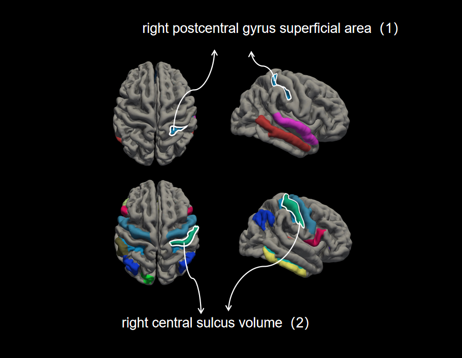
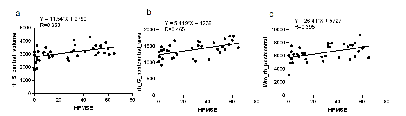
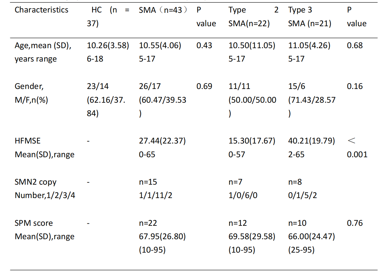
Table 1 Demographic characteristics of all study subjects
43 SMA patients and 37 HCs were finally involved. The 43 SMA patients included 22 type 2 patients and 23 type 3 patients. The age range of the enrolled participants was 5-17 years, with an average age of 10 years. Age and gender were comparable between the two groups.HFMSE, SMN2 copy number, and SPM score were comparable between type 2 and type 3 patients. (HC, healthy controls; SMA, spinal muscular atrophy; SD, standard deviation; M, male; F, female; SPM score, Raven's Standard Progressive Matrices score.)
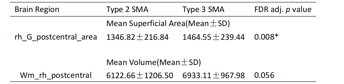
Table 2 The significant differences in means of region of interest stats between the type 2 and type 3 spinal muscular atrophy patients (type 2 SMA, type 3 SMA).
Right postcentral gyrus superficial area and right postcentral gyrus volumetric stats are significantly higher in Type 2 SMA patients than in type 3 SMA patients
(FDR false discovery ratio, adj adjusted. p values adjusted for false discovery ratio with the Benjamini–Hochberg method; * p values are statistically significant; wm, white matter segmentation volumetric stats.)