3352
Lifestyle determinants of Hippocampal Volume: An AI-enabled Volumetric Analysis on a Large Healthy Cohort.1Voxelwise Imaging Technology Inc, Vancouver, BC, Canada, 2Prenuvo, Vancouver, BC, Canada, 3Division of Neurosurgery, Vancouver General Hospital, Vancouver, BC, Canada
Synopsis
Keywords: Gray Matter, Aging, Machine Learning, Artificial Intelligence
The volume of the hippocampus and temporal horn of the lateral ventricle provides insight into the state of neurological health and the changes in volume are associated with conditions such as traumatic brain injury1, Alzheimer's Disease 2 and treatment response. It remains an open question what are the normal age-related changes over time in hippocampal and temporal horn volumes in an average healthy population cohort. We seek to establish these baseline parameters via AI-based MRI brain segmentation and quantification techniques applied to a large cohort of screened healthy patients.Introduction
The hippocampus plays essential roles such as spatial navigation, consolidation of verbal and visual memory. Problems in the hippocampus have been linked to traumatic brain injury1, Alzheimer's Disease2 and dementia etc. It is well known that the hippocampus is asymmetric but this asymmetry is poorly understood3. While several other comparison-control studies have demonstrated correlations between various pathologic states and characteristic hippocampal changes, the healthy control groups in those studies typically involve a relatively small number of healthy subjects.In this study we evaluate the possibility of using AI-based brain segmentation and volume quantification techniques from T1-weighted MR-images. Our goal was to determine the range of normal hippocampal volumes, temporal horn volumes, and their respective contralateral symmetries/asymmetries in a large cohort of healthy patients who underwent Whole-Body MRI as part of a preventive health screening program. The cohort was further stratified by gender, smoking, and sedentary versus physically active lifestyle histories, to further differentiate potential volume correlations within these population subsets.
Methods
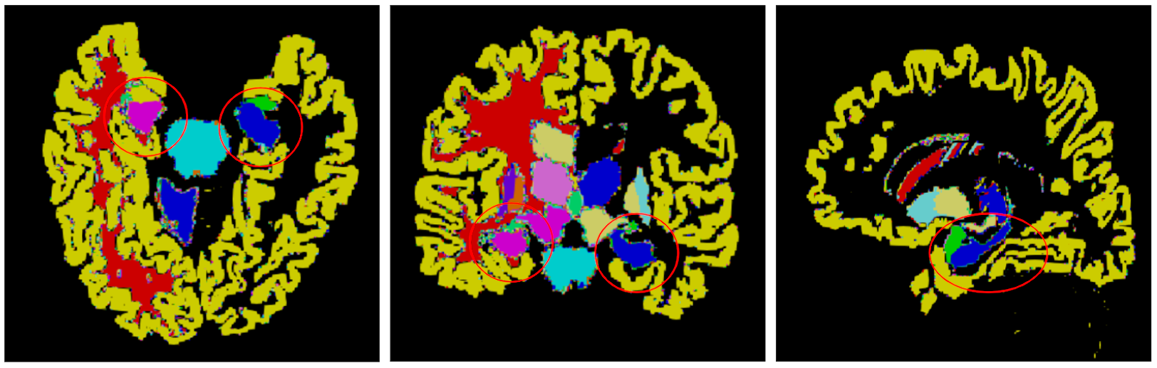
The study dataset comprised of 8119 subjects who underwent preventive health screening whole body-MRI, utilizing a protocol that included dedicated T1-weighted MPRAGE volumetric brain MRI acquisitions which served as the substrate for the present study. Of the total cohort, 4298 are males (mean age = 52.83y, std=13.07y), 3821 are females (mean age=53.74y, std=12.85y); 2860 individuals (35.2%) self-reported a history of smoking and 4269 individuals (52.5%) reported engaging in vigorous physical activity at least once per week. The hippocampus and temporal horn (inferior lateral ventricle) is automatically segmented using a state-of-the-art FastSurfer4-like deep network that was trained on our dataset. The network segments 96 different regions of the brain including hippocampus.The left and right hippocampus and temporal horn are segmented separately that were used to measure asymmetry. The asymmetric index (AI) of region of interest (ROI) was computed using equation$$AI=100\times\frac{|v_{left} -v_{right}|}{v_{left} + v_{right}}$$
A lower value of mean AI indicates the volume of left and right are similar. Further, we computed slopes using linear regression analysis to measure the rate of reduction with age and looked into the association of smoking, physical exercise on the ROI volume in each gender separately.
Results
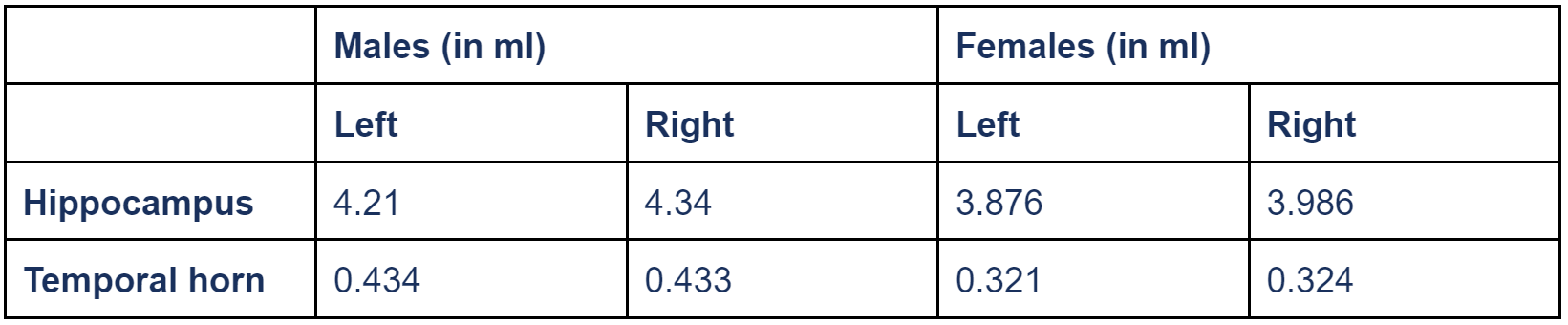
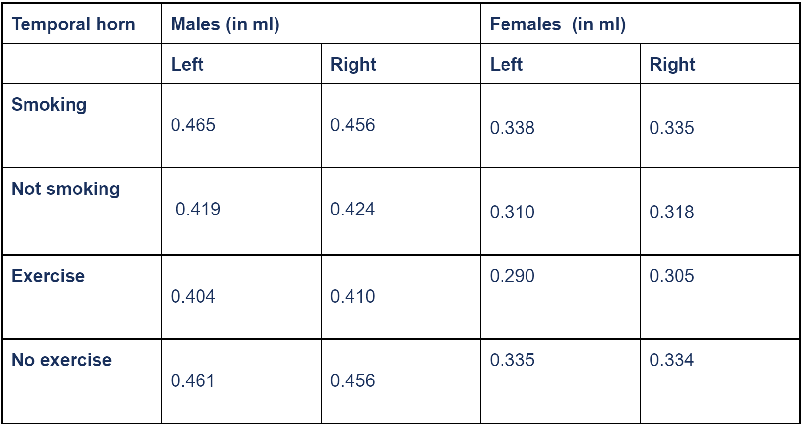
Firstly, when comparing the mean volume of the left and right hippocampus over the whole population, the left hippocampus had smaller volume (4.056ml, std=0.43ml) compared to the right hippocampus (4.175ml, std=0.44ml). The difference in left and right hippocampus volume is statistically significant (p<0.05) using t-test. Similarly, the left and right temporal horn volume was measured to be 0.381ml (std=0.26ml) and 0.382ml (std=0.25ml), respectively, although statistical significance was not demonstrated (p=0.767).When looking at the gender differences, males were found to have larger volumes compared to females as described in Table 1. For males the mean AI=2.28 and for females it was slightly smaller 2.18. These numbers are similar to what are reported in another study5.
Further, we fit a linear regression model using age as independent variable and hippocampus volume as the dependent variable. The slope was computed and found that males have a slope of -0.30%, -0.28% for left and right hippocampus whereas females have a slope of -0.24% and -0.23% suggesting female decline is relatively smaller as compared to males. In comparison, the whole brain declined at a rate of -0.24% in both genders.
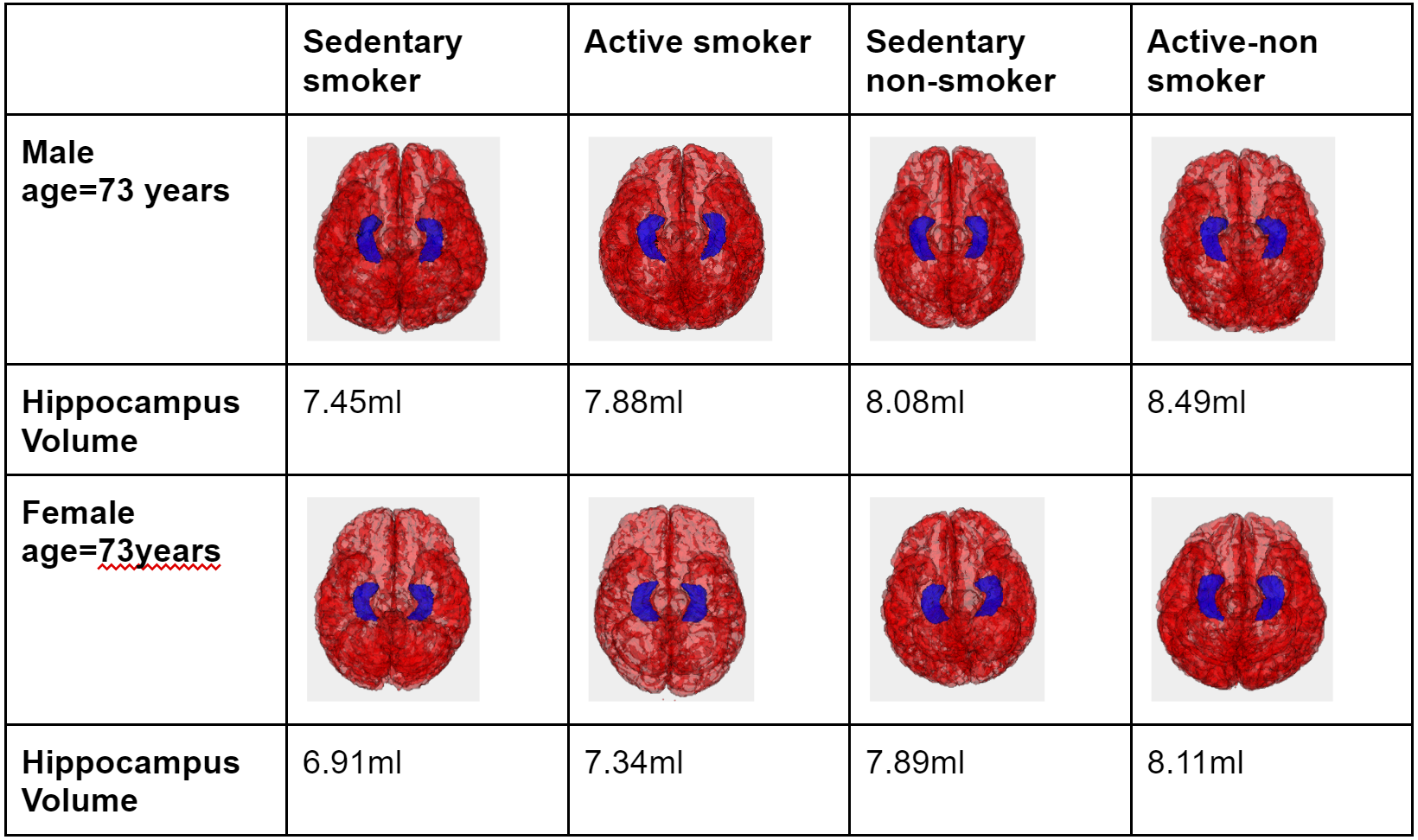
We also performed multiple linear regression, after regressing out age, to measure the effect of smoking and exercise (independent variables) on the ROI volume (dependent variable). For smoking males, regression coefficients were -3.30 (left hippocampus) and -4.05 (right hippocampus); for physically active males, regression coefficients were +7.34 (left hippocampus) and +7.02 (right hippocampus). Similar trends, although slightly smaller in magnitude, were seen in females. For smoking females, regression coefficients were -1.10 (left hippocampus) and -2.10 (right hippocampus); for physically active females, regression coefficients were +5.38 (left hippocampus) and +5.80 (right hippocampus).
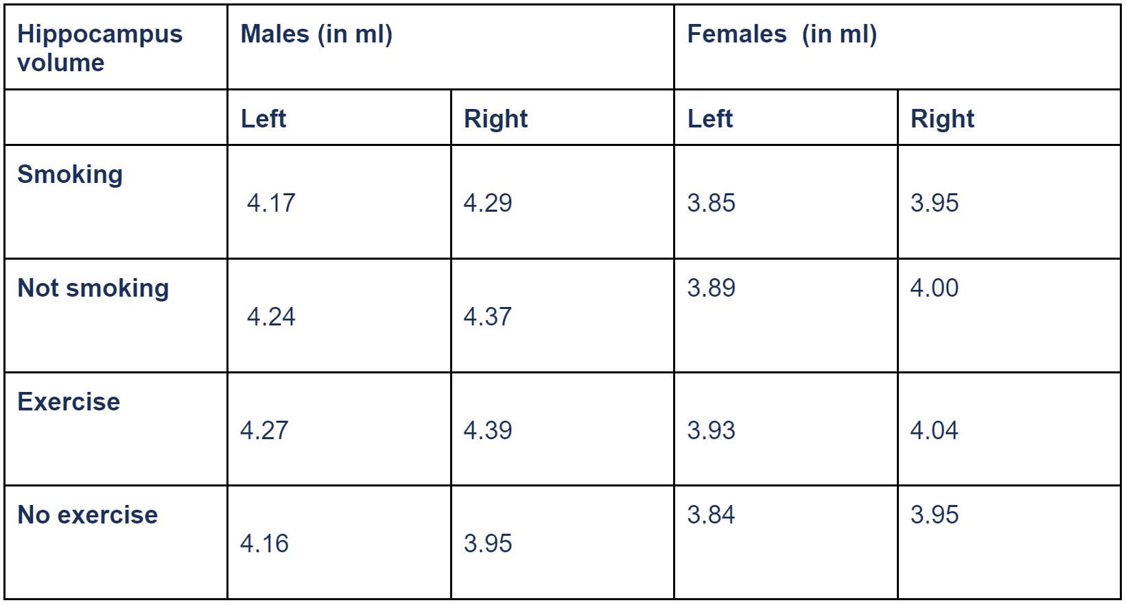
When looking at the left temporal horn smokers, the temporal horn was 11% larger in males and 9.2% larger in females when compared to the respective gender that did not smoke. The difference was a bit smaller at 7.6% and 5.2% for right temporal horn in males and females, respectively.
Discussion
Hippocampal volumes were observed to decrease with aging and a normative curve for expected rate of volume loss with aging is demonstrated. The smoker subgroup demonstrated relatively accelerated hippocampal volume loss, and the physically active subgroup demonstrated relative hippocampal volume preservation with aging. These findings may suggest enabling detection of hippocampal volume loss deviating from the normal aging curve prompting early behavioral intervention of modifiable risk factors. In particular, smoking was observed as a risk-factor for accelerated hippocampal atrophy, while physical activity was observed as a potentially protective-factor. Similar observations have been reported elsewhere from a smaller less generalized cohort of middle aged smokers6.Conclusion
We show the normative volumes of the hippocampus on a large dataset representative of the general population, sub-stratified for gender, smoking history, and physically active versus sedentary lifestyle.Acknowledgements
We would like to acknowledge the efforts of MRI Technologists who helped us with the data collection without which this study would not have been possible.References
[1] Jorge, R. E., Acion, L., Starkstein, S. E., & Magnotta, V. (2007). Hippocampal volume and mood disorders after traumatic brain injury. Biological psychiatry, 62(4), 332-338.
[2] Ewers, M., Cheng, X., Zhong, Z., Nural, H. F., Walsh, C., Meindl, T., ... & Hampel, H. (2011). Increased CSF-BACE1 activity associated with decreased hippocampus volume in Alzheimer's disease. Journal of Alzheimer's Disease, 25(2), 373-381.
[3] Woolard, A. A., & Heckers, S. (2012). Anatomical and functional correlates of human hippocampal volume asymmetry. Psychiatry Research: Neuroimaging, 201(1), 48-53.
[4] Henschel, L., Conjeti, S., Estrada, S., Diers, K., Fischl, B., & Reuter, M. (2020). Fastsurfer-a fast and accurate deep learning based neuroimaging pipeline. NeuroImage, 219, 117012.
[5] Sarica, A., Vasta, R., Novellino, F., Vaccaro, M. G., Cerasa, A., Quattrone, A., & Alzheimer's Disease Neuroimaging Initiative. (2018). MRI asymmetry index of hippocampal subfields increases through the continuum from the mild cognitive impairment to the Alzheimer's disease. Frontiers in Neuroscience, 12, 576.
[6] Durazzo, T. C., Meyerhoff, D. J., & Nixon, S. J. (2013). Interactive effects of chronic cigarette smoking and age on hippocampal volumes. Drug and alcohol dependence, 133(2), 704-711.
Figures