3349
Abnormal brain function changes in chronic tinnitus with hearing loss: A Resting-State Functional Magnetic Resonance Imaging Study1Henan Provincial People's Hospital, Zhengzhou, China
Synopsis
Keywords: Brain Connectivity, Brain Connectivity
To investigate the differences of spontaneous brain activity and brain functional connectivity between chronic tinnitus patients with hearing loss and healthy control group. Resting state functional magnetic resonance imaging (rs-fMRI) was performed to obtain the low-frequency fluctuation amplitude (ALFF), regional homogeneity (ReHo) and functional connectivity (FC). In this study, compared with healthy controls, chronic tinnitus patients with hearing loss showed aberrant brain activity increased in the temporal lobe, frontal lobe, hippocampus, precuneus, amygdala and cingulate cortex. It provided additional evidence to understand the neuropathophysiological mechanism of chronic tinnitus with hearing loss.Abstract
INTRODUCTION: Tinnitus and hearing loss often occur concurrently and the underlying neurophysiological mechanism remains unclear. This study aimed to explore the differences of spontaneous brain activity and brain functional connectivity between chronic tinnitus patients with hearing loss and healthy control group.METHODS: In this study, 30 chronic tinnitus patients with hearing loss and 30 healthy controls matched in age, sex and education level were included. Resting state functional magnetic resonance imaging (rs-fMRI) was performed to obtain the low-frequency fluctuation amplitude (ALFF) and regional homogeneity (ReHo). Left primary auditory cortex (PAC) were selected as seed regions to obtain the intrinsic functional connectivity (FC) with the whole brain.
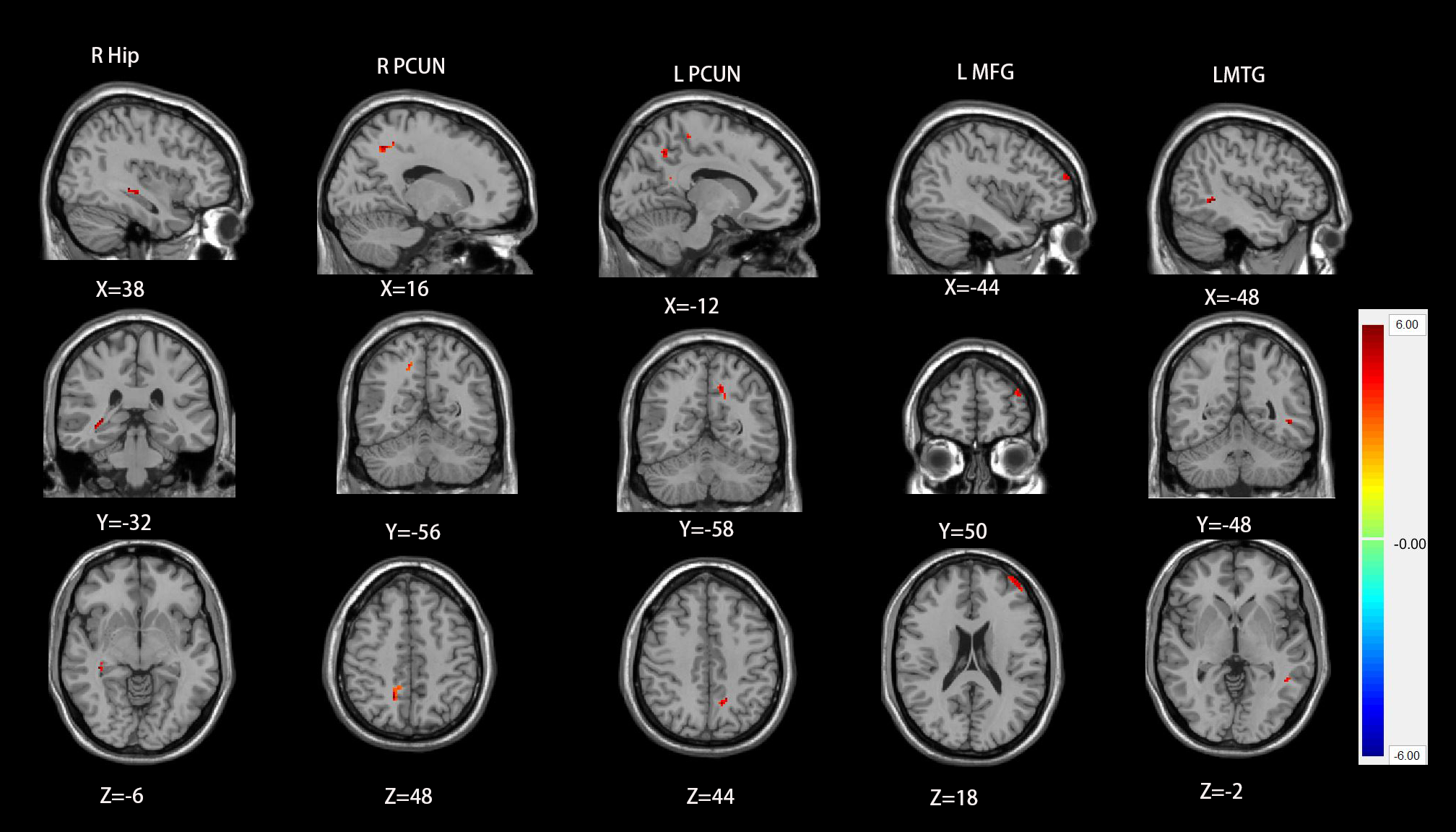
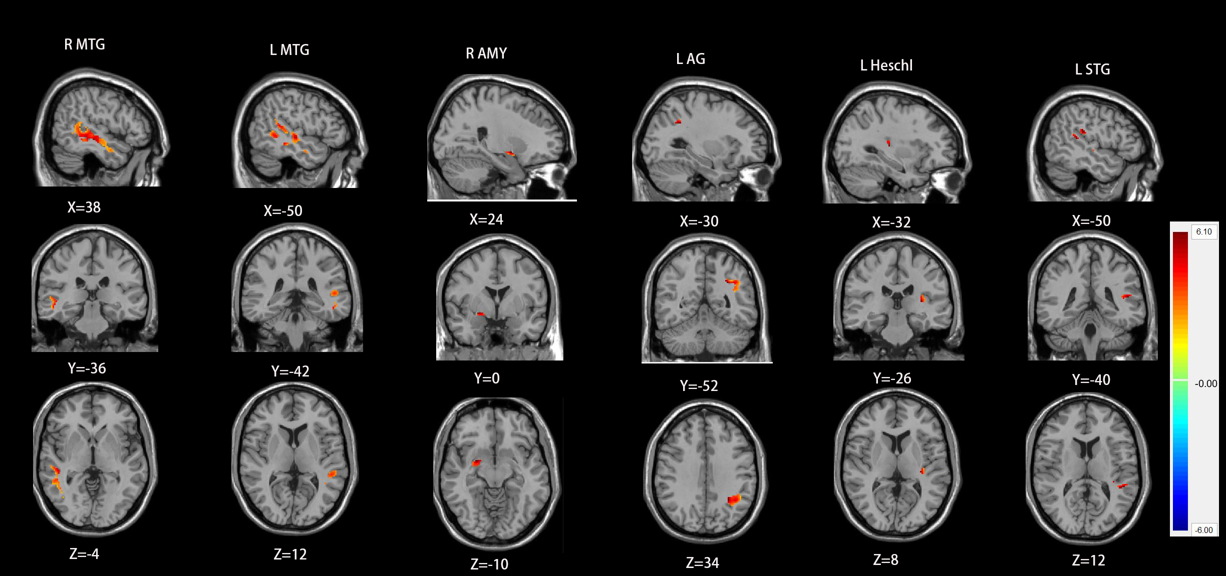
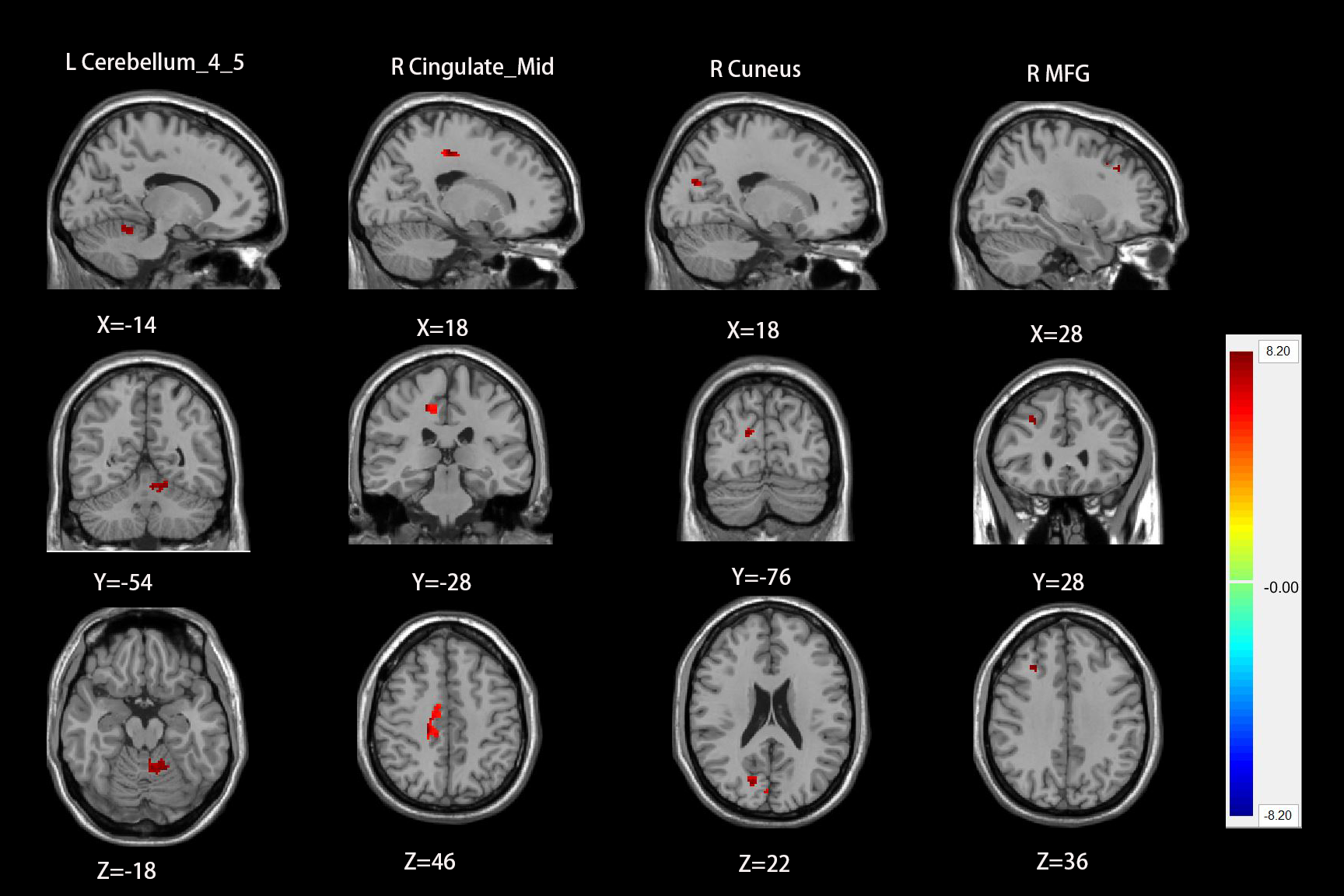
RESULTS: Compared with healthy controls, chronic tinnitus patients with hearing loss showed a significantly increased ALFF values in the right hippocampus, left middle temporal gyrus (MTG), left middle frontal gyrus (MFG), and bilateral precuneus, and a significantly increased ReHo values in left PAC, bilateral MTG, left superior temporal gyrus (STG), right amygdala and left angular gyrus (AG). Based on FC analysis, the left PAC showed reduced functional connectivity with non-auditory brain regions including the right MFG, right cingulate cortex, right cuneus and left cerebellum in the groups of chronic tinnitus with hearing loss compared with healthy controls.
DISCUSSION: Tinnitus is a common hearing disorder characterized by abnormal perception of sound in the absence of an external sound source, with about 10-15% of people suffering from persistent tinnitus1. Tinnitus and hearing loss often occur concurrently and the underlying neuroimaging mechanism is still poorly understood2, 3. Some studies revealed neural network alterations in tinnitus patients, including the auditory system, limbic system and default mode network4, 5. In consistent with our study, we found abnormal changes in several brain networks in chronic tinnitus patients with hearing loss, including the auditory network, limbic network, default network, visual network and cerebellum. The alteration of auditory network plays an important role in the occurrence and development of tinnitus. Our study found ALFF increased in left MTG and ReHo increased in left PAC, bilateral MTG, left superior temporal gyrus (STG). Chen et al6 detected spontaneous brain activity based on ALFF methods in acute tinnitus patients with hearing loss, and they found a significantly increased ALFF values in the left MTG and bilateral MFG. Therefore, It is possible that MTG and MFG has abnormal brain activity in the early stage of tinnitus. Tinnitus also accompanies some mood disorders like depression and anxiety7. The limbic system and frontal lobe are the main brain areas that participate in emotion processing. Our study found ALFF increased in right hippocampus left MFG and ReHo increased in right amygdala. Previous studies have shown increased FC between the left auditory cortex and the right hippocampus increased 8, and decreased FC between amygdala and the superior and middle temporal gyrus7. The default mode network (DMN) also changes in tinnitus. Schmidt et al found that decreased FC between DMN and the precuneus was related to long-term tinnitus9. However, we found ALFF increased in bilateral precuneus. This may be due to the heterogeneity of the tinnitus patients. The left PAC showed reduced FC with non-auditory brain regions including the right MFG, right cingulate cortex, right cuneus and left cerebellum in the groups of chronic tinnitus with hearing loss compared with healthy controls. Zhou et al noted that tinnitus showed aberrant FC within DMN, the cerebellum network, and visual network compared with healthy controls2. Zhou et al indicated the disrupted connectivity the major components in DMN and VN10. In conclusion, tinnitus is the result of both auditory and non-auditory networks.
CONCLUSION: This study indicated that there are abnormal ALFF, ReHo and FC in chronic tinnitus patients with hearing loss, indicating abnormal brain activity in both auditory and non-auditory networks. Our findings may provide additional evidence to understand the neuropathophysiological mechanism of chronic tinnitus with hearing loss.
Acknowledgements
The National Key R&D Program of China (2017YFE0103600), the National Natural Science Foundation of China (81720108021 and 31470047), the Zhongyuan Thousand Talents Plan Project - Basic Research Leader Talent (ZYQR201810117), and the Zhengzhou Collaborative Innovation Major Project (20XTZX05015).References
1. Tunkel DE, Bauer CA, Sun GH, et al. Clinical practice guideline: tinnitus[J]. Otolaryngol Head Neck Surg, 2014. 151(2 Suppl):S1-S40.
2. Zhou GP, Li WW, Chen YC, et al. Disrupted intra- and inter-network connectivity in unilateral acute tinnitus with hearing loss[J]. Front Aging Neurosci, 2022. 14(833437):1-10.
3. Zhou GP, Chen YC, Li WW, et al. Aberrant functional and effective connectivity of the frontostriatal network in unilateral acute tinnitus patients with hearing loss[J]. Brain Imaging Behav, 2021.
4. Hu J, Cui J, Xu JJ, et al. The Neural Mechanisms of Tinnitus: A Perspective From Functional Magnetic Resonance Imaging[J]. Front Neurosci, 2021. 15:621145.
5. Kok TE, Domingo D, Hassan J, et al. Resting-state Networks in Tinnitus : A Scoping Review[J]. Clin Neuroradiol, 2022. 6. Chen J, Fan L, Cai G, et al. Altered Amplitude of Low-Frequency Fluctuations and Degree Centrality in Patients with Acute Subjective Tinnitus: A Resting-State Functional Magnetic Resonance Imaging Study[J]. J Integr Neurosci, 2022. 21(4):116.
7. Chen YC, Bo F, Xia W, et al. Amygdala functional disconnection with the prefrontal-cingulate-temporal circuit in chronic tinnitus patients with depressive mood[J]. Prog Neuropsychopharmacol Biol Psychiatry, 2017. 79(Pt B):249-257.
8. Chen YC, Xia W, Chen H, et al. Tinnitus distress is linked to enhanced resting-state functional connectivity from the limbic system to the auditory cortex[J]. Hum Brain Mapp, 2017. 38(5):2384-2397. 9. Schmidt SA, Carpenter-Thompson J, and Husain FT. Connectivity of precuneus to the default mode and dorsal attention networks: A possible invariant marker of long-term tinnitus[J]. Neuroimage Clin, 2017. 16:196-204.
10. Zhou GP, Shi XY, Wei HL, et al. Disrupted Intraregional Brain Activity and Functional Connectivity in Unilateral Acute Tinnitus Patients With Hearing Loss[J]. Front Neurosci, 2019. 13:1010.
Figures