3348
Altered Brain Activity and Functional Connectivity in transition from recent-onset to chronic tinnitus, a rs-fMRI study
1Department of Radiology, First Affiliated Hospital of Soochow University, Suzhou, China, Suzhou city, China, 2Philips Healthcare, Shanghai, China, Shanghai, China
Synopsis
Keywords: Brain Connectivity, fMRI (resting state), tinnitus
As a common disorder, the development of tinnitus deserves the attention of neuroscientists. The present study combined fractional amplitude of low-frequency fluctuations and functional connectivity to explore brain functional abnormalities in transition from recent-onset to chronic tinnitus. Abnormal intraregional neural activity and functional connectivity were observed in the left middle frontal gyrus and left dorsolateral superior frontal gyrus during the development of chronic tinnitus, and these regions are major components of attention network and executive control network. These findings provide us with a better understanding of the aberrant brain changes and neuropathophysiological mechanisms of the progression of tinnitus.
Introduction
Tinnitus, as a common disorder, is the perceived sensation of sound in the absence of a corresponding external acoustic stimulus1,2. The aberrant brain changes and neuropathophysiological mechanisms underlying the development and progression of tinnitus remain largely undetected. The fractional amplitude of low-frequency fluctuations (fALFF) and functional connectivity (FC) analysis have been proven to be reliable methods for calculating local activity and internal connectivity in the brain3,4. We aimed to investigate differences in fALFF and FC in the brains of patients with recent-onset tinnitus (ROT), patients with chronic tinnitus and healthy controls (HCs), and to explore their relationship with the different features of tinnitus.Methods
Twenty-seven ROT patients, 33 chronic tinnitus patients, and 30 HCs were recruited and performed audiological tests and assessment of tinnitus (only for tinnitus patients), ran magnetic resonance imaging (MRI) scans. Hearing thresholds were measured by pure tone audiometry, and patients with tinnitus were tested for pitch and loudness matching of tinnitus. Other characteristics of tinnitus are assessed by the Tinnitus Functional Index (TFI)5.The MRI examinations were performed using a 3.0T MRI scanner (Ingenia, Philips Healthcare, Best, the Netherlands) with a 15-channel head coil. Resting-state functional MRI images were obtained with the following parameters: TR/TE = 2000/30 ms, slices = 30, thickness = 4 mm, gap = 0.4 mm, FOV = 240 mm ×240 mm, acquisition matrix = 64 × 64, and FA = 90 degrees. In total 250 functional volumes were obtained. High-resolution 3D T1-weighted anatomical images were collected for normalization, with the following parameters: TR/TE =7.0/3.1 ms, FA = 8 degrees, acquisition matrix = 256 × 240 × 185, and FOV = 256 mm × 256mm × 185mm. Resting-State fMRI data were preprocessed using DPABI (http://rfmri.org/dpabi)6, which is based on SPM12 (http://www.fil.ion.ucl.ac.uk/spm) and the MATLAB R2019a (Mathworks,Natick, MA, USA) platform.
For the analysis of fALFF and FC, we refer to the article by Jiawei Chen7. Based on the fALFF finding in the brain regions between groups, the orbital part of the left Middle frontal gyrus (MFG) was defined as the seed region of interest for seed-based FC analysis.
An analysis of covariance (ANCOVA) based on the DPABI toolbox was used to compare the standardized fALFF and zFC maps between ROT patients, chronic tinnitus patients and HCs, with age and gender used as covariates to remove their effects. Post-hoc comparisons were made using the Bonferroni test. To test the clinical correlations, the DPABI software was used to extract the mean normalized fALFF and FC values of the different brain regions between the three groups. Then we correlated mean fALFF values and FC values from the regions with result of audiological test and assessment of tinnitus by partial correlation analysis, controlling for age and gender.
Results
There were no significant differences in age (F=0.295, p=0.745) and gender (F=2.044, p=0.360) between these three groups (Table1). Hearing thresholds in both the chronic tinnitus and ROT groups were slightly higher than that in the HC group (F=7.979, p=0.001). And for the cognitive complaints subscale score of TFI scale (TFI-C), chronic tinnitus patients have a significant reduction compared with the ROT patients (Table2). Comparisons of fALFF in three groups show that chronic tinnitus patients had significantly increased fALFF values in the left MFG, orbital parts, compared to the HCs and ROT patients (Figure1). Comparisons of FC maps show that chronic tinnitus patients had significantly increased FC between the left MFG and the left dorsolateral superior frontal gyrus (DLSFG) (Figure2). In addition, the duration of tinnitus was positively correlated with the fALFF value of the left MFG as well as the FC value of the left DLSFG. While, the loudness of the tinnitus was negatively correlated with the fALFF value of the left MFG and the FC value of the left DLSFG. The TFC-C score in tinnitus patients showed a negative correlation with the FC value of the left DLSFG. (Figure3).Discussion
This study examined the hearing and tinnitus characteristics of tinnitus patients in two different periods and detected brain functional abnormalities in the left MFG and DLSFG by fALFF and FC analysis. The MFG, a region of the ventral lateral prefrontal cortex that mediates the interaction between the dorsal and ventral attentional networks, to which it is functionally connected, has been considered an essential brain region for attentional processing8,9.And the DLSFG is located in the dorsolateral prefrontal cortex, which is a critical node in the central executive network and plays a key role in regulating attention, working memory, and decision-making10. In summary, abnormal Increased local brain activity and FC observed in the frontal cortex might reflect an involvement of emotion and attention in the transition from recent-onset to chronic tinnitus.Conclusion
Our findings further suggest that the emergence and development of tinnitus is a dynamic process involving abnormalities in local neural activity and abnormal functional connections in brain.Acknowledgements
No acknowledgement found.References
1. Jastreboff PJ. Phantom auditory perception (tinnitus): mechanisms of generation and perception. Neurosci Res. 1990;8:221–254.
2. McCormack A, Edmondson-Jones M, Somerset S, Hall D. A systematic review of the reporting of tinnitus prevalence and severity. Hear Res. 2016;337:70–79.
3. Zou Q-H, Zhu C-Z, Yang Y, Zuo X-N, Long X-Y, Cao Q-J, Wang Y-F, Zang Y-F. An improved approach to detection of amplitude of low-frequency fluctuation (ALFF) for resting-state fMRI: Fractional ALFF. J Neurosci Methods. 2008;172:137–141.
4. Fox MD, Zhang D, Snyder AZ, Raichle ME. The Global Signal and Observed Anticorrelated Resting State Brain Networks. J Neurophysiol. 2009;101:3270–3283.
5. Meikle MB, Henry JA, Griest SE, Stewart BJ, Abrams HB, McArdle R, Myers PJ, Newman CW, Sandridge S, Turk DC, et al. The tinnitus functional index: development of a new clinical measure for chronic, intrusive tinnitus. Ear Hear. 2012;33:153–176.
6. Yan C-G, Wang X-D, Zuo X-N, Zang Y-F. DPABI: Data Processing & Analysis for (Resting-State) Brain Imaging. Neuroinformatics. 2016;14:339–351.
7. Chen J, Hu B, Qin P, Gao W, Liu C, Zi D, Ding X, Yu Y, Cui G, Lu L. Altered Brain Activity and Functional Connectivity in Unilateral Sudden Sensorineural Hearing Loss. Neural Plast. 2020;2020:9460364.
8. Song P, Lin H, Liu C, Jiang Y, Lin Y, Xue Q, Xu P, Wang Y. Transcranial Magnetic Stimulation to the Middle Frontal Gyrus During Attention Modes Induced Dynamic Module Reconfiguration in Brain Networks. Front Neuroinform. 2019;13:22.
9. Beevers CG, Clasen PC, Enock PM, Schnyer DM. Attention Bias Modification for Major Depressive Disorder: Effects on Attention Bias, Resting State Connectivity, and Symptom Change. J Abnorm Psychol. 2015;124:463–475.
10. Miller EK, Cohen JD. An integrative theory of prefrontal cortex function. Annu Rev Neurosci. 2001;24:167–202.
Figures
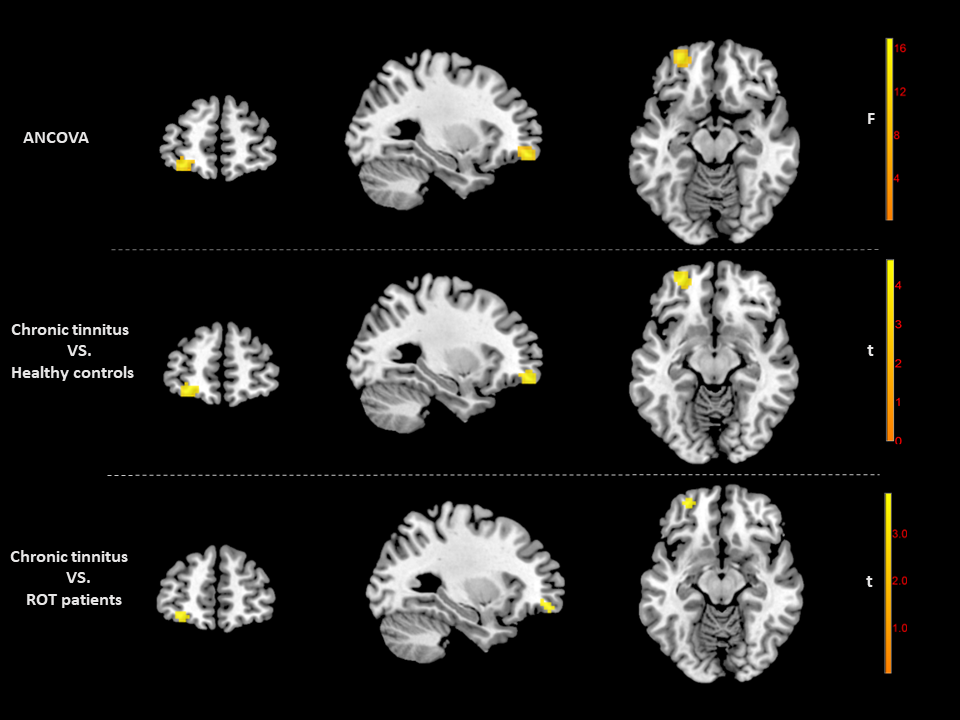
Figure1. fALFF differences between chronic tinnitus patients, recent-onset tinnitus patients and healthy controls.The comparison of fALFF between the three groups. ANCOVA of standardized fALFF maps between the three groups showed significant differences in fALFF values in the left MFG, orbital parts (warm colors).
ANCOVA, analysis of covariance; ROT, recent-onset tinnitus; fALFF, fractional amplitude of low-frequency fluctuations; MFG, middle frontal gyrus.
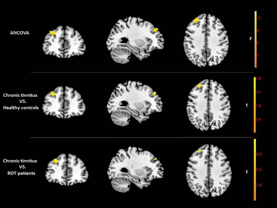
Figure 2 | Regions showing seed-based FC differences between three groups.
Note: ANCOVA of standardized FC maps between the three groups showed significant differences in fALFF values in the left DLSFG (warm colors).
ANCOVA, analysis of covariance; ROT, recent-onset tinnitus; fALFF, fractional amplitude of low-frequency fluctuations; MFG, middle frontal gyrus; DLSFG, superior frontal gyrus, dorsolateral.
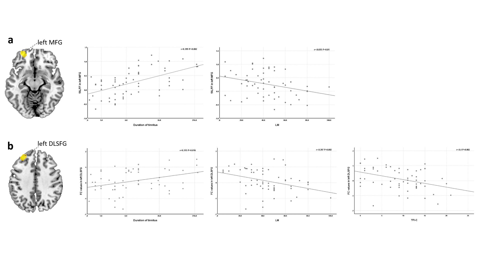
Figure 3 | Correlation coefficients between altered fALFF, FC values, and different tinnitus characteristics.
Note: fALFF, fractional amplitude of low-frequency fluctuations; FC, functional connectivity; MFG, middle frontal gyrus; DLSFG, superior frontal gyrus, dorsolateral; LM, loudness matching; TFI-C, cognitive complaints.

Table 1 | Demographic and clinical characteristics of chronic tinnitus patients, ROT patients and HCs.
Note: χ2/F values were obtained using chi-square test and Kruskal-Wallis test respectively for comparisons between three groups, and the corresponding P values were derived. P2 were obtained using a Bonferroni multiple comparison for determining statistically significant changes between each group. P* < 0.05 indicates a significant difference. ROT, recent-onset tinnitus; HCs, healthy controls; PTA, Pure-tone audiometry.

Table 2 | Tinnitus characteristics of chronic tinnitus patients and ROT patients
Note: P* < 0.05 indicates a significant difference. ROT, recent-onset tinnitus; PM, pitch matching; LM, loudness matching; VAS, visual analogue scales; TFI, Tinnitus Functional Index; TFI-I, Intrusiveness of tinnitus; TFI-SC, sense of control, TFI-C, cognitive complaints; TFI-SL, sleep disturbance; TFI-A, auditory difficulties; TFI-R, relaxation; TFI-Q, quality of life relaxation; TFI-E, emotional distress.