3336
Cognitive Brain Networks in Adult Fetal Alcohol Syndrome: Analyses of Functional Connectivity with a Higher Criticism Approach
Benedikt Sundermann1,2,3, Reinhold Feldmann4, Christian Mathys1,3, Johanna Rau5, Stefan Garde2,6, Anna Braje2, Josef Weglage4, and Bettina Pfleiderer2
1Institute of Radiology and Neuroradiology, Evangelisches Krankenhaus Oldenburg, Medical Campus University of Oldenburg, Oldenburg, Germany, 2Clinic of Radiology, Medical Faculty, University of Münster, Münster, Germany, 3Research Center Neurosensory Science, University of Oldenburg, Oldenburg, Germany, 4Department of General Pediatrics, University Hospital Münster, Münster, Germany, 5Department of Neurology with Institute of Translational Neurology, University Hospital Münster, Münster, Germany, 6Bergman Clinics Augenklinik Universitätsallee Bremen, Bremen, Germany
1Institute of Radiology and Neuroradiology, Evangelisches Krankenhaus Oldenburg, Medical Campus University of Oldenburg, Oldenburg, Germany, 2Clinic of Radiology, Medical Faculty, University of Münster, Münster, Germany, 3Research Center Neurosensory Science, University of Oldenburg, Oldenburg, Germany, 4Department of General Pediatrics, University Hospital Münster, Münster, Germany, 5Department of Neurology with Institute of Translational Neurology, University Hospital Münster, Münster, Germany, 6Bergman Clinics Augenklinik Universitätsallee Bremen, Bremen, Germany
Synopsis
Keywords: Brain Connectivity, fMRI (resting state)
Functional connectivity (FC) between and within a majority of cognition-related brain networks is altered in young adults with Fetal Alcohol Syndrome. Findings in a network-level analysis of resting-state fMRI data with a Higher Criticism approach were most obvious within a dorsal attention subnetwork and to a lesser extent in a salience / ventral attention subnetwork. When analyzing these effects further by looking at single connections, no individual FC alteration was statistically significant when adjusting for multiple comparisons. The finding of a wide distribution of FC alterations across networks might help resolve partially contradictory results in previous studies.Introduction
Fetal Alcohol Syndrome (FAS) describes the full clinical picture related to prenatal alcohol exposure, which has a profound negative impact on cognitive functions 1. Many of these functions are subserved by a limited number of functional brain networks 2,3. Functional connectivity (FC) within and between these networks is studied by resting state functional MRI (rs-fMRI) 4. There are few studies on resting state FC in children with FAS adopting related FC analyses: While all three main studies reported altered FC in FAS, the specific cognitive networks affected, their interactions and directionalities of findings have been contradictory 5-7.Purpose of this study was to assess FC in cognition-related networks in young adults with FAS. Hypotheses: (1) FC between all brain regions constituting cognition-related brain networks is altered in FAS (omnibus test), (2) FC within and (3) between these individual networks is altered in FAS.
Methods
The study was approved by the local ethics committee. 39 patients with FAS (age: 22.2 ± 3.4 years, 43.6 % female) and 44 healthy controls (age: 20.9 ± 3.4, 54.5 % female) could be included in the final analyses. MRI data were acquired at 3 Tesla: 9:45 min rsfMRI (234 volumes, repetition time: 2500 ms, echo time: 35 ms, spatial resolution 3.6 x 3.6 x 3.6 mm), 3D T1 (inversion-prepared turbo field echo). Preprocessing was carried out with fmriprep 8 followed by denoising with fmridenoise 9. FC analyses are based on a cortical atlas with 400 regions assigned to 17 functional networks 10. Ten cognition-related components out of these 17 networks (comprising 243 regions) were selected. FC was calculated as linear correlation between these atlas regions 11.Basis for subsequent modelling were multiple two-tailed two-sample t-tests (one test per pair of regions) comparing FC between the groups. Results were subjected to a hierarchical statistical testing approach, first determining whether there is any alteration of FC in FAS in the full cognitive connectome and subsequently resolving these findings to the level of either FC within each network or between-network connectivity, and finally to individual connections. The overall tests (full connectome and separate networks) are based on the Higher Criticism (HC) approach 12,13 in order to test, whether there are any non-zero effects within a large number of individual tests. Under a null-hypothesis of only zero-effects in parallel tests, an equal distribution of p-values is expected. HC statistics test a joint hypothesis of an excess of low p-values as evidence of non-zero effects 12,14. HC is suitable to identify the existence of rare and weak effects in high-dimensional data 12 such as rsfMRI 15. To determine which networks are affected by within-network FC differences between FAS and controls, we carried out equivalent HC tests separately for these 10 networks. A methodologically similar between-network analysis was also conducted. In a third level we wanted to identify single connections (pairwise between regions) exhibiting FC differences between FAS (FDR-adjusted) and controls.
Results
We observed significantly altered FC of cognition-related brain regions in FAS compared with non-exposed controls in the global analysis.FC was altered within in 7 out of 10 networks based on the HC joint hypothesis tests. This effect was most obvious in the dorsal attention A sub-network followed by the salience / ventral attention A subnetwork. Findings also spanned sub-components of the fronto-parietal control and default mode networks (Figure 1). None of the single FC alterations within these networks could be identified as statistically significant (multiple comparison correction).
Between-network connectivity was altered (based on HC). Single between-network alterations were not significant. Descriptively, strongest relative decreases in FAS were observed between the salience / ventral attention B and fronto-parietal control C sub-networks, strongest relative increases between the dorsal attention B and fronto-parietal control sub-networks as well as between the default mode C sub-network with other parts of the default mode network and fronto-parietal control network.
Discussion
High-dimensional FC analyses in infrequent disorders such as FAS carry the risk to report only a “tip of the iceberg” of true underlying alterations due low statistical power (see a related discussion 16,17). This might in part explain contradictory results in children with FAS 5-7. Our approach with HC-based joint hypothesis tests 12,13 at the network level might help avoid this shortcoming without sacrificing information from individual connections. Compared with conventional fMRI analyses it avoids selectively reporting and interpreting few selected results passing a multiple-comparison-threshold and which might not well represent the true underlying effect in a low to medium-power setting.The wide distribution of findings across cognition-related networks reflects the similarly wide range of cognitive deficits in FAS 1. It thus suggests a rather distributed neural basis of such deficits rather than strongly localized alterations. The observation of more obvious effects in attention-related systems compared with networks underlying other cognitive functions highlights the importance of attentional deficits in FAS. However, this is limited by the infeasibility of a direct quantitative comparison between networks as well as reverse inference.
Conclusion
FC in cognition-related brain networks is altered in young adults with FAS. These alterations are widely distributed across different cognition-related networks rather than confined to few individual connections or networks.Acknowledgements
No acknowledgement found.References
1. Davis KM, Gagnier KR, Moore TE, et al. Cognitive aspects of fetal alcohol spectrum disorder. Wiley Interdiscip Rev Cogn Sci 2013;4(1):81-92.2. Niendam TA, Laird AR, Ray KL, et al. Meta-analytic evidence for a superordinate cognitive control network subserving diverse executive functions. Cogn Affect Behav Neurosci 2012;12(2):241-268.
3. Menon V. Large-scale brain networks and psychopathology: a unifying triple network model. Trends Cogn Sci 2011;15(10):483-506.
4. van den Heuvel MP, Hulshoff Pol HE. Exploring the brain network: a review on resting-state fMRI functional connectivity. Eur Neuropsychopharmacol 2010;20(8):519-534.
5. Fan J, Taylor PA, Jacobson SW, et al. Localized reductions in resting-state functional connectivity in children with prenatal alcohol exposure. Hum Brain Mapp 2017;38(10):5217-5233.
6. Little G, Reynolds J, Beaulieu C. Altered Functional Connectivity Observed at Rest in Children and Adolescents Prenatally Exposed to Alcohol. Brain Connect 2018;8(8):503-515.
7. Ware AL, Long X, Lebel C. Functional connectivity of the attention networks is altered and relates to neuropsychological outcomes in children with prenatal alcohol exposure. Dev Cogn Neurosci 2021;48:100951.
8. Esteban O, Markiewicz CJ, Blair RW, et al. fMRIPrep: a robust preprocessing pipeline for functional MRI. Nat Methods 2019;16(1):111-116.
9. Finc KC, M.; Bonna, K. fMRIDenoise: automated denoising, denoising strategies comparison, and functional connectivity data quality control. 2020.
10. Schaefer A, Kong R, Gordon EM, et al. Local-Global Parcellation of the Human Cerebral Cortex from Intrinsic Functional Connectivity MRI. Cereb Cortex 2018;28(9):3095-3114.
11. Chao-Gan Y, Yu-Feng Z. DPARSF: A MATLAB Toolbox for "Pipeline" Data Analysis of Resting-State fMRI. Front Syst Neurosci 2010;4:13.
12. Donoho D, Jin J. Higher Criticism for Large-Scale Inference, Especially for Rare and Weak Effects. Statistical Science 2015;30(1):1-25.
13. Donoho D, Jin J. Higher criticism for detecting sparse heterogeneous mixtures. The Annals of Statistics 2004;32(3):962-994.
14. Breheny P, Stromberg A, Lambert J. p-Value Histograms: Inference and Diagnostics. High Throughput 2018;7(3).
15. Gerlach AR, Karim HT, Kazan J, et al. Networks of worry-towards a connectivity-based signature of late-life worry using higher criticism. Transl Psychiatry 2021;11(1):550.
16. Yarkoni T. Big Correlations in Little Studies: Inflated fMRI Correlations Reflect Low Statistical Power-Commentary on Vul et al. (2009). Perspect Psychol Sci 2009;4(3):294-298.
17. Cremers HR, Wager TD, Yarkoni T. The relation between statistical power and inference in fMRI. PLoS One 2017;12(11):e0184923.
Figures
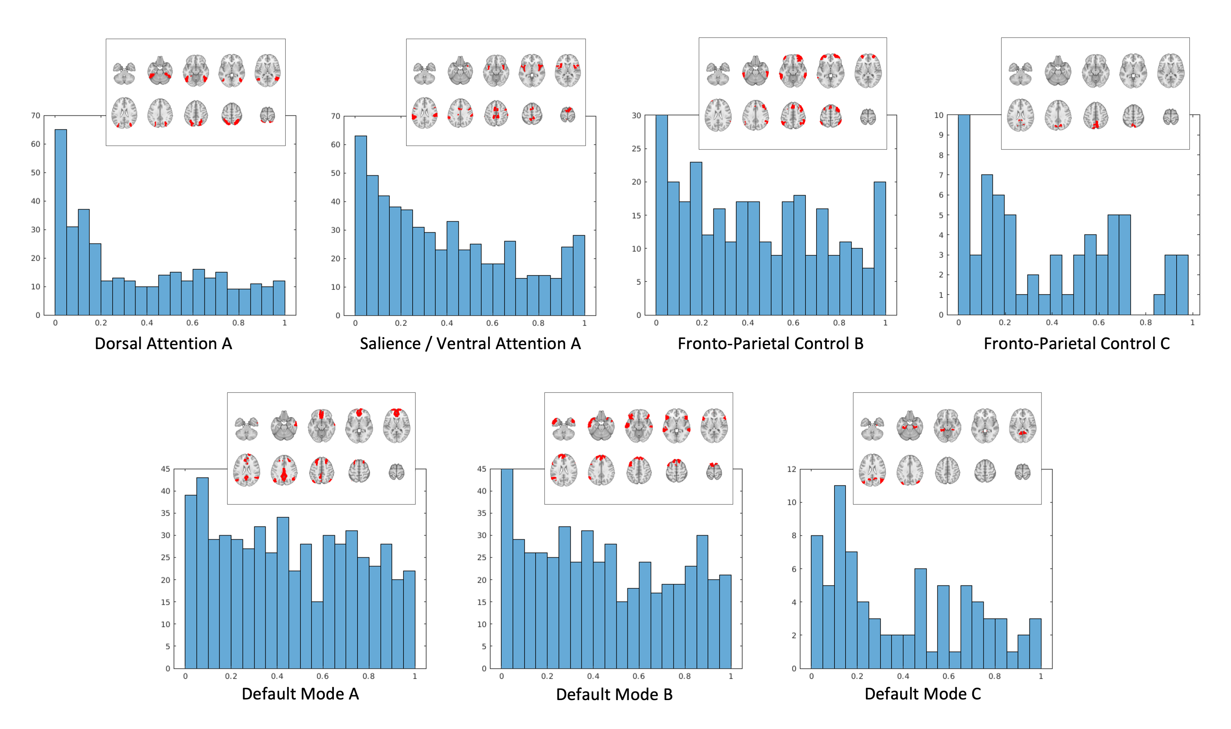
Figure 1 – Within-network FC in cognition-related brain networks in FAS was altered in these 7 out of 10 subnetworks (overall extent instead of individual regions shown for illustrative purposes). P-value histograms of parallel pairwise FC-comparisons (t-tests) between subregions underlying the HC-based joint hypothesis tests. Under the null-hypothesis of equal FC in both groups, equal numbers of p-values are expected in each histogram bin. The histograms show an excess of low p-values (left).
DOI: https://doi.org/10.58530/2023/3336