3333
Altered Resting-State Network Connectivity in Essential Tremor1Institute of Biomedical Engineering and Nanomedicine, National Health Research Institutes, Miaoli, Taiwan, 2Department of Neurology, Taoyuan General Hospital, Ministry of Health and Welfare, Taoyuan, Taiwan, 3Department of Biomedical Engineering and Environmental Sciences, National Tsing Hua University, Hsinchu, Taiwan, 4Department of Radiology, Taoyuan General Hospital, Ministry of Health and Welfare, Taoyuan, Taiwan, 5Graduate Institute of Biomedical Materials and Tissue Engineering, College of Biomedical Engineering, Taipei Medical University, Taipei, Taiwan, 6Institute of Medical Device and Imaging, National Taiwan University College of Medicine, Taipei, Taiwan
Synopsis
Keywords: Brain Connectivity, fMRI (resting state), essential tremor
We aim to characterize the functional connectivity in essential tremor to portray the resting-state network organization and its correlation to tremor features. Reduced functional connectivity between default mode network and ventral attention network was found in essential tremor subjects. The inter-network connectivity and intra-network connectivity were also shown to be associated with tremor features. Our result suggest that analysis of resting-state network is a potential approach in detecting the cerebral alterations in ET.Introduction
Essential tremor (ET) is a common neurological disorder with a wide spectrum of clinical characteristics 1. In addition to motor related symptoms, non-motor features including cognitive and psychiatric alterations have been suggested 2. Although decades of efforts have been done on investigating ET, the origins and causes are not well recognized 3. As a movement disorder, tremor-related studies mainly focus on the function of cerebellum and cerebellar-associated circuits. Since the cognitive and psychiatric alterations in ET have been suggested 2, we aim to extend the scope from cerebellum to cerebrum and to explore tremor from network aspect. To our knowledge, there was limited number of studies emphasizing on the connectivity within and between various resting state networks (RSN) of tremor. In this study, we aim to characterize the common RSNs with the hubs summarized by Power et al. 4, providing a more comprehensive analysis on the network connectivity of both normal control and ET patients. The inter-network connectivity and intra-network connectivity will be evaluated to portray the network organization, together with its correlation to tremor features.Methods
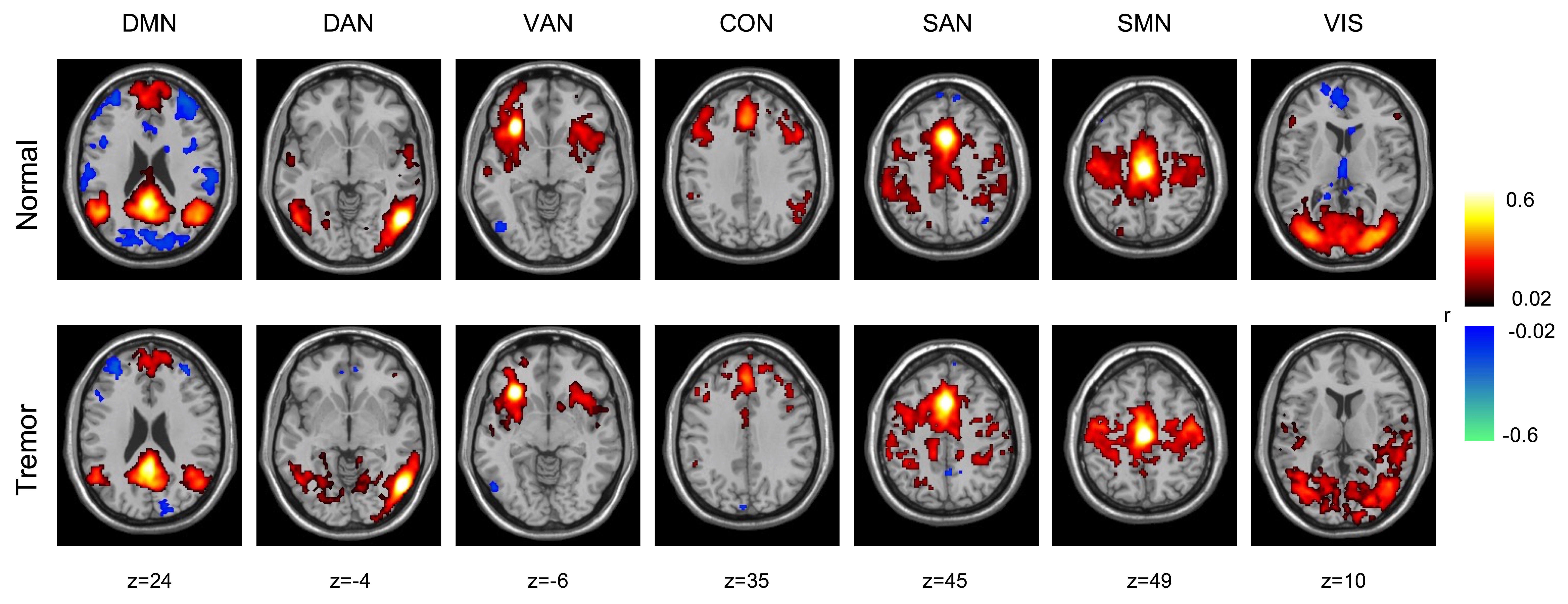
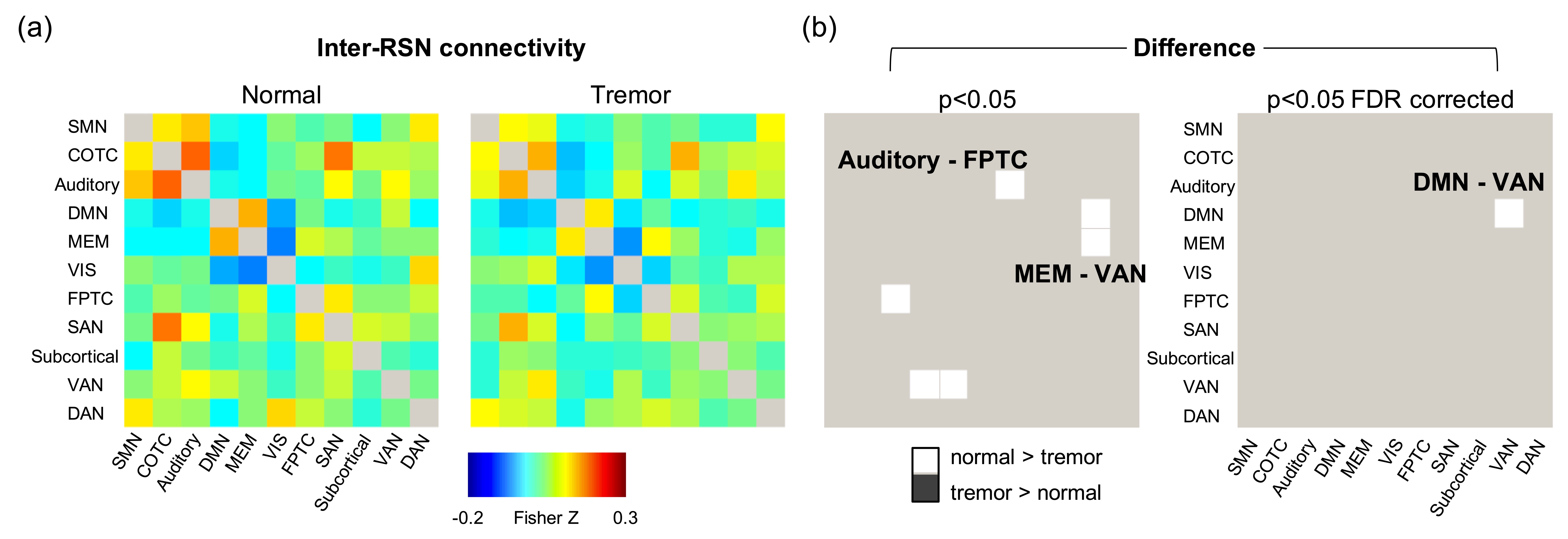
A total of 17 patients with ET (10 females) and 19 healthy volunteers (12 females) were recruited under approved IRB. The WHIGET score was evaluated by neurologist (YCH). MRI data were obtained on Philips Ingenia 1.5T with a 20-channel head coil. T1WI was acquired by 3D gradient echo with FOV = 25.6 × 25.6 × 16.5 cm3, matrix size = 400×400×150, TR/TE = 7.51/3.49 ms, FA = 8°. Resting-state fMRI were obtained by gradient echo EPI with 26 interleaved-transversal slices. Scanning parameters were: FOV = 19.2 × 19.2 cm2, matrix size = 64 × 64, TR/TE = 2500/40 ms, FA = 90°, total time points = 156. Each subject was asked to stay relax but awake with eye closed during scanning. Data were pre-processed via CONN toolbox version 19.c (www.nitrc.org/projects/conn) 5 with standard steps provided in CONN. After spatial processing, common nuisance signal components were removed and linear detrend and despike were performed, followed by a band-pass filter of 0.01 - 0.08 Hz. The Power 264 atlas was used and the regions engaged in the following 11 common RSNs were selected: sensory-motor network (SMN), cingulo-opercular task control network (COTC), auditory, default mode network (DMN), memory retrieval (MEM), visual network (VIS), fronto-parietal task control network (FPTC), salience network (SAN), subcortical, ventral attention network (VAN), dorsal attention network (DAN). A total of 232 spherical regions (10-mm diameter) were used to extract the signal and to evaluate the Pearson correlation coefficient matrix via DPARSF toolbox 6. The intra-network connectivity and inter-network connectivity were then calculated based on the Fisher’s z transformed correlation coefficient, as used in literature 7. Sphere seeds in 12-mm diameter were used to generate the voxel-wise RSN maps for visualization (Figure 1).Results
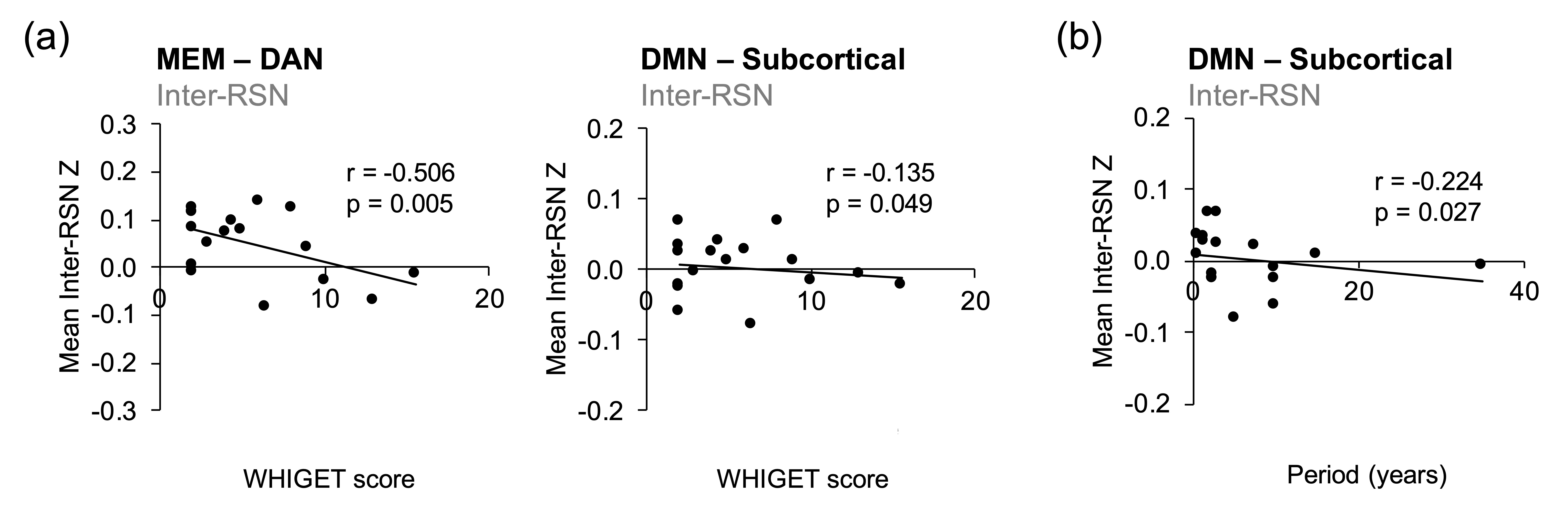
The inter-network connectivity among 11 RSNs was depicted in matrices (Figure 2). Specifically, decreased inter-network connectivity in auditory–FPTC, MEM–VAN, and DMN–VAN was found in tremor group comparing to normal control. The difference in DMN–VAN connectivity holds after FDR correction. Figure 3 shows the correlations among functional connectivity and tremor features with p-value < 0.05. Inter-RSN connectivity of MEM–DAN and DMN–subcortical were negatively correlated with WHIGET score. A negative correlation was found between period and the inter-RSN connectivity of DMN–subcortical. Intra-RSN connectivity showed no significant correlation to either WHIGET score or disease period.Discussion
Inter-network connectivity of auditory–FPTC, MEM–VAN, and DMN–VAN were shown to be reduced in ET patients in this study, which have not been reported before. Particularly, difference in DMN–VAN connectivity persists after correction for multiple comparisons. It has been reported that ET patients have worse cognitive performance than that of healthy control, including attention, executive function, and language 8. Although the cognitive performances were not assessed here, the altered DMN–VAN connectivity may be associated with the dysregulation of attention, or the abnormality between the transition from default mode to attention-demanded task. Researches in volumetric MRI also indicates the gray matter volume loss of ET group within superior temporal gyrus and inferior frontal gyrus 9, which are engaged in the VAN network. Other studies also suggested altered gray matter volume in precuneus 10, which is one of the hubs of DMN. These evidences may provide the structural basis of the disruption in DMN–VAN connectivity. A moderate correlation (r= -0.506) was found in MEM–DAN. As DAN- and DMN-affiliated regions are important in memory retrieval 11, these evidences may imply the disturbance in memory-associated networks. On the other hand, DMN–subcortical connectivity was shown to be negatively correlated with both WHIGET score and disease period. Although the correlation was mild, these findings might also imply the altered thalamo-cortical circuit. Overall, this study has explored the network-level functional connectivity in ET patients. Other analyzing approaches such as amplitude of low frequency fluctuations and graph-theory evaluations can be carried out to assess the interconnections of functional hubs and modules in the future.Conclusion
Reduced functional connectivity between default mode network and ventral attention network was identified in essential tremor subjects, and the connectivity measures were also shown to be associated with tremor features. Analysis of resting-state network is a potential approach in detecting the cerebral alterations in ET.Acknowledgements
We acknowledge the grants supported by Ministry of Health and Welfare (MOHW 109-HSO-M-211-000001), National Science and Technology Council (NSTC 111-2221-E-400 -001-MY2, 111-2811-E-400-002-MY2), and National Health Research Institutes (NHRI BN-111-PP-06).References
1. Bhatia, K.P., et al. Consensus Statement on the classification of tremors. from the task force on tremor of the International Parkinson and Movement Disorder Society. Mov Disord 33, 75-87 (2018).
2. Chandran, V. & Pal, P.K. Essential tremor: beyond the motor features. Parkinsonism Relat Disord 18, 407-413 (2012).
3. Louis, E.D. & Faust, P.L. Essential tremor pathology: neurodegeneration and reorganization of neuronal connections. Nat Rev Neurol 16, 69-83 (2020).
4. Power, J.D., et al. Functional network organization of the human brain. Neuron 72, 665-678 (2011).
5. Whitfield-Gabrieli, S. & Nieto-Castanon, A. Conn: a functional connectivity toolbox for correlated and anticorrelated brain networks. Brain Connect 2, 125-141 (2012).
6. Yan, C.-G. & Zhang, Y.-F. DPARSF: A MATLAB Toolbox for "Pipeline" Data Analysis of Resting-State fMRI. Front Syst Neurosci 4, 13 (2010).
7. Brier, M.R., et al. Loss of intranetwork and internetwork resting state functional connections with Alzheimer's disease progression. J Neurosci 32, 8890-8899 (2012).
8. Benito-Leon, J., et al. Altered Functional Connectivity in Essential Tremor: A Resting-State fMRI Study. Medicine (Baltimore) 94, e1936 (2015).
9. Cameron, E., Dyke, J.P., Hernandez, N., Louis, E.D. & Dydak, U. Cerebral gray matter volume losses in essential tremor: A case-control study using high resolution tissue probability maps. Parkinsonism Relat Disord 51, 85-90 (2018).
10. Cao, H., et al. A Voxel-Based Magnetic Resonance Imaging Morphometric Study of Cerebral and Cerebellar Gray Matter in Patients Under 65 Years with Essential Tremor. Med Sci Monit 24, 3127-3135 (2018).
11. Stawarczyk, D., Jeunehomme, O. & D'Argembeau, A. Differential Contributions of Default and Dorsal Attention Networks to Remembering Thoughts and External Stimuli From Real-Life Events. Cereb Cortex 28, 4023-4035 (2018).
Figures