3327
Accelerating 3T MRI standardized protocol for detection of Amyloid Related Imaging Abnormality (ARIA) using Philips SmartSpeed
Sandeep Ganji1,2, Brian Johnson3,4, Spencer Waddle1, Johannes Peeters5, Laszlo Mechtler6, and Nandor Pinter6,7
1Philips, Rochester, MN, United States, 2Mayo Clinic, Rochester, MN, United States, 3Philips, Gainesville, FL 32608, FL, United States, 4University of Texas Southwestern Medical Center, Dallas, TX, United States, 5Philips, Eindhoven, Netherlands, 6Dent Neurologic Institute, Buffalo, NY, United States, 7Department of Neurosurgery, University at Buffalo, Buffalo, NY, United States
1Philips, Rochester, MN, United States, 2Mayo Clinic, Rochester, MN, United States, 3Philips, Gainesville, FL 32608, FL, United States, 4University of Texas Southwestern Medical Center, Dallas, TX, United States, 5Philips, Eindhoven, Netherlands, 6Dent Neurologic Institute, Buffalo, NY, United States, 7Department of Neurosurgery, University at Buffalo, Buffalo, NY, United States
Synopsis
Keywords: Alzheimer's Disease, Alzheimer's Disease
Amyloid Related Imaging Abnormality (ARIA) was reported in 40% of patients treated with anti-amyloid beta drugs in phase 3 trials. It is expected to be a significant factor in the clinical application of new Alzheimer’s Disease (AD) modifying therapies and requires standardized and practical imaging. By employing Compressed-SENSE (CS-SENSE) and newer deep learning based SmartSpeed to accelerate acquisition we created a robust MRI protocol that can be completed in under ten minutes without loss of any image quality. This accelerated protocol can provide a clinically feasible strategy for scanning large populations.Introduction
Amyloid Related Imaging Abnormality (ARIA) was reported in 40% of patients treated with anti-amyloid beta drugs in phase 3 trials. The US Food and Drug Administration (FDA) mandates a baseline and two follow-up MRI scans to identify and track various kinds of ARIA during the start of aducanumab treatment. ARIA is expected to occur with newer disease modifying therapies as well. To maximize data acquisition consistency between time points and sites, MRI methods must be a) standardized; b) high resolution to identify anomalies with high sensitivity; and c) quick to ease workload and guarantee patient compliance. Based on Benzinger et. al. (AAN 2022 abstract1) vendor-neutral protocol suggestion, we recommend particular settings for 3.0T Philips systems employing Compressed-SENSE (CS-SENSE)3 and newer deep learning based SmartSpeed4,5 to accelerate acquisition.Methods
The recommended ARIA MRI protocol1,2 includes 3D T2-weighted fluid-attenuated inversion recovery FLAIR), 2-D T2*-weighted gradient-recalled echo, diffusion-weighted imaging (DWI), and 3-D T1-weighted imaging (Figure 1). Bleeding in the brain parenchyma or on the pial surface (ARIA-H) and brain edema or sulcal effusion (ARIA-E) are assessed and monitored using 3D FLAIR and 2-D T2* GRE sequences, respectively. DWI is advised for differential diagnosis, while 3-D T1-weighted imaging is advised for post-processing and tracking the development of diseases. Additionally, a multi echo 3D susceptibility weighted imaging (SWI) sequence can also be used for microhemorrhage visualization. The protocol was developed on a 3.0 T Philips Elition X scanner, using 15 channel and 32-channel head coils. We used the clinically available CS-SENSE3 and newer deep learning based SmartSpeed4,5 capabilities. CS-SENSE3 was also used on the DWI acquisition to further improve the quality using the capabilities of the CS-SENSE3 wavelet de-noising. Quality of the images was assessed by a radiologist.Results
Recommended ARIA MRI protocol typically takes about 18 minutes (Fig. 1) of scan time (which is the gradient ON time), which can be result in a total exam time of 25 minutes combined with patient setup and acquisition of references scans. By using the CS-SENSE acceleration, the MRI protocol can be reduced to under 10 minutes (gradient ON time, Fig. 2), and whole study can be performed under 15 minutes. By using the Philips SmartSpeed on top of the CS-SENSE, the image quality can be further improved and it is potentially possible to further reduce the scan time in patients with less compliance (such as severely claustrophobic). There was no noticeable degradation of the image quality based in the acquisitions with CS-SENSE and Smartspeed (Fig. 3 and Fig. 4). The developed MRI protocols are intended for use of clinical and research community and are made available at https://github.com/nandorkpinter/ARIA. Any trained MR technician or physician who is familiar with MRI acquisition can implement the protocol.Conclusion
Our accelerated and enhanced protocol is suitable for 3T Philips systems with either a 15- or 32-channel head coil, with CS-SENSE and newer SmartSpeed capabilities. The suggested C-SENSE settings keep image quality while cutting down on exam duration. While using the SmartSpeed allows for achieving improved SNR, image quality, and potentially further reducing the scan time. Both the patient experience and workflow can be improved by this. Also, the greater sensitivity of SWI can deliver more precise data and better thresholds for therapeutic monitoring in Alzheimer’s Disease. For accurate diagnoses, effective treatment, and the decrease of variability in real-world evidence, quick and good quality MRI collection is crucial. The authors hope that this protocol will be applied widely for ARIA and will become the standard ARIA protocol for Philips users.Acknowledgements
References
- Benzinger T., et al. Defining A Standardized MRI Acquisition Protocol to Be Proposed to ICARE AD-US Sites for Baseline and ARIA Monitoring. The Journal of Prevention of Alzheimer's Disease - JPAD. Volume 8, Supplement 1, 2021.
- Ketter N, et al. Central Review of Amyloid-Related Imaging Abnormalities in Two Phase III Clinical Trials of Bapineuzumab in Mild-To-Moderate Alzheimer's Disease Patients. J Alzheimers Dis. 2017;57(2):557-573. doi: 10.3233/JAD-160216. PMID: 28269765.
- Geerts-Ossevoort L., et al. Speed done right. Every time. Philips WhitePaper (2020).
- Pezzotti N., et al., An Adaptive Intelligence Algorithm for Undersampled Knee MRI Reconstruction. IEEE Access, vol. 8, pp. 204825-204838,2020, doi: 10.1109/ACCESS.2020.3034287.
- Peeters
H., et al. Philips SmartSpeed No compromise. Philips WhitePaper (2022).
Figures
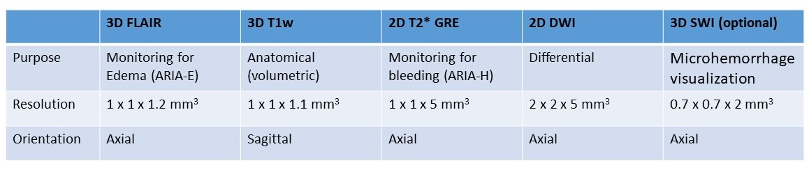
Fig. 1. Standardized MRI acquisition
protocol to be proposed to ICARE AD-US SITES for baseline and ARIA monitoring.
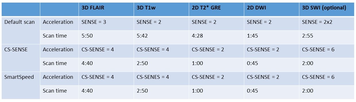
Fig. 2. Scan time for the Standardized
MRI acquisition protocol versus the protocol built with Philips CS-SENSE and SmartSpeed.
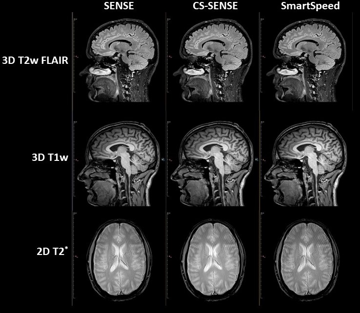
Fig. 3. 3D T2w FLAIR,
3D T1w and
2D GRE T2* images from
the Standardized MRI acquisition protocol compared with images acquired with
Philips CS-SENSE and SmartSpeed.
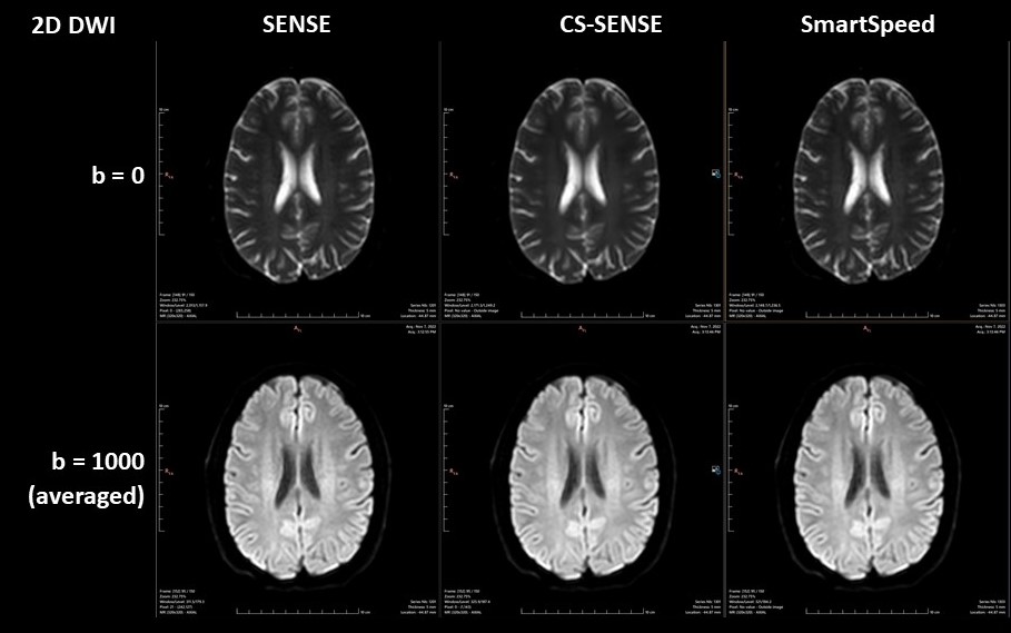
Fig. 4. 2D DWI images from the
Standardized MRI acquisition protocol compared with images acquired with
Philips CS-SENSE and SmartSpeed.
DOI: https://doi.org/10.58530/2023/3327