3325
Neuroimaging and Cognitive Testing in Healthy Aging Adults using a Portable Low-Field MRI Scanner and Web-Based Assessment
Sean Deoni1, Phoebe Burton2, Jennifer Beauchemin2, Rosa Cano-Lorente2, Matthew De Both3, Megan Johnson3, Lee Ryan4, and Mathew Huentleman3
1MNCH D&T, Bill & Melinda Gates Foundation, Seattle, WA, United States, 2Advanced Baby Imaging Lab, Providence, RI, United States, 3TGen, Pheonix, AZ, United States, 4University of Arizona, Tucson, AZ, United States
1MNCH D&T, Bill & Melinda Gates Foundation, Seattle, WA, United States, 2Advanced Baby Imaging Lab, Providence, RI, United States, 3TGen, Pheonix, AZ, United States, 4University of Arizona, Tucson, AZ, United States
Synopsis
Keywords: Dementia, Aging, Remote Neuroimaging
In this study we sought to (1) Determine the feasibility of collecting remote MRI and cognitive data in adults and elderly individuals; and (2) Replicate previously reported population-based associations between regional brain volumes and cognitive performance with an established cognitive assessment, PAL. Overall, this initial report of at-home MRI shows that MRI data collection on a portable low-field MRI system at a participant's home is possible and offers time efficiency, convenience, and accessibility to participants who might otherwise not be able to participate.INTRODUCTION
Consumer health wearables, internet-based health and cognitive assessments, and at-home biosample (e.g., saliva and capillary blood) collection kits increasingly allow the recruitment and follow-up of large study populations without intensive in-person study visits. By reducing participant time and travel burden, remote data collection allows the inclusion of individuals who live long distances from research centers and those who have limited time, mobility, or access to transportation.Unfortunately, studies that include magnetic resonance neuroimaging are challenged by the infrastructure requirements of MRI scanners. As consequence, they often omit socially, economically, and educationally disadvantaged individuals. Portable lower magnetic field strength systems offer the potential to perform neuroimaging at a participant’s home and convenience, but their utility remains to be demonstrated. Paired with web-based cognitive assessments, portable neuroimaging could offer new opportunities for neuroscience research and the communities that can be included. Here we report our experience using a low-field MRI “scan van” with an online assessment of paired-associate learning (PAL) to examine associations between brain morphometry and verbal memory performance.
METHODS & MATERIALS
75 healthy individuals (42 female), 18-94 (mean 54.2±19.7) years of age were enrolled in this study. 67 (39 female) completed MRI and a web-based paired associated learning (PAL) task on mindcrowd.org. The PAL task involves presentation of 12 word-pairs (e.g., apple | green, swim | suit) with each pair presented for 2 seconds. Following, participants are presented with the first word of each pair and asked to fill in the missing paired word. This learning-recall procedure is repeated for two additional trials of 12 pairs (3 trials in total). Word-pairs are presented in different random orders during each learning and each recall phase. The same word pairs and order of presentation is used for all participants. Total PAL score is the number of correct word pairs entered across the 3 trials (with 36 being perfect).MRI was performed on a 64mT Hyperfine Swoop installed in a modified Ford Transit “scan van” [1] at the participant’s residence (Fig. 1). T2-weighted 3D FSE images were acquired in the three orthogonal orientations and super-resolution was used to reconstruct a (1.5 x 1.5 x 1.5)mm3 anatomical image [1]. White and gray matter maps were calculated [2] and spatially registered to a custom study template generated from 26 random study participants aligned to the MNI template. Results were corrected for the effects of the warping and subtly blurred with a 4mm Gaussian kernel.
VBM analysis [3] was performed to identify associations between PAL scores and gray matter density, controlling for sex, age, and years of educational attainment. Analysis was performed using the Randomise tool of the FMRIB Software Library (FSL) [4]. Threshold-free cluster enhancement (TFCE) [5] was used to control for multiple comparisons.
RESULTS
Figure 2 displays brain areas with significant (p<0.05 FWE) associations between gray matter density and PAL test scores predominately within left hemisphere regions, including hippocampus, parahippocampal gyrus, inferior temporal gyrus, thalamus, putamen, frontal pole and orbital cortex, caudate, and Broca’s area. Right hemisphere precentral gyrus was also identified.DISCUSSION & CONCLUSIONS
Our results highlight the potential to perform participant screening, enrollment, and at-home study visits, even in the context of neuroimaging studies. Findings agree with brain regions previously associated with memory performance, including hippocampus, thalamus, caudate, and putamen), left parahippocampal gyrus, frontal pole, and Broca’s area (Brodmann area 44), and right precentral gyrus (Brodmann area 4). These results carry important implications for future directions in neuroscience research. The combination of at-home neuroimaging and web-based cognitive assessments presents an important new opportunity to engage individuals from socially, economically, and educationally disadvantaged communities that are often under-represented in clinical and public health research. By bringing the scanner to them at home or work, and allowing cognitive assessments to be performed on their time, involving these important cohorts may be less challenging.Acknowledgements
References
1. Deoni, S.C.L., et al., Simultaneous high-resolution T2 -weighted imaging and quantitative T2 mapping at low magnetic field strengths using a multiple TE and multi-orientation acquisition approach. Magn Reson Med, 2022. 88(3): p. 1273-1281. 2. Avants, B.B., et al., An open source multivariate framework for n-tissue segmentation with evaluation on public data. Neuroinformatics, 2011. 9(4): p. 381-400. 3. Ashburner, J. and K.J. Friston, Voxel-based morphometry--the methods. Neuroimage, 2000. 11(6 Pt 1): p. 805-21. 4. Jenkinson, M., et al., Fsl. Neuroimage, 2012. 62(2): p. 782-90. 5. Smith, S.M. and T.E. Nichols, Threshold-free cluster enhancement: addressing problems of smoothing, threshold dependence and localisation in cluster inference. Neuroimage, 2009. 44(1): p. 83-98.Figures
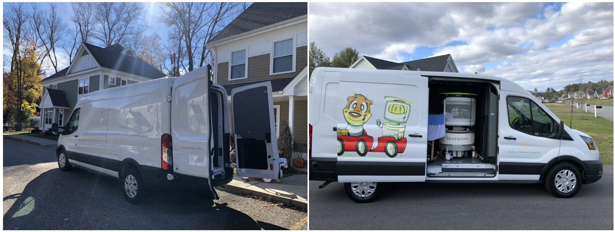
Figure 1. Example photos of the Scan-Van at participant homes. Participants enter the van from the rear doors. The side door can be opened to reduce claustrophobia, allow extra cooling, or just improve the general participant experience
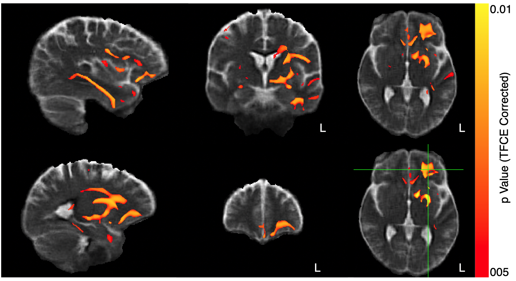
Figure 2. Exploratory voxel-based morphometry analysis examining associations between gray matter density and PAL score, controlling for subject age, biological sex, and education attainment. Highlighted regions denote significant associations (corrected for multiple comparisons using threshold-free cluster enhancement).
DOI: https://doi.org/10.58530/2023/3325