3321
Associations between CSF Biomarkers of Alzheimer's Disease and Subcortical Volume in Healthy Aging1Department of Neurology, Emory University, Atlanta, GA, United States, 2Department of Radiology and Imaging Sciences, Emory University, Atlanta, GA, United States, 3Goizueta Alzheimer’s Disease Research Center, Emory University, Atlanta, GA, United States, 4Joint Department of BioMedical Engineering, Emory University and Georgia Institute of Technology, Atlanta, GA, United States
Synopsis
Keywords: Alzheimer's Disease, Aging, CSF biomarker; Aβ; Tau; Subcortical; Volume
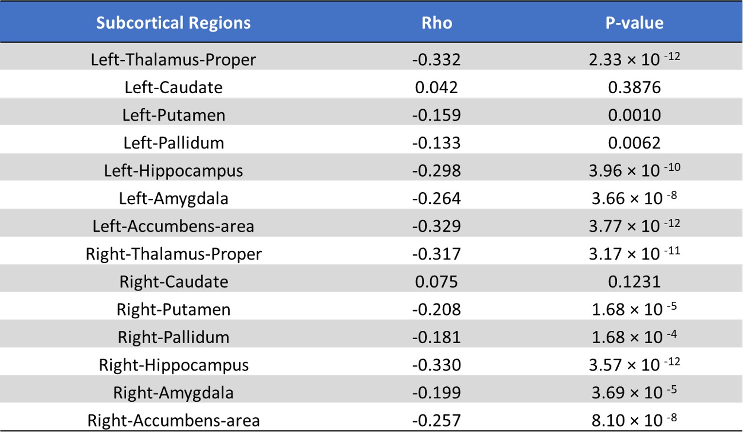
This study aims to evaluate the association between subcortical volumes and cerebrospinal fluid (CSF) biomarkers of Alzheimer’s Disease (AD) in a large group of healthy aging from a single center. CSF samples were obtained and quantitative levels of amyloid-β and Tau were measured. Subcortical tissue segmentations were performed on T1-weighted MPRAGE scans and the volume of each subcortical structure were calculated. Significant correlations were found between CSF biomarkers of AD and volume of the right and the left caudate. This may indicate that the volume of caudate are associated with CSF AD biomarker in healthy aging participants.Introduction
Accumulation of the β-amyloid peptide (Aβ) and neurofibrillary tangles (Tau) are the pathological hallmarks of Alzheimer's disease (AD)(Jack et al., 2013). Many previous studies have demonstrated that Aβ and Tau depositions are associated with neuronal damage and cortical and subcortical atrophy in both symptomatic AD and preclinical AD (Fortea et al., 2014). Cerebrospinal fluid (CSF) Aβ and Tau have been established as sensitive biomarkers for AD diagnosis and early AD pathology in healthy elderly(Mattsson et al., 2009). Subcortical volume measurements derived from non-invasive structural MRI have been widely used to detect atrophy in healthy aging and AD. In this study, we aim to study whether subcortical volumes were associated with the level of CSF biomarkers of AD in a large group of healthy old participants.Materials and Methods
Participants: A total cohort of 425 cognitively normal healthy old participants (HO) (age: 63.2 ± 6.5; 117 males) were included from the ongoing Emory Healthy Brain Study. MRI acquisitions: MRI data were acquired on a Siemens Magnetom Prisma 3T scanner with a 32-channel phased-array head coil. 3D T1-weighted (T1w) images were acquired using an MPRAGE sequence with the following parameters: TR = 2300 ms, TE = 2.96 ms, TI = 900 ms, flip angle = 9°, 208 sagittal slices with slice thickness = 1 mm, in-plane matrix size = 256 × 240, isotropic voxel size. CSF biomarker Collection: Lumbar punctures were performed in the HO participants to obtain CSF samples, from which amyloid-β 1-42(Aβ) and total tau (T-tau) were measured. Data analysis: Subcortical tissue segmentation was performed on the T1w images using the Freesurfer pipeline followed by visual inspection to minimize the segmentation errors. Then the volumes of 14 subcortical structures (7 regions for each hemisphere) and the total intracranial volume (TIV) were calculated for further statistical analysis. Statistical analysis: To access the age effect on subcortical volume, we utilized a generalized linear model (GLM) on 14 subcortical structures with controlling gender and TIV as covariates and followed a false discovery rate correction (FDR) with p<0.05. Then we perform GLM to estimate the associations between the level of CSF biomarkers and subcortical volumes with controlling age, gender and TIV as covariates, FDR correction with p < 0.05.Results
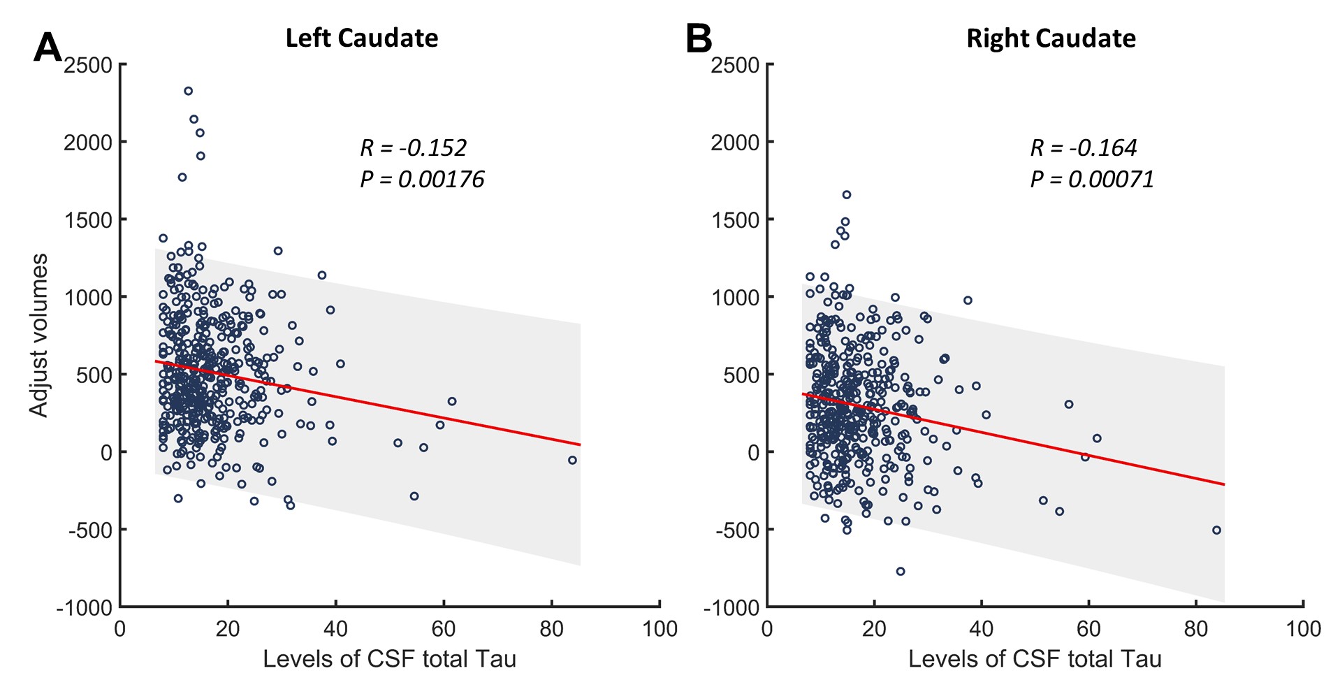
Significant age effects were found in most of the subcortical regions except left and right caudate nuclei (all p< 0.05, FDR corrected) (Figure.1). Further correlation analysis revealed that the volume of left caudate and right caudate were significantly negatively correlated with level of CSF total Tau (p < 0.05 FDR corrected, Figure. 2).Discussion & Conclusion
In this study, we combine CSF biomarkers of AD and subcortical volume derived from the structural MRI imaging to investigate the association between CSF biomarkers of AD and subcortical in a large group of healthy aging participants from a single center. A significant AD pathology-related effect was found in the volume of left caudate and right caudate, while no significant age effect was found in these two regions. Previous positive emission tomography (PET) studies have demonstrated depositions of Aβ and Tau in the caudate areas in AD patients (Chetelat et al., 2013; Rowe et al., 2008). These results may indicate that CSF AD pathology is associated with changes in structural properties in caudate regions.Acknowledgements
National Institutes of Health Grants: P30AG066511, R01AG072603, R01AG070937 and R21AG064405.References
Chetelat, G., La Joie, R., Villain, N., Perrotin, A., de La Sayette, V., Eustache, F., & Vandenberghe, R. (2013). Amyloid imaging in cognitively normal individuals, at-risk populations and preclinical Alzheimer's disease. Neuroimage Clin, 2, 356-365. doi:10.1016/j.nicl.2013.02.006
Fortea, J., Vilaplana, E., Alcolea, D., Carmona-Iragui, M., Sanchez-Saudinos, M. B., Sala, I., . . . Alzheimer's Disease Neuroimaging, I. (2014). Cerebrospinal fluid beta-amyloid and phospho-tau biomarker interactions affecting brain structure in preclinical Alzheimer disease. Ann Neurol, 76(2), 223-230. doi:10.1002/ana.24186
Jack, C. R., Jr., Knopman, D. S., Jagust, W. J., Petersen, R. C., Weiner, M. W., Aisen, P. S., . . . Trojanowski, J. Q. (2013). Tracking pathophysiological processes in Alzheimer's disease: an updated hypothetical model of dynamic biomarkers. The Lancet. Neurology, 12(2), 207-216. doi:10.1016/S1474-4422(12)70291-0
Mattsson, N., Zetterberg, H., Hansson, O., Andreasen, N., Parnetti, L., Jonsson, M., . . . Blennow, K. (2009). CSF biomarkers and incipient Alzheimer's disease in patients with mild cognitive impairment. JAMA, 302(4), 385-393. doi:10.1001/jama.2009.1064
Rowe, C. C., Ackerman, U., Browne, W., Mulligan, R., Pike, K. L., O'Keefe, G., . . . Villemagne, V. L. (2008). Imaging of amyloid beta in Alzheimer's disease with 18F-BAY94-9172, a novel PET tracer: proof of mechanism. The Lancet. Neurology, 7(2), 129-135. doi:10.1016/S1474-4422(08)70001-2
Figures