3305
Feasibility of Performing High-Resolution Multi-delay PCASL Imaging with Intravascular Signal Suppression Within 7 Minutes1University of Minnesota, Minneapolis, MN, United States
Synopsis
Keywords: Data Processing, Brain, ASL, cerebral perfusion or blood flow
To ensure satisfactory perfusion signal-to-noise ratio (SNR) for ASL imaging, intravascular perfusion signals are typically not suppressed (e.g., high-resolution multi-delay SMS/MB-EPI PCASL imaging for Human Connectome Projects) but can induce significant bias for CBF quantification if not addressed sufficiently. Recently proposed image denoising methods may be able to improve ASL imaging quality and SNR, having the potential to making it practical to perform ASL imaging with intravascular signal suppression. Our preliminary study results suggested that with image denoising, high-resolution multi-delay SMS/MB-EPI PCASL imaging with intravascular suppression could be achieved within 7 minutes.Purpose
Arterial spin labeling (ASL) (1) imaging, as a non-contrast enhanced and non-invasive approach, has been widely applied to measure brain perfusion. For example, the SMS/MB-EPI PCASL imaging method (2) has been applied for diverse brain imaging studies, such as the Human Connectome Projects (HCP) to study evolutions of CBF in children and elderly populations. The current widely applied study protocol of SMS/MB-EPI PCASL imaging method (3) uses five different post-labeling delays (PLDs) to facilitate the estimation of arterial transit time (ATT) to improve the accuracy of cerebral blood flow (CBF) quantification, which is critical for studies with participants potentially having largely varied ATT.To ensure satisfactory perfusion signal-to-noise ratio (SNR) for SMS/MB-EPI PCASL imaging with a limited/practical acquisition time (e.g., ~ 5 minutes and 30 seconds for HCP studies), currently, the intravascular perfusion signals are not suppressed. The intravascular perfusion signals can be quite prominent, especially for the data acquired with short PLDs, and induce significant bias for CBF quantification if not sufficiently addressed.
Recently, denoising methods based on principle component analysis (PCA) and random matrix theory (4-5) have been demonstrated effective for improving timeseries in functional and diffusion imaging studies (6-9). However, it remains unclear as to how such denoising could improve ASL imaging. Here we propose to denoise ASL timeseries in the complex domain using a PCA-based denoising algorithm and demonstrate its utility for promoting the image quality of ASL perfusion imaging. Our preliminary results show that it is feasible to perform high-resolution, whole-brain multi-delay SMS/MB-EPI PCASL imaging at 3T under 7 minutes with intravascular suppression.
Methods
Studies with healthy volunteers were performed on a Siemens 3T Prisma MRI scanner under an IRB approved protocol with written informed consent. The body coil was used for RF transmission and a 32-channel phased array head coil for signal reception. ASL perfusion studies were performed using the single-shot 2D SMS/MB-EPI PCASL imaging method (3) with and without an applied bipolar crushing gradient. This bipolar crushing gradient was played out after the excitation RF pulse and before the EPI readout echo train with an encoding velocity equal to 2.0 mm per second. Data sets were acquired from each volunteer by using the following major parameters: TE = 29 ms; FOV = 215 x 215; matrix size = 86 x 86; slice thickness/gap = 2.3 mm/10%; partial Fourier = 6/8; MB factor = 6 with 24 reference lines and FOV shift factor 3; labeling duration/post-labeling delay (PLD) = 1500/{200, 700, 1200, 1700, 2200} ms; the number of label and control images per PLD = 80; and 2 M0 images were acquired at the end of the scan for each PLD with a 8 second TR. In addition to high-resolution anatomic images, as before, to facilitate SNR analysis, 200 noise images were also acquired (3) and to facilitate image distortion correction, field map was obtained by acquiring spin-echo EPI images with opposite phase-encoding directions and ASL-imaging-matched protocols. PLD-specific minimal TRs were used for best time efficiency. Basically, the major parameters of the performed studies matched those for HCP projects except some specific parameters that had to be adapted to accommodate the added bipolar crushing gradients. For each volunteer, data was also acquired using fewer and varied number of label and control images for five PLDs (similar to those used for HCP studies) with and without the applied crushing gradient.To demonstrate the usefulness of our denoising method, we considered two scenarios. In scenario 1, half of the entire dataset, comprising 20 pairs of label-control images plus 2 M0 images per PLD were denoised and the results were compared to other data quantities without denoising. In scenario 2, a smaller dataset having PLD-dependent pairs of label-control images plus 2 M0 images for each PLD was denoised and the results were compared against the same, doubled, and tripled data without denoising. Data-driven denoising was applied to the complex-valued time series obtained by merging the channel-combined magnitude and phase dicom images exported from the MR scanner. Noise mapping necessary for data scaling in the subsequent singular value manipulation was done using MPPCA (10) and optimal singular value shrinkage (11) was used to reduce the impact of thermal noise. The image phases were also stabilized as in NORDIC (7) for improved denoising performances.
Data processing, including motion correction, was performed using SPM. The ATT maps were generated based on the weighted delay (WD) approach (12-13), and CBF quantification employed the single compartment perfusion model that considered the estimated ATT.
Results and Discussions
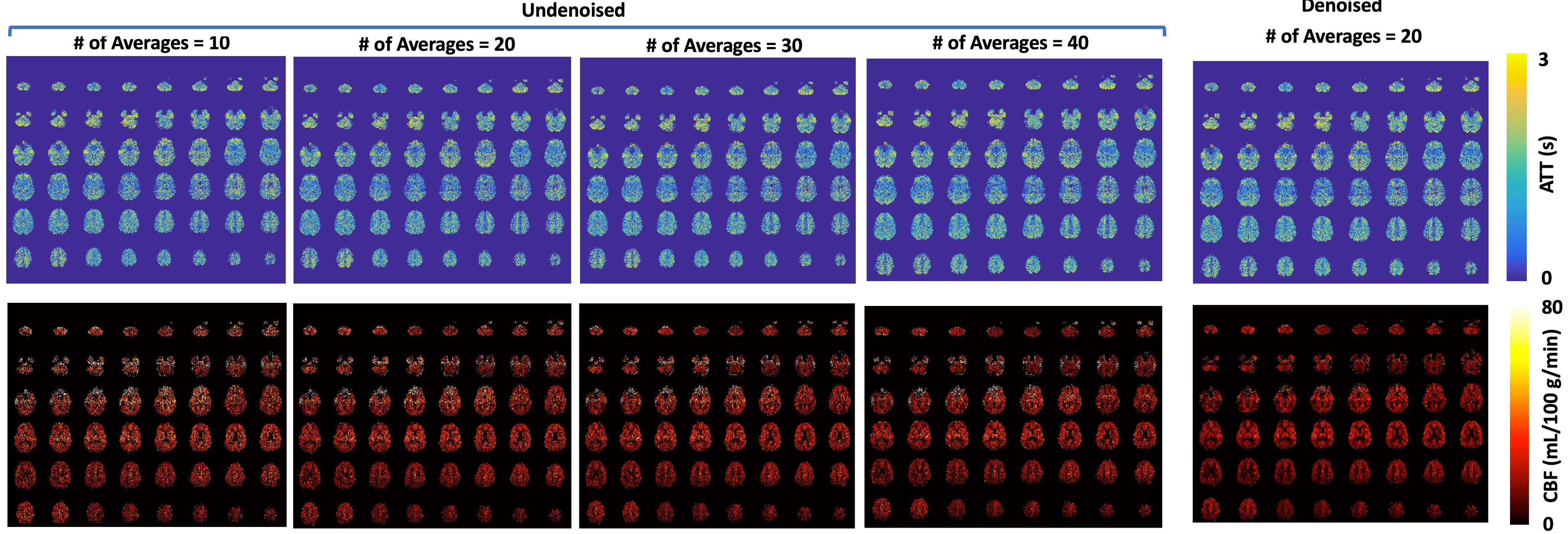
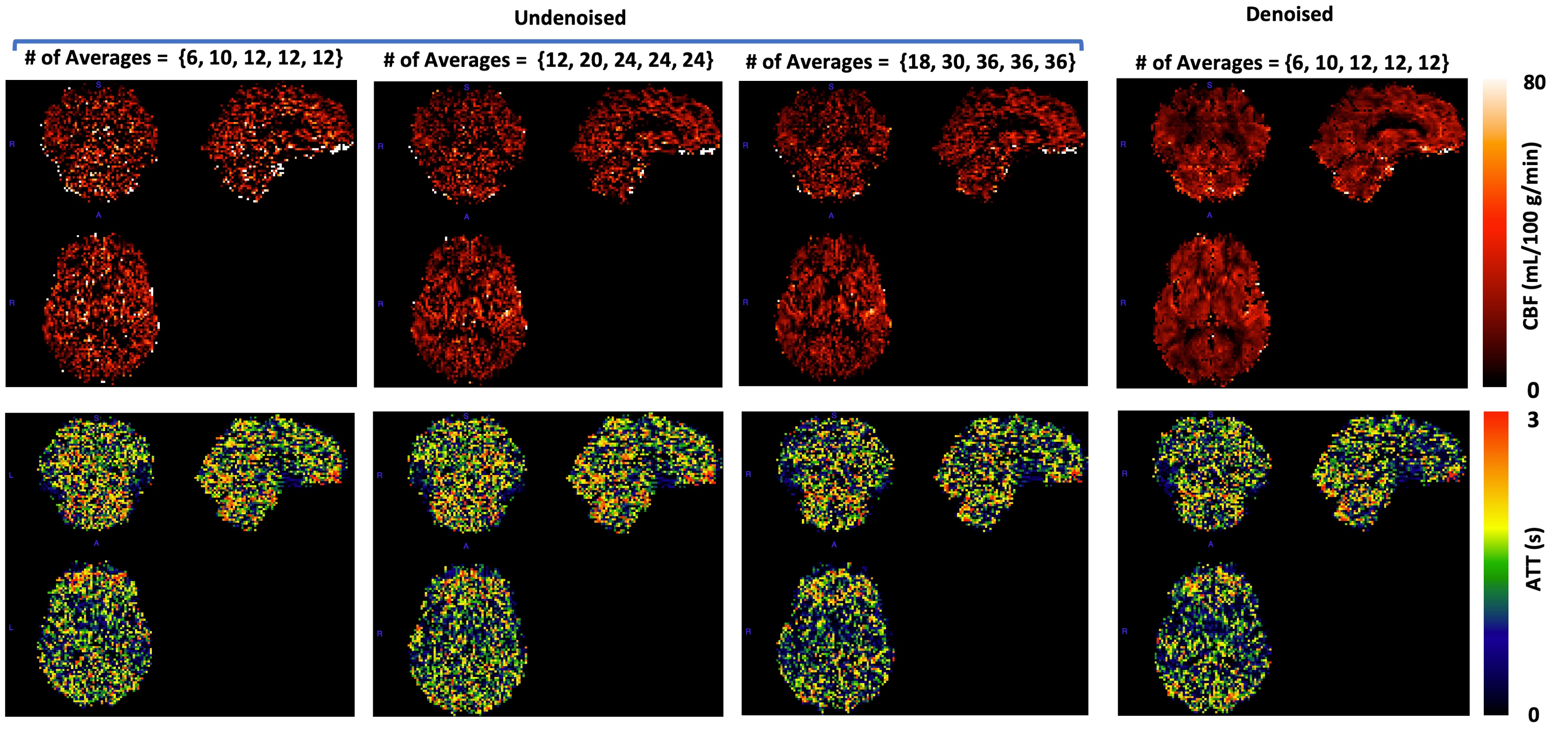
In scenario 1, the use of denoising appeared to largely improve perfusion image quality and SNR (Figure 1), producing image quality comparable to or better than what was attainable with the entire data (i.e., doubled quantity) without denoising. Likewise, when considering PLD-specific label-control pairs, the use of denoising was found effective for increasing SNR (Figure 2), enabling ASL imaging within 7 minutes to only acquire 6, 10, 12, 12 and 12 pairs of label-control images for the five PLDs under consideration.Conclusions
Denoising can improve ASL imaging quality and high-resolution multi-delay SMS/MB-EPI PCASL imaging with intravascular signal suppression could be achieved within 7 minutes.Acknowledgements
This study was supported by National Institute of Health R56EB033365, P41 EB027061 and U01 EB025144.References
1. Detre JA, Leigh JS, Williams DS, Koretsky AP (1992) Perfusion imaging. Magn Reson Med 23:37-45.
2. Li X, Wang D, Auerbach EJ, Moeller S, Ugurbil K, et al. Theoretical and experimental evaluation of multi-band EPI for high-resolution whole brain pCASL Imaging. Neuroimage. 2015 Feb 1;106:170-81.
3. Harms MP, Somerville LH, Ances BM, Andersson J, Barch DM, Bastiani M, Bookheimer SY, Brown TB, Buckner RL, Burgess GC, Coalson TS, Chappell MA, Dapretto M, Douaud G, Fischl B, Glasser MF, Greve DN, Hodge C, Jamison KW, Jbabdi S, Kandala S, Li X, Mair RW, Mangia S, Marcus D, Mascali D, Moeller S, Nichols TE, Robinson EC, Salat DH, Smith SM, Sotiropoulos SN, Terpstra M, Thomas KM, Tisdall MD, Ugurbil K, van der Kouwe A, Woods RP, Zöllei L, Van Essen DC, Yacoub E. Extending the Human Connectome Project across ages: Imaging protocols for the Lifespan Development and Aging projects. Neuroimage. 2018 Dec;183:972-984.
4. Manjon, J.V., Coupe, P., Concha, L., Buades, A., Collins, D.L., Robles, M., 2013. Diffusion Weighted Image Denoising Using Overcomplete Local PCA. PLOS ONE 8.
5. Veraart, J., Novikov, D.S., Christiaens, D., Ades-aron, B., Sijbers, J., Fieremans, E., 2016b. Denoising of diffusion MRI using random matrix theory. NeuroImage 142, 394-406.
6. Ma, X., Ugurbil, K., Wu, X., 2020. Denoise magnitude diffusion magnetic resonance images via variance-stabilizing transformation and optimal singular-value manipulation. NeuroImage 215, 116852.
7. Moeller, S., Pisharady, P.K., Ramanna, S., Lenglet, C., Wu, X., Dowdle, L., Yacoub, E., Uğurbil, K., Akçakaya, M., 2021. NOise reduction with DIstribution Corrected (NORDIC) PCA in dMRI with complex-valued parameter-free locally low-rank processing. NeuroImage 226, 117539.
8. Vizioli, L., Moeller, S., Dowdle, L., Akcakaya, M., De Martino, F., Yacoub, E., Ugurbil, K., 2021. Lowering the thermal noise barrier in functional brain mapping with magnetic resonance imaging. Nat Commun 12, 5181.
9. Zhu, W., Ma, X.D., Zhu, X.H., Ugurbil, K., Chen, W., Wu, X.P., 2022. Denoise Functional Magnetic Resonance Imaging With Random Matrix Theory Based Principal Component Analysis. IEEE Transactions on Biomedical Engineering 69, 3377-3388.
10. Veraart, J., Fieremans, E., Novikov, D.S., 2016a. Diffusion MRI noise mapping using random matrix theory. Magnetic Resonance in Medicine 76, 1582-1593.
11. Gavish, M., Donoho, D.L., 2017. Optimal Shrinkage of Singular Values. Ieee Transactions on Information Theory 63, 2137-2152.
12. Dai W, Robson PM, Shankaranarayanan A, Alsop DC. Reduced resolution transit delay prescan for quantitative continuous arterial spin labeling perfusion imaging. Magn Reson Med. 2012 May;67(5):1252-65.
13. Wang DJ, Alger JR, Qiao JX, Gunther M, Pope WB, Saver JL, Salamon N, Liebeskind DS; UCLA Stroke Investigators. Multi-delay multi-parametric arterial spin-labeled perfusion MRI in acute ischemic stroke - Comparison with dynamic susceptibility contrast enhanced perfusion imaging. Neuroimage Clin. 2013 Jul 6;3:1-7.
Figures