3299
Optimization of T1-Weighted DANTE-SPACE for Intracranial Vessel Wall Imaging at 7T1Department of Radiology, Mayo Clinic, Jacksonville, FL, United States, 2Siemens Medical Solutions USA, Inc., Jacksonville, FL, United States
Synopsis
Keywords: Blood vessels, High-Field MRI, DANTE, SPACE, 7T
MR vessel-wall-imaging (VWI) is used clinically for noninvasive characterization of vessel wall pathology. The DANTE preparation module has been combined with conventional 3D-T1W-SPACE to achieve better slow-flow-suppression. 7T could further improve VWI by its increased SNR and spatial-resolution. Here we sought to optimize DANTE preparation module for T1W-SPACE at 7T. Simulations were done to determine the optimal parameters for the DANTE module. Human scans showed that the optimized T1W- DANTE-SPACE at 7T efficiently suppressed slow-blood-flow in both intracranial arteries and veins, and in turn improved wall contrast compared with conventional T1W-SPACE.Introduction
MR vessel-wall-imaging (VWI) is used clinically for noninvasive characterization of vessel wall pathology. Unlike luminal imaging techniques, such as CT angiogram and DSA, VWI can provide additional characterization of the vessel pathology, such as differentiation of atherosclerotic disease from vasculitis, both of which can produce similar luminal patterns of stenosis. 3D VWI is often preferred due to its higher SNR and isotropic resolution for MPR. 7T could further enhance VWI by its increased SNR and spatial-resolution if 7T-specific challenges, such as the increased specific-absorption-rate (SAR) and B1 inhomogeneity, can be mitigated. SPACE, a 3D variable-flip-angle turbo-spin-echo (TSE) sequence, has been developed to reduce SAR and blurring at higher field and has effectively replaced the classic constant-flip-angle TSE readout. Though SPACE has excellent flow suppression in the readout direction, VWI using T1W SPACE is still challenged by slow-flow-related artifacts in the other two encoding directions, which results in hyperintense blood signals in pre- and post-contrast images and hinder the diagnostic value for lesion evaluation. The delay alternating with nutation for tailored excitation (DANTE)1 preparation module has been combined with SPACE in order to achieve better slow-flow suppression2. In this study we sought to optimize DANTE preparation module for T1W SPACE at 7T to effectively suppress slow arterial blood flow and venous flow and in turn achieve sharp depiction of the vessel wall.Purpose
To evaluate the feasibility and optimization of 3D T1W DANTE-SPACE sequence in high-resolution intracranial VWI at 7T.Method
The magnetization signals of vessel wall and blood were simulated using MATLAB (MathWorks, Natick, Massachusetts). Simulations were done across DANTE pulse flip angles (FA) of 8°,10°,12°, and 14°, with flow-encoding gradient strengths set at 25 mT/m and parallel to blood velocity. Simulations were then performed across flow-encoding gradient strengths of 20, 25, 30, and 35 mT/m, with DANTE pulse flip angle set at 12°. The rest of the simulation parameters were shown in Table 1.In compliance with institutional regulations, 3D T1W SPACE and 3D T1W DANTE-SPACE scans, which were both prototype sequences, were performed on a total of three healthy volunteers on the investigational pTx part of a Siemens 7T MAGNETOM Terra (Siemens Healthcare, Erlangen, Germany) with an investigational Nova 8Tx/32Rx head coil (Nova Medical Inc., Wilmington, MA, USA). The imaging parameters were shown in Table 2. Note that the DANTE flow-encoding gradients were only set in the phase and slice encoding directions, since the SPACE’s inherent flow suppressing capability already performed well in the read-out direction. The signal-to-noise ratio (SNR) were calculated in the thalamus and the contrast ratio between vessel wall and lumen were calculated in the internal carotid artery (ICA) for T1W-SPACE and T1W-DANTE-SPACE sequences for each of the three volunteers. Two MR scientists scored the image quality blindly and independently based on subjective wall visualizations of the two VWI methods using a 4-point scale followed by inter-rater reproducibility analysis.
Results
As the simulation has shown in Figure 1A, the blood signal in intracranial vessels decreased with increasing DANTE flip angle and number of DANTE pulses, while the vessel wall signal was only minorly affected. As shown in Figure 1B, for the assumed blood velocity (0.1 m/s), the best set of DANTE pulse number and flow-encoding gradient strength were 150 and 25 mT/m. In volunteer scans, the optimized DANTE module parameters were confirmed, and the preferred parameters were set to be DANTE pulse number=150, FA=14°, and flow-encoding gradient strength=25 mT/m. In the human scans, the thalamus SNR of the T1W SPACE and T1W DANTE-SPACE is 66.22±12.56 vs. 56±13.81 (p >0.05). The contrast-ratio between vessel wall and lumen is 5.57±0.95 vs. 12.57±5.85 (p<0.001). Subjective wall visualization-score of T1W DANTE-SPACE is significantly higher than T1W SPACE (3.65±0.23 vs. 2.16±0.35). The two MR scientists’ scores had excellent agreement, evidenced by the intraclass-correlation-coefficient (ICC) values being higher than 0.79 (p<0.001).Discussion
According to the simulation results of DANTE pulse, the larger the flip angle and numbers of DANTE pulses, the lower the signal of flowing blood, but with decreasing contrast and signal-to-noise ratio. Due to the SAR restriction, our tests showed DANTE pulse numbers over 200 cannot be achieved even with FA of 8°. Furthermore, longer DANTE module will have impact on image contrast and SPACE TR may need to be increased to retain the desired T1 contrast, which will result in a prolonged scan time. Our results suggested the ideal DANTE pulse scheme for T1W SPACE is FA=14°, DANTE pulse number=150, and flow-encoding gradient amplitude=25mT/m in the phase and slice encoding directions. With the optimized DANTE module, the commonly seen hyperintense signal in the C3 segment of ICA in T1W SPACE was sufficiently suppressed with DANTE (Figure 2B). The blood signal in the superior sagittal sinus was also greatly suppressed with DANTE (Figure 2D).Conclusions
T1W SPACE at 7T with DANTE preparation module can efficiently suppress slow blood flow in both intracranial arteries and veins. The optimization of DANTE module offered improved 7T vessel wall contrast compared with conventional T1W SPACE. The dark blood resulted from T1W DANTE-SPACE will also benefit post contrast images and allow improved differentiation of enhancing lesions and wall thickening from slow-flow artifact in blood vessels in post-contrast T1W images.Acknowledgements
No acknowledgement found.References
1. Li L, Chai JT, Biasiolli L, Robson MD, Choudhury RP, Handa AI, Near J, Jezzard P. Black-blood multicontrast imaging of carotid arteries with DANTE-prepared 2D and 3D MR imaging. Radiology. 2014 Jun 11;273(2):560-9.
2. Xie Y, Yang Q, Xie G, Pang J, Fan Z, Li D. Improved black‐blood imaging using DANTE‐SPACE for simultaneous carotid and intracranial vessel wall evaluation. Magnetic resonance in medicine. 2016 Jun;75(6):2286-94.
3. Viessmann O, Li L, Benjamin P, Jezzard P. T2‐weighted intracranial vessel wall imaging at 7 Tesla using a DANTE‐prepared variable flip angle turbo spin echo readout (DANTE‐SPACE). Magnetic resonance in medicine. 2017 Feb;77(2):655-63.
Figures
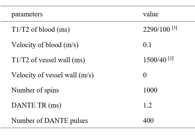
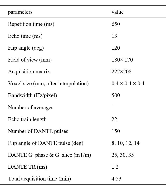
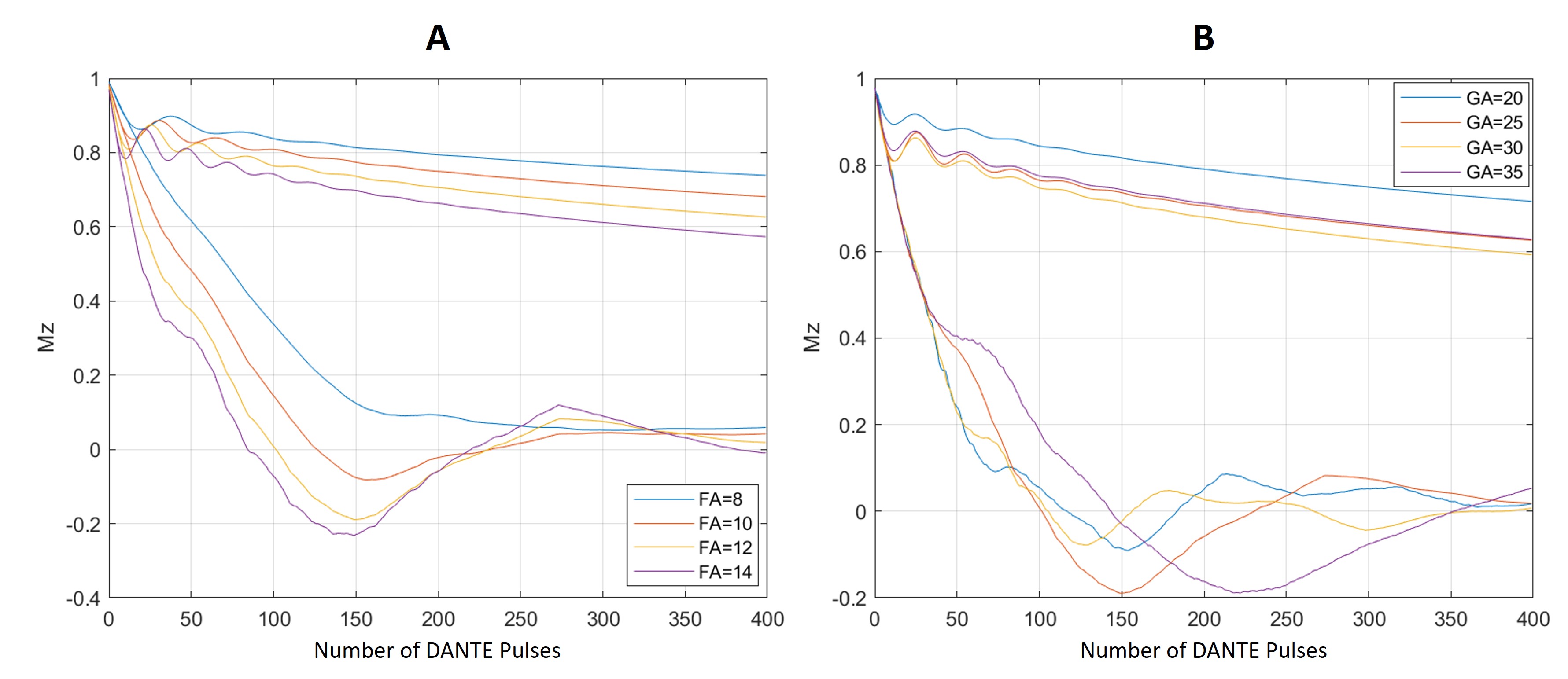
Figure 1: Vessel wall (upper group) and blood (lower group) signals simulated across DANTE pulse flip angles (A) and across DANTE flow encoding gradient strengths (B). DANTE flip angle=14° and number of DANTE pulses=150 is the best for reducing blood signal. For the assumed blood velocity (0.1 m/s), the best set of DANTE pulse number and gradient strength were 150 and 25 mT/m. Vessel wall signals were only minimally decreased.
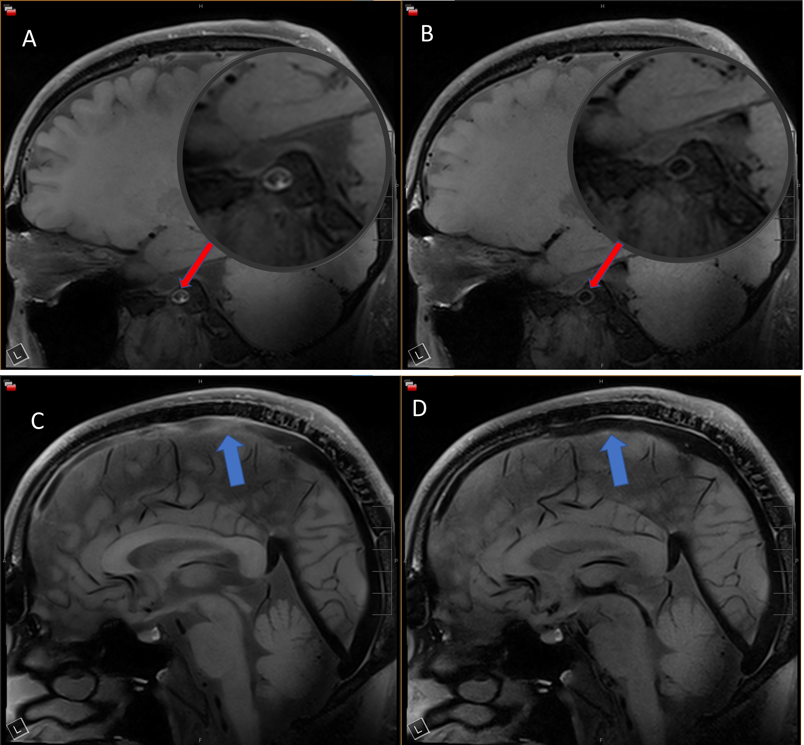
Figure 2: A&C: T1W SPACE; B&D: Optimized T1W DANTE-SPACE. Blood suppression in the internal carotid artery C3 (red arrow) was much improved with the optimized DANTE preparation module (A vs. B). The slow venous blood flow in the superior sagittal sinus (blue arrow) was suppressed with the optimized DANTE preparation module (C vs.D).