3256
Amide proton transfer weighted and diffusion kurtosis imaging in evaluating lymphovascular space invasion of endometrial carcinoma1Department of Radiology, the First Affiliated Hospital of Dalian Medical University, Dalian, China
Synopsis
Keywords: Uterus, Cancer
Lymphovascular space invasion(LVSI)is one of the important factors for poor prognosis of endometrial carcinoma(EC).Quantitative parameters of amide proton transfer weighted(APTw)and diffusion kurtosis imaging(DKI) is a novel MRI tool to evaluating LVSI in EC. The APT and MK values of EC with LVSI were higher than those without LVSI.The AUC after combination was higher than that of MK alone.Both APTw and DKI can effectively evaluate EC LVSI.APTw combined DKI application can improve the evaluation efficiency .Objective
To explore the value of amide proton transfer weighted (APTw) and diffusion kurtosis imaging (DKI) quantitative parameters in evaluating lymphovascular space invasion (LVSI) in endometrial carcinoma (EC).Materials and Methods
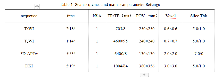
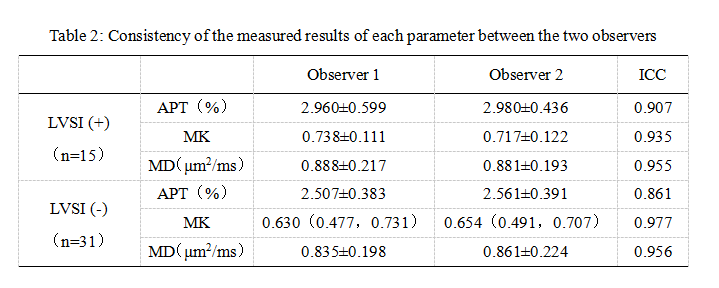
The data of 46 patients with EC confirmed by surgery and pathology were analyzed retrospectively, including 15 patients with LVSI and 31 patients without LVSI. The preoperative MR scanning sequence included APTw and DKI. After post-processing, APT, mean kurtosis (MK) and mean diffusion (MD) images were obtained. The APT, MK and MD values of the two groups of cases were measured by two observers respectively. The consistency of the measurement results of the parameters of the two groups of cases by two observers was tested by intra-class correlation coefficients (ICC). The differences of the parameters of the two groups were compared by independent sample t-test or Mann Whitney U test. ROC curve was used to evaluate the parameters with statistical differences and to evaluate the effectiveness of evaluating LVSI after combination. The difference of area under curve (AUC) was compared by Delong test.Results
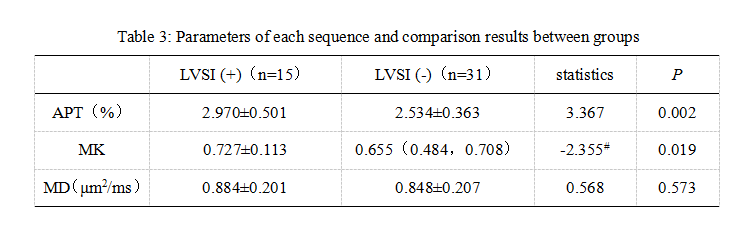
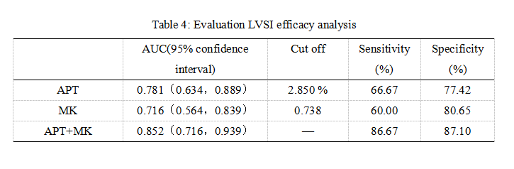
The consistency of the data measured by the two observers was good (ICC > 0.75). The APT, MK and MD values of EC with LVSI were 2.970±0.501 %, 0.727±0.113 and 0.884±0.201 μm2/ms, respectively. The above parameter values of EC without LVSI were 2.534±0.363 %, 0.655(0.484, 0.708) and 0.848±0.207 μm2/ms, respectively. The APT and MK values of EC with LVSI were higher than those without LVSI (P<0.05), and there was no difference in MD values between the two groups (P>0.05). The AUC of APT, MK and APT+MK values evaluating LVSI were 0.781, 0.716 and 0.852 respectively. The AUC after combination was higher than that of MK alone (P<0.05).Conclusion
Both APTw and DKI can effectively evaluate EC LVSI. The combined application can improve the evaluation efficiency and has a certain clinical value.Acknowledgements
noneReferences
[1] Harris KL, Maurer KA, Jarboe E, et al. LVSI positive and NX in early endometrial cancer: Surgical restaging (and no further treatment if N0), or adjuvant ERT? Gynecol Oncol, 2020, 156(1):243-250.
[2] Veade AE, Foote J, Ehrisman J, et al. Associations between lymphovascular space invasion, nodal recurrence, and survival in patients with surgical stage I endometrioid endometrial adenocarcinoma. World J Surg Oncol, 2019, 17(1):80.
[3] Ironi G, Mapelli P, Bergamini A, et al. Hybrid PET/MRI in Staging Endometrial Cancer: Diagnostic and Predictive Value in a Prospective Cohort. Clin Nucl Med, 2022, 47(3):e221-e229.
[4] Kumar S, Bandyopadhyay S, Semaan A, et al. The role of frozen section in surgical staging of low risk endometrial cancer. PLoS One, 2011, 6(9):e21912.[6] Jia X, Wang W, Liang J, et al. Risk stratification of abdominal tumors in children with amide proton transfer imaging. Eur Radiol, 2022, 32(4):2158-2167.
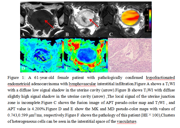
Figures