3225
A preliminary study of radiological characteristics of diffuse hemispheric gliomas, H3 G34-mutant1Radiology, Fudan University, Huashan Hospital, Shanghai, China, 2Neurosurgery, Fudan University, Huashan Hospital, Shanghai, China
Synopsis
Keywords: Tumors, Tumor, H3 G34-mutant, glioma
Diffuse hemispheric glioma, H3 G34-mutant (H3G34-DHG) is a rare newly proposed entity in 2021 WHO classification of central nervous system tumors. We retrospectively analysis characteristics of 10 cases of H3G34-DHG and found some certain characteristics in clinical and radiological manifestations in this entity. In most cases, absent or focally faint contrast enhancement initially suggested another diagnosis than a high-grade glioma. However, increased tumor vessels still suggested abundant tumor blood supply. Moreover, hyperintensity on CT and restricted diffusion reflected high density of tumor cells. MRS also showed extremely high metabolism active in the tumor.Introduction
Diffuse hemispheric glioma, H3 G34-mutant (H3G34-DHG) is a rare newly proposed entity in 2021 WHO classification of central nervous system tumors1,2, which is is still poorly understood by clinicians and radiologists. The aims of this study were to describe the clinical and radiological characteristics of H3G34-DHG.Method
The characteristics of 10 cases of H3G34-DHG in our institution were retrospectively analyzed.Result
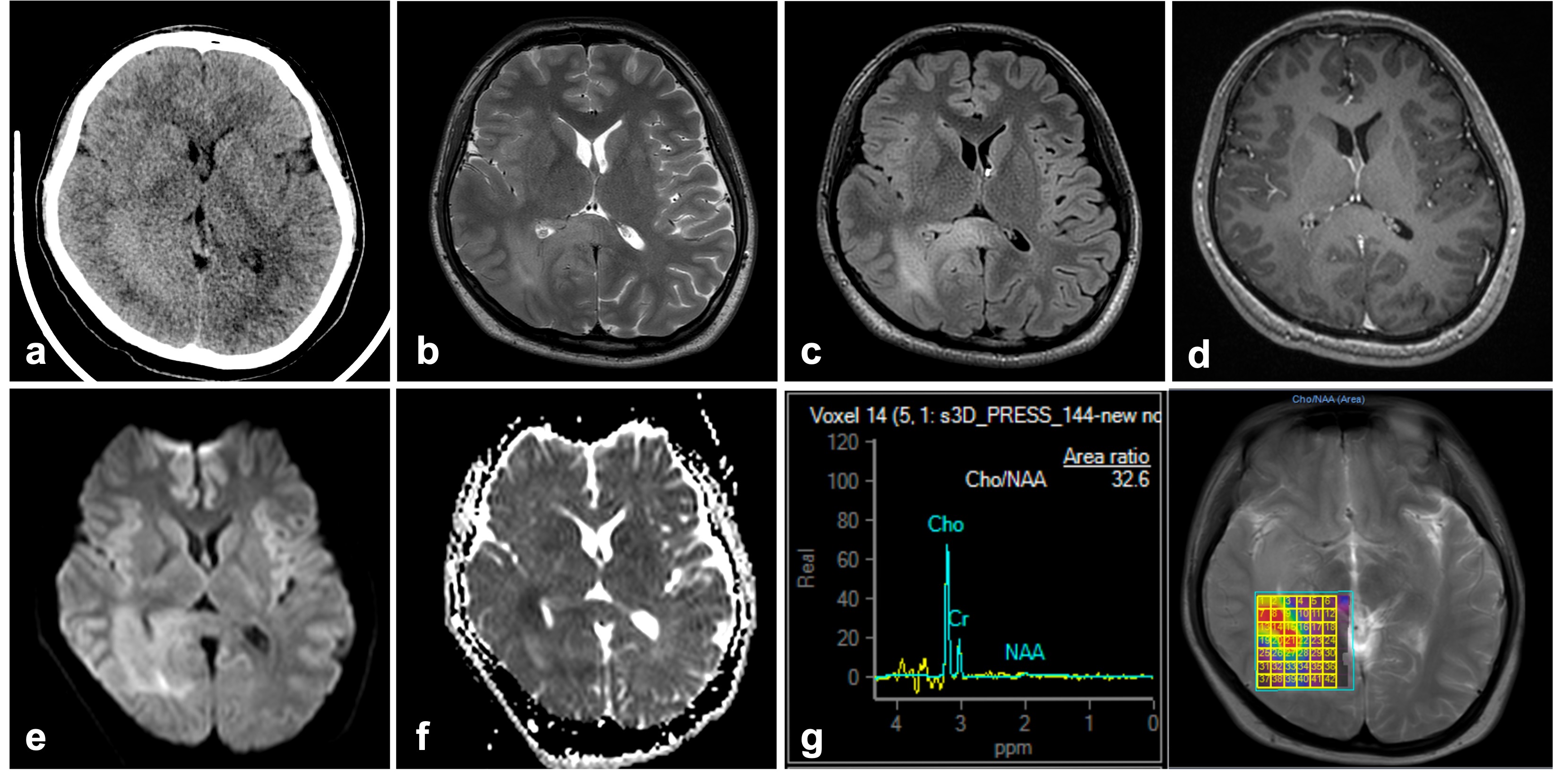
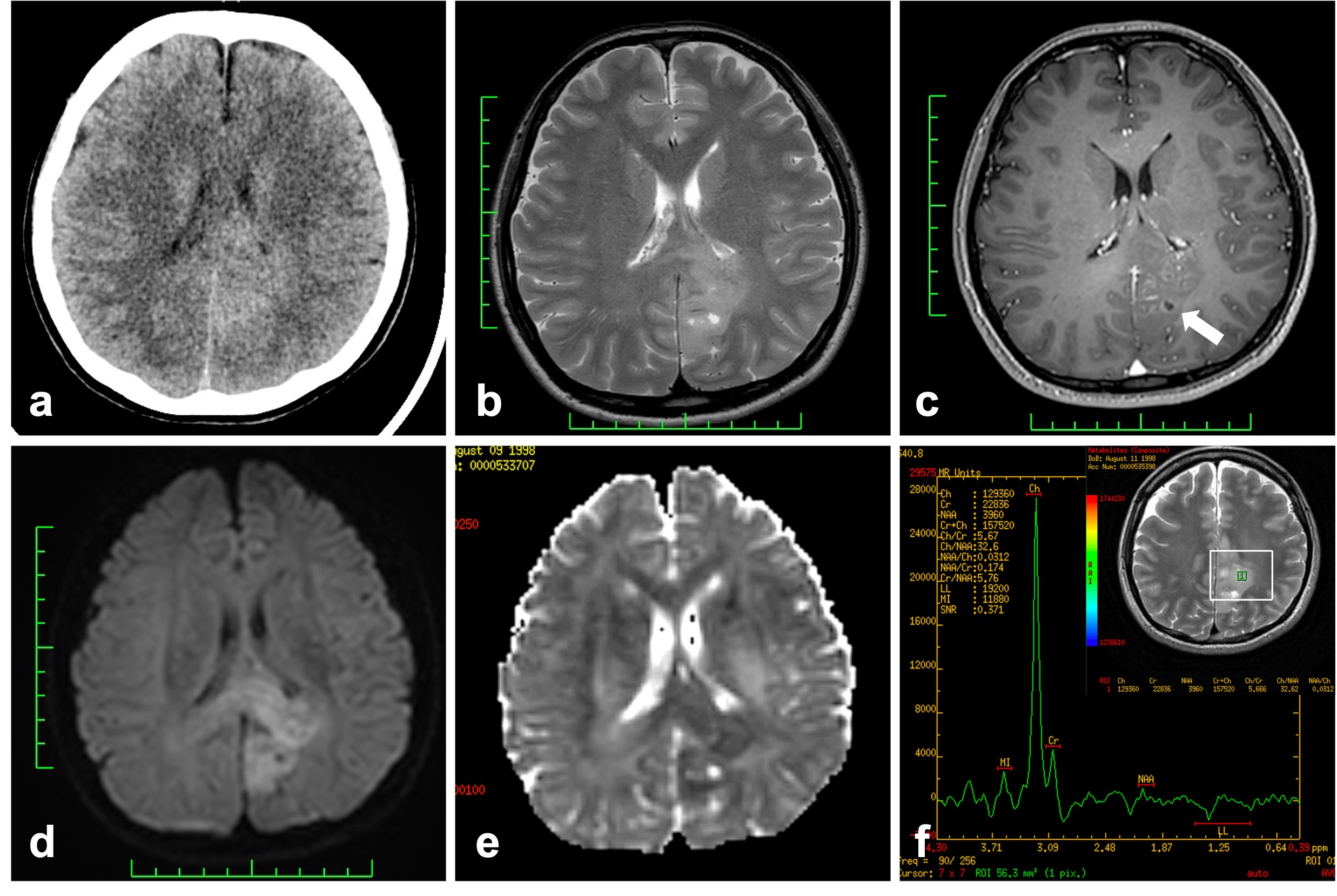
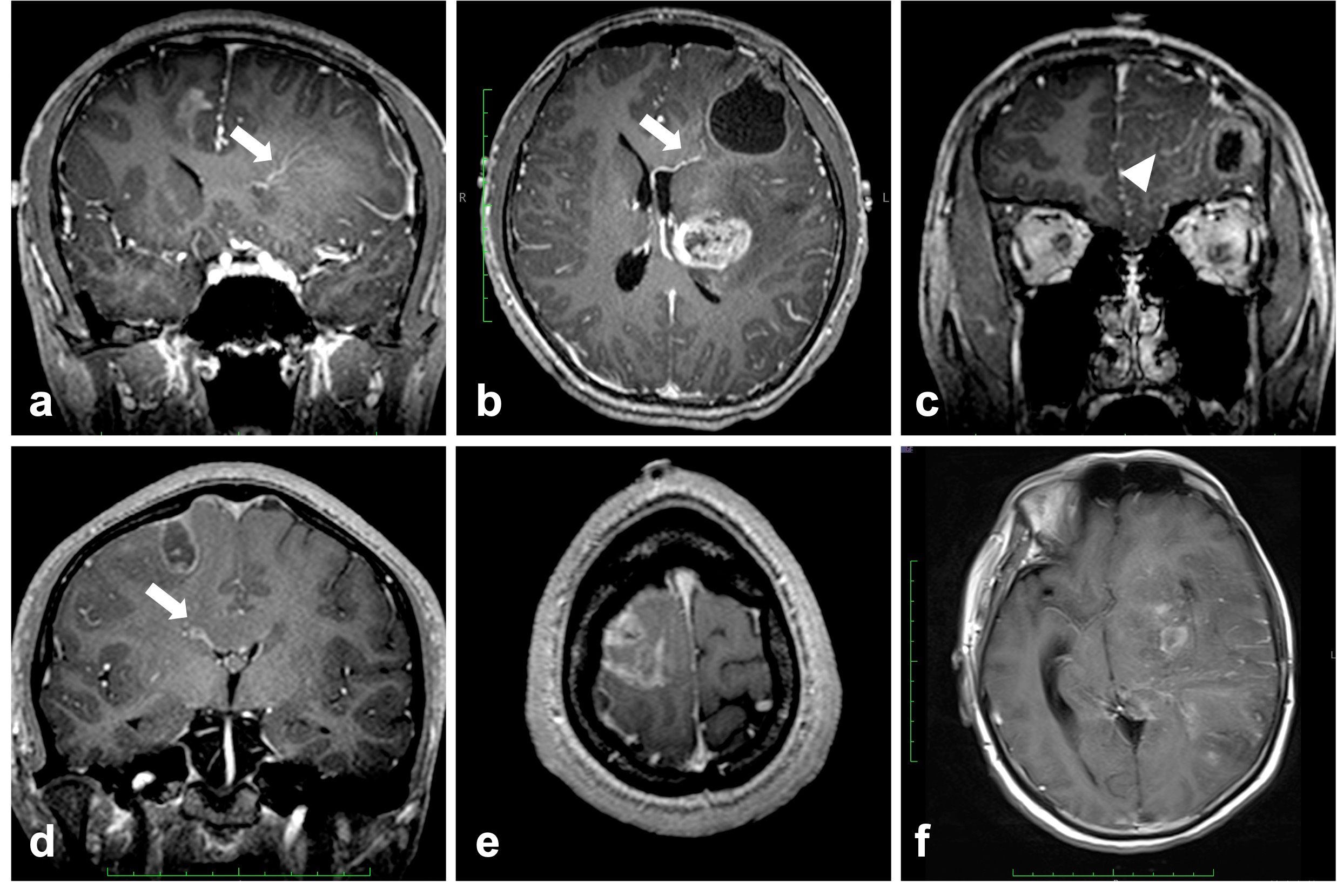
Median age at diagnosis of H3G34-DHG was 25 years (range: 15–42). Male: female=6:4. All tumors were hemispheric with brain stem and cerebellum involved in only one case. Cyst (9/10) and hemorrhage (7/10) were common, while necrosis (3/10)and calcification (1/10) were rare. For all tumors, the solid component was hyperintensity on CT. Most (7/8) of tumors demonstrated restricted diffusion on DWI and ADC. For 7 patients (7/9, 78%), absent or focal contrast enhancement were seen, while increased tumor vessels were observed. Leptomeningeal and/or ependymal enhancement was observed in more than half cases (6/9, 67%). MR-spectroscopy showed increased Choline peak and decreased NAA peak indicating abnormally exuberant metabolism in tumors.Discussion
H3G34-DHG is a subtype of pediatric-type diffuse high-grade gliomas, also seen in younger adult, slightly more common in male. The tumor arises in the cerebral hemisphere, occasionally spread to midline structure. Histopathologically, the tumor cells are dense, some of them are uniform small round cells3. Correspondingly the tumors are mainly hyperintensity solid on CT and restricted diffusion on DWI. In most cases, absent or focally faint contrast enhancement initially suggested another diagnosis than a high-grade glioma4. However, increased tumor vessels still suggested abundant blood supply of the tumor. It is presumed that the relative integrity of blood-brain barrier leads to less leakage of contrast agent, and perfusion MR may be helpful to evaluate the blood supply of tumor. Leptomeningeal and /or ependymal enhancement was commonly observed in our study, and the tumor cells infiltrating subarachnoid space and perivascular space could be seen in histopathology, suggesting that this subtype is prone to cerebrospinal fluid dissemination. Some studies have reported H3G34-DHG cases with cerebrospinal fluid dissemination5,6. MR spectroscopy demonstrated significantly decreased NAA peak and abnormally increased Choline peak, and the Cho/NAA ratio was extremely increased up to 42.8, which indicated active metabolism in the, in line with the characteristic of high-grade gliomas. Since the sample size is limited in our study, further research on large size sample and more multiple modality imaging techniques is still needed.Conclusion
H3G34-DHG showed some distinct characteristics in the clinical and radiological manifestations.Acknowledgements
No acknowledgement.References
1. Louis, D. N. et al. The 2021 WHO Classification of Tumors of the Central Nervous System: a summary. Neuro-oncology 23, 1231–1251 (2021).
2. Korshunov, A. et al. Histologically distinct neuroepithelial tumors with histone 3 G34 mutation are molecularly similar and comprise a single nosologic entity. Acta Neuropathol 131, 137–146 (2016).
3. Wang, W. et al. [High-grade gliomas with H3 G34R mutation: a clinicopathological study]. Zhonghua Bing Li Xue Za Zhi Chin J Pathology 49, 1267–1271 (2020).
4. Picart, T. et al. Characteristics of diffuse hemispheric gliomas, H3 G34-mutant in adults. Neuro-oncology Adv 3, vdab061- (2021).
5. Roux, A. et al. High-grade gliomas in adolescents and young adults highlight histomolecular differences from their adult and pediatric counterparts. Neuro-oncology 22, 1190–1202 (2020).
6. Vettermann, F. J. et al. Characterization of Diffuse Gliomas With Histone H3-G34 Mutation by MRI and Dynamic 18F-FET PET. Clin Nucl Med 43, 895–898 (2018).
Figures