3214
Dynamic characteristics of functional network activity in brains with left-frontal glioma
Siqi Cai1,2, Yuchao Liang3, Yinyan Wang3, Yufei Liu4, Fanfan Chen4, Chunxiang Jiang1,2, Lei Wang3, and Lijuan Zhang1,2
1Shenzhen Institute of Advanced Technology, Chinese Academy of Sciences, Shenzhen, China, 2University of Chinese Academy of Science, Beijing, China, 3Beijing Tiantan Hospital of Capital Medical University, Beijing, China, 4Shenzhen Second People's Hospital, Shenzhen, China
1Shenzhen Institute of Advanced Technology, Chinese Academy of Sciences, Shenzhen, China, 2University of Chinese Academy of Science, Beijing, China, 3Beijing Tiantan Hospital of Capital Medical University, Beijing, China, 4Shenzhen Second People's Hospital, Shenzhen, China
Synopsis
Keywords: Tumors, fMRI (resting state)
This study investigated the temporal features of the brain network activity with dynamic fractional amplitude of low-frequency fluctuation (dfALFF) and sliding window approach. The time-varying brain activity was clustered into two distinct states. Patients’ brains featured high occurrence of the weak state, low occurrence of the strong state, and network dALFF that decreased but spatially extended with the tumor malignancy. The dfALFF variance of networks was attenuated in HGGs but not LGGs. These malignancy-specific alterations indicate a diverse neuropathological profile of gliomas, and highlight the importance of temporal features of brain activity in the disease characterization of glioma.Introduction
The temporal coordination of neural activity may be impaired in brains with glioma1,2. Dynamic characteristics of the time-varying brain activity are expected to provide additional references for disease characterization of glioma. In this study, we aim to investigate the temporal properties of the functional network activity in brains with glioma based on the resting-state functional MRI (rs-fMRI).Participants and Methods
This study was approved by the local institutional review board. A total of 49 subjects with histologically confirmed glioma located in the left frontal lobe (LF) were consecutively recruited, among which 29 were categorized as low-grade glioma (LGG, 14 females, aged 34.83 ± 10.30 years) and 20 as high-grade glioma (HGG, 7 females, aged 47.60 ± 12.41 years). Rs-fMRI data was acquired using gradient echo-planar imaging sequence with a 20-channel phased-array head coil (3.0T, Siemens Prisma, Germany). The major imaging parameters were TR/TE 2000/30ms, FOV 210mm × 210mm, matrix 70 × 70, 30 slices with a thickness of 3.0mm, 210 volumes. In addition, the rs-fMRI data of 50 healthy subjects were obtained as controls (HC, 26 females, aged 39.70 ± 12.81 years).The rs-fMRI data were preprocessed using the SPM8 and DPABI toolbox with the following steps: discarding the first 10 scans; slice time correction; realignment; images co-registering; spatial normalization and smoothing; linear detrend; regression of nuisance variables. Subjects with apparent head movement were excluded from this study. Subsequently, the functional network mapping was performed at the individual-level with each hemisphere segmented into 17 resting-state networks (RSN) based on subject-specific margin3, minimizing the effect of cortical reorganization and functional remodeling due to glioma progression.
The fractional amplitude of low-frequency fluctuation (fALFF) combined with the sliding window approach (window size = 50 TRs and steps = 5 TRs) was employed to characterize the dynamic fluctuation of local brain activity. The network-level dynamic fALFF (dfALFF) matrix across windows was constructed for each subject, followed by calculation of the mean strength and temporal variance to quantify the dynamic variability of network activity. Two-sample t-test was utilized to estimate the intergroup difference in these dynamic features. The significance level was set at P < 0.05 with the false discovery rate (FDR) corrected.
K-means clustering analysis was conducted to extract the reoccurring network activity states from all fALFF matrices at network-level. The optimal cluster number was determined by the silhouette statistics. Fractional window (the proportion of time spent in each state), mean dwell time (the average number of consecutive windows assigned in each state) and the number of state transitions were employed to quantify the temporal properties of these states. The difference in the temporal properties among groups of HCs, LGGs, and HGGs were estimated using Kruskal-Wallis test, followed by Dunn’s multiple comparisons test for pairwise comparison.
Results
The group-averaged dfALFF mean strength and temporal variance of functional networks were shown in Figure 1. As compared to HCs, LF-LGGs showed comparable dfALFF variance of all RSNs and a moderate decline of dfALFF strength in bilateral visual (VN), somatomotor (SMN), dorsal attention (DAN) and the limbic network (P < 0.05, FDR corrected). The functional network activity of LF-HGGs was characterized by the decline of dfALFF mean strength across functional networks and lower dfALFF variance of the bilateral VN.A, Limbic.A, DMN.C and left DMN.B, as compared to HCs. The network-level fALFF matrices of all subjects were classified into two states by k-means clustering. State-1 featured low fALFF of all RSNs and state-2 featured high fALFF across networks. The fractional window and mean dwell time were significantly increased in state-1 and decreased in state-2 for glioma patients, as compared to HCs (P < 0.05) (Figure 2A-B). No intergroup difference in the state-transition number was identified among HCs, LGGs and HGGs (P = 0.48) (Figure 2C).Discussion and Conclusions
Malignancy-specific dynamics of the time-varying brain activity were identified in brains with left frontal glioma. Brain activity was clustered into two distinct states. Patients’ brains featured high occurrence of the weak state and low occurrence of the strong state. The dfALFF strength of network activity decreased but spatially extended with the tumor malignancy. The temporal variance of network activity was attenuated in brains with HGG but not LGG. These malignancy-specific alterations highlight the important role of dynamic brain activity in the disease characterization of glioma and the inference of functional remodeling.Acknowledgements
This work was partially supported by NSFC (92159101, 81627901) and the Key Laboratory for Magnetic Resonance and Multimodality Imaging of Guangdong Province (2020B1212060051, 2021A1515010193 ).References
1. Vidaurre D, Smith SM, Woolrich MW. Brain network dynamics are hierarchically organized in time. Proceedings of the National Academy of Sciences. 2017;114:12827.
2. Savarraj JP, Kelly KC, DeCoster MA. Early glioma is associated with abnormal electrical events in cortical cultures. Med Biol Eng Comput. 2019;57(8):1645-1656.
3. Cui W, Wang Y, Ren J, et al. Personalized fMRI Delineates Functional Regions Preserved within Brain Tumors. Annals of Neurology. 2022;91:353-366.
Figures
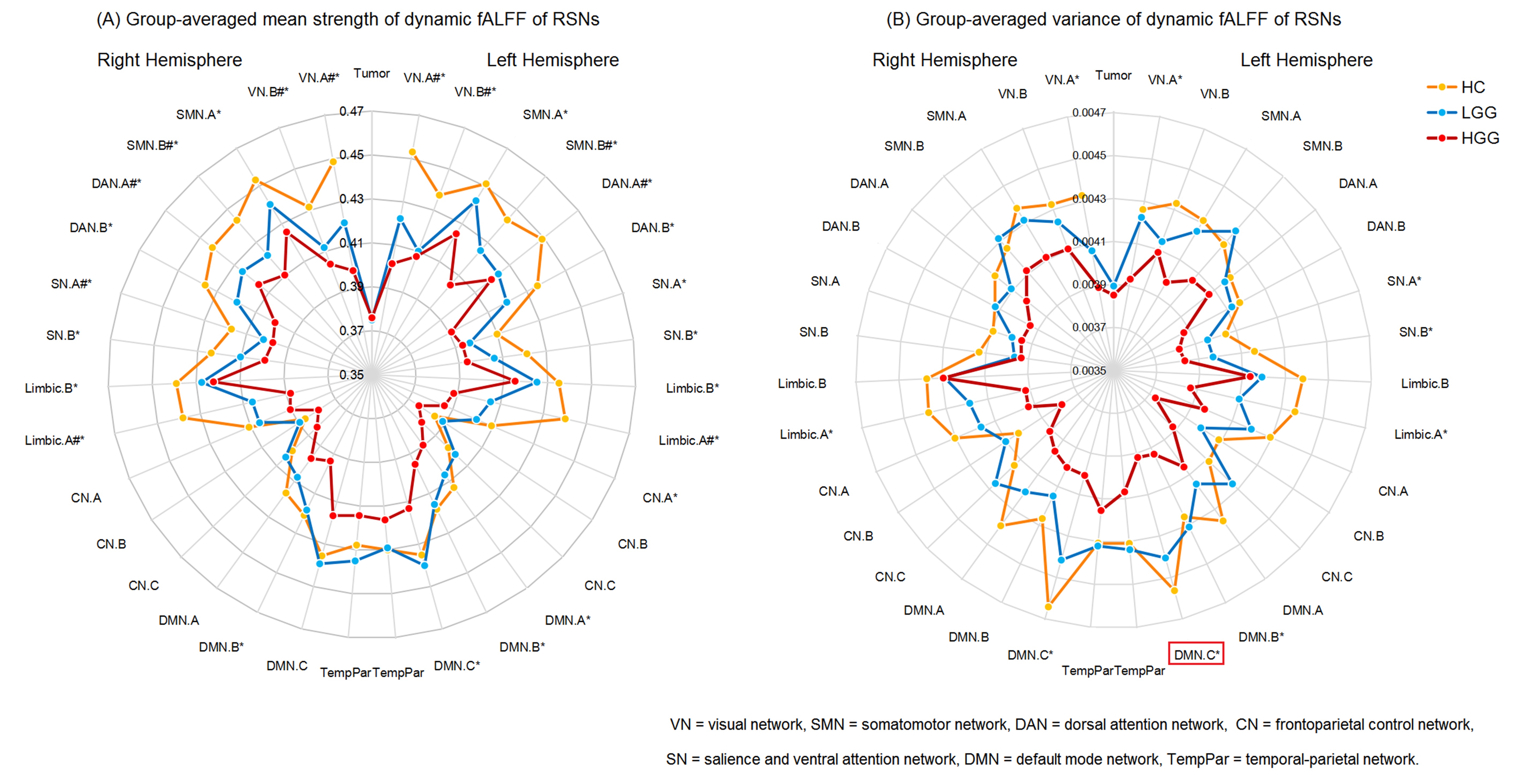
Figure 1. Group-averaged mean strength (A) and temporal variance (B) of dynamic fALFF for functional networks. # and * represent significant HC-LGG and HC-HGG difference (P < 0.05,FDR corrected), respectively. The network labeled with red box represents significant LGG-HGG difference.
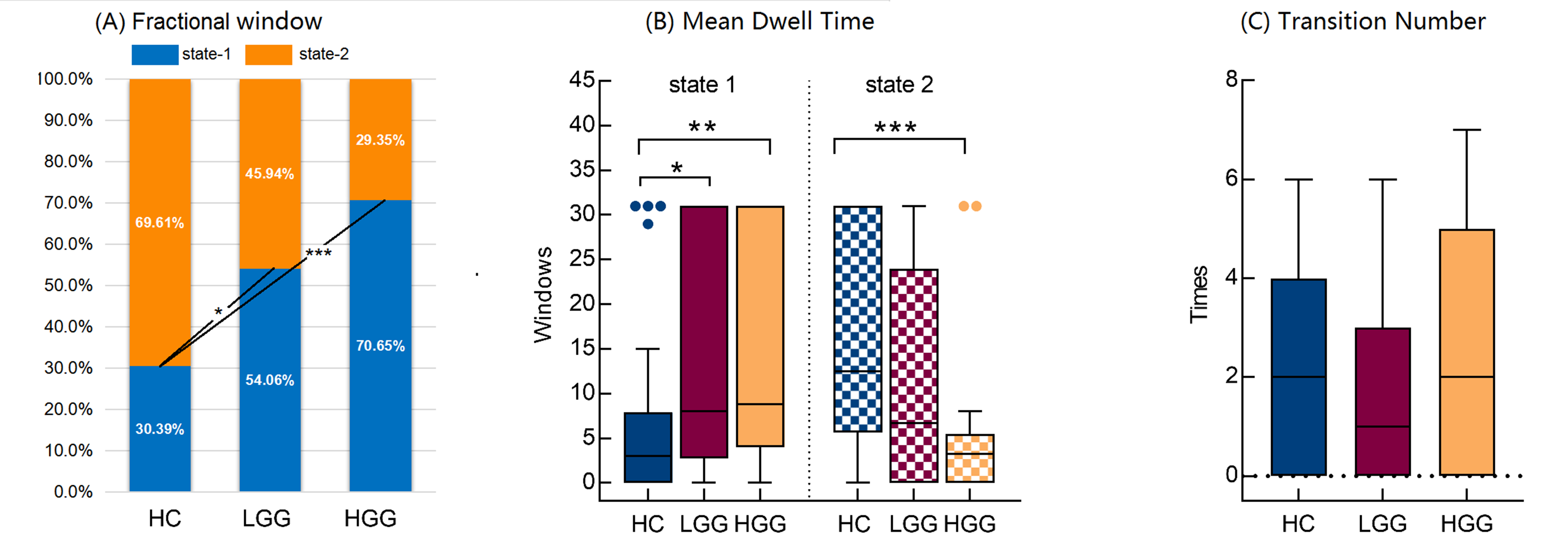
Figure 2. Statistical analysis of the temporal properties of network activity states. *, **, and *** represent P < 0.05, P < 0.01, and P <0.001, respectively.
DOI: https://doi.org/10.58530/2023/3214