3185
Concurrent measurement of perfusion parameters related to small blood and lymphatic vessels in the human brain
Di Cao1,2,3, Yuanqi Sun1,2,3, Su Pan4, Jay J. Pillai5,6,7, Ye Qiao5, Hanzhang Lu1,2,3, Peter C.M. Van Zijl1,2, Linda Knutsson1,2,8, and Jun Hua1,2
1F.M. Kirby Research Center for Functional Brain Imaging, Kennedy Krieger Institute, Baltimore, MD, United States, 2Russell H. Morgan Department of Radiology and Radiological Science, Johns Hopkins University, Baltimore, MD, United States, 3Department of Biomedical Engineering, The Johns Hopkins University School of Medicine, Baltimore, MD, United States, 4School of Medicine, University of Maryland, Baltimore, MD, United States, 5Neuroradiology, Department of Radiology, Johns Hopkins University, Baltimore, MD, United States, 6Department of Neurosurgery, Johns Hopkins University, Baltimore, MD, United States, 7Neuroradiology, Mayo Clinic, Rochester, MN, United States, 8Department of Medical Radiation Physics, Lund University, Lund, Sweden
1F.M. Kirby Research Center for Functional Brain Imaging, Kennedy Krieger Institute, Baltimore, MD, United States, 2Russell H. Morgan Department of Radiology and Radiological Science, Johns Hopkins University, Baltimore, MD, United States, 3Department of Biomedical Engineering, The Johns Hopkins University School of Medicine, Baltimore, MD, United States, 4School of Medicine, University of Maryland, Baltimore, MD, United States, 5Neuroradiology, Department of Radiology, Johns Hopkins University, Baltimore, MD, United States, 6Department of Neurosurgery, Johns Hopkins University, Baltimore, MD, United States, 7Neuroradiology, Mayo Clinic, Rochester, MN, United States, 8Department of Medical Radiation Physics, Lund University, Lund, Sweden
Synopsis
Keywords: Neurofluids, DSC & DCE Perfusion
Accumulating evidence has indicated the importance of studying the interaction between the microvascular and lymphatic systems in the brain. To date, most imaging methods can only measure blood or lymphatic vessels separately. This study proposes an MRI approach for concurrent measurement of perfusion parameters related to small blood and lymphatic vessels within one single scan. A dual-echo TSE sequence was optimized for the measurement of gadolinium(Gd)-induced blood and CSF signal changes. The proposed method showed consistent results in human brains as previous studies using separate methods. Signal changes from small blood vessels occurred faster than lymphatic vessels after intravenous Gd-injection.INTRODUCTION:
The cerebrovascular system and CSF circulation system are two important components in the brain. Cerebral vessels with typical endothelial markers found in lymphatic vessels in other organs have been identified in several parts of the brain. These cerebral lymphatic vessels may communicate with the glymphatic system and other routes for CSF circulation and are believed to play a crucial role in the drainage of CSF from brain tissues to cervical lymph nodes. Accumulating evidence has indicated the interaction between the cerebrovascular and lymphatic systems in the brain. To date, most imaging methods can only measure blood or lymphatic vessels separately. An approach that can measure the two systems in a single scan will offer the advantage of shortened scan time, less confounding effects from physiological variations between scans, and halved dosage of contrast media needed for contrast enhanced MRI methods. Dynamic-susceptibility-contrast (DSC) MRI is a perfusion technique routinely performed in the clinics. To date, DSC MRI has been optimized primarily for detecting Gd-induced contrast changes in blood vessels. Recently, we developed an MRI approach(1) for the detection of Gd-based signal changes in the CSF and cerebral lymphatic vessels, namely DSC-MRI-in-the-CSF (cDSC MRI)(1). Building upon the DSC and cDSC methods, we propose a new MRI method for concurrent measurement of perfusion parameters related to small blood and lymphatic vessels in the brain within one single scan. A dual-echo turbo-spin-echo (TSE) sequence was optimized for the measurement of Gd-induced blood and CSF signal changes using a short and a long TE, respectively.METHODS:
Simulation and optimization: A temporal resolution of 2s and a spatial resolution of 1x1x2mm3 were chosen to balance the requirements between blood and CSF signals. The first-echo (TE1=80ms) is intended to measure the blood signals similar to DSC-MRI. The second-echo (TE2=500ms) measures predominantly the CSF signals with parenchyma and blood signals suppressed. MRI experiments were performed in healthy human subjects to evaluate the proposed combined approach by comparing it with existing separate methods. The following scans were performed on 3T for each subject: a) FLAIR similar to previous studies(2); b) the proposed dual-echo TSE sequence continuously before and after Gd injection; c) a second FLAIR scan (post-Gd injection). Data from existing studies were used for cDSC-MRI for lymphatic vessels(1) and standard DSC-MRI for blood vessels(3). Note that the spatial resolution in standard DSC MRI was lower (2.8x2.8x5mm3). Data analysis: The short-TE images and the DSC scans were analyzed using DSCoMAN, from which CBV/CBF/MTT were calculated in GM. The long-TE images and cDSC scans were analyzed using in-house Matlab code, from which ∆S/S, onset time (Tonset), time to peak (TTP), and Gd concentration [Gd] were computed(1).RESULTS:
Fig. 1 shows the main simulation results for the proposed sequence. The short-TE was chosen around the maximum blood contrast. The CSF contrast shows a positive signal change, and gradually decreases with increasing TE. The long-TE was chosen when blood signals are completely suppressed (~0) to minimize partial volume effects from small blood vessels. Human results-blood vessels: The short-TE images from the proposed method and standard DSC-MRI data showed consistent parametric maps of CBV, CBF, and MTT (not shown). Human results-lymphatic vessels: Fig. 2 shows typical images acquired using the proposed sequence at long-TE and cDSC-MRI. As an example, only results from the ROI around dural sinuses (superior sagittal sinus) identified on FLAIR were shown. CSF signal changes after Gd injection were detected in FLAIR and in the proposed sequences. Maps of several parameters including Tonset, TTP, ΔS/S, and Gd concentration in the CSF ([Gd]) extracted from the dynamic signal changes are shown. Time courses from blood and lymphatic vessels: Fig. 3a-f compares the time courses from blood vessels in GM (a,b), dural (c,d) and olfactory (e,f) lymphatic vessels measured by the proposed combined method and corresponding separate methods. Both the original (left) and normalized (by peak values, right) time courses are shown. In general, the time courses showed a consistent temporal pattern from the combined and separate methods for both blood and lymphatic vessels. Fig. 3g,h compares the time courses from blood and lymphatic vessels measured using the proposed method simultaneously in the same subjects. Tonset and TTP were both shorter in blood than in CSF. The blood time courses quickly returned to baseline whereas the CSF time courses did not return to baseline for the duration of our experiments. The time of return for blood signals was comparable to Tonset for CSF signals, and was significantly shorter than TTP for CSF signals.DISCUSSION & CONCLUSION:
We demonstrated a dual-echo-MRI approach to measure perfusion parameters related to both small blood and lymphatic vessels concurrently after i.v. injection of Gd contrast in healthy human subjects. Interestingly, signal changes from small blood vessels occurred much faster than those from the CSF and cerebral lymphatic vessels. To the best of our knowledge, this may be the first study in which such interaction was measured and reported in the same human subjects. We believe that the proposed MRI approach may provide a useful tool for studies on these two systems in the healthy brain and various brain diseases.Acknowledgements
No acknowledgement found.References
(1)Cao, D, et al. Magn Reson Med 2020;84:3256. (2)Absinta, M, et al. Elife 2017;6: (3)Su, P, et al. NMR Biomed 2020;33:e4202.Figures
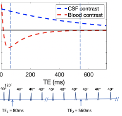
Pulse sequences and simulation results for the proposed dual-echo TSE sequence. MR signals in blood and CSF with and without Gd are simulated and the contrast between pre- and post-Gd signals in blood and CSF are shown. The fractional MR signals (Mz/M0) and the fractional MR signal changes before and after Gd injection (ΔMz/M0=(Mz_post_Gd-Mz_pre_Gd)/M0) are displayed as functions of TE. The vertical dashed lines indicate the TEs in respective sequences.
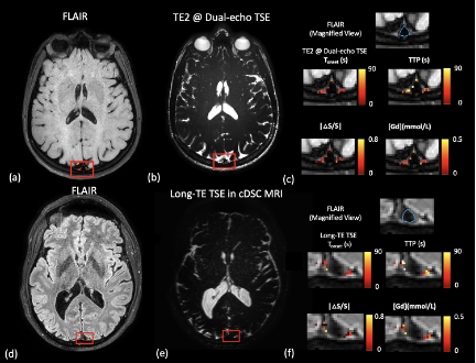
Typical results for the measurement of dynamic signal changes in the CSF. The ROI (red boxes) around the dural sinuses (DS) that contains the meningeal lymphatic vessels is manually drawn. (a) FLAIR to confirm the location. (b) The raw long TE image in the proposed sequence. (c) A magnified FLAIR in the ROI, and maps of the key parameters extracted from the time courses. For comparison, results from the previous cDSC MRI: (d) FLAIR, (e) raw cDSC image, and (f) parametric maps.
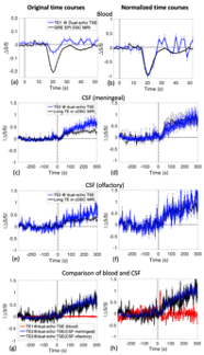
Time courses detected in blood and CSF before and after Gd injection.
DOI: https://doi.org/10.58530/2023/3185