3148
Deep Learning Reconstruction for Abdomen Diagnosis: Improvement of Diagnostic Performance with higher Spatial or Temporal Resolution1Department of Medical Imaging, National Taiwan University Hospital and National Taiwan University College of Medicine, Taipei, Taiwan, 2Department of Medical Imaging, National Taiwan University Cancer Center and National Taiwan University College of Medicine, Taipei, Taiwan, 3GE Healthcare, Taipei, Taiwan
Synopsis
Keywords: Liver, Machine Learning/Artificial Intelligence, Deep learning reconstruction, Abdomen diagnosis
We have previously validated the SNR improvement of abdominal MRI by Deep Learning Reconstruction (DLRecon). In this study, we further investigate the improvement of image quality and diagnostic performance when trading the SNR with higher spatial and temporal resolution imaging setting. In the result, the clinical scoring of images with high-speed or high-resolution settings in DLRecon was superior to that of the images with conventional setting and reconstruction.INTRODUCTION
In clinical settings, MRI with a 5-6 mm slice thickness is commonly used for abdominal imaging. Higher resolution can lead to better delineation of the lesion and its relationship with adjacent structures. However, better spatial resolution would prolong the data acquisition time and lead to serious motion artefact, lower signal-to-noise ratio (SNR), contrast-to-noise ratio (CNR), and uncomfortable scanning [1-5]. Therefore, one method that can maintain or improve SNR while acquiring with higher spatial resolution setting would greatly benefit the diagnosis in abdomen and pancreas diseases. Recent advances in deep learning reconstruction (DLRecon) for MRI is capable of increasing the quality of diagnostic images. Our previous study validated the improvement of image quality by DLRecon, and DLRecon shows its potential power in clinical diagnosis of abdomen images [6]. It may be possible to leverage DLRecon to overcome classical MRI trade-off resolution, SNR, and scan time. With the validated improvement in image quality by DLRecon, this study aims to investigate the feasibility of DLRecon to the 2D axial T2-weighted magnetic resonance cholangiopancreatography with high-speed setting and high-resolution setting.METHODS
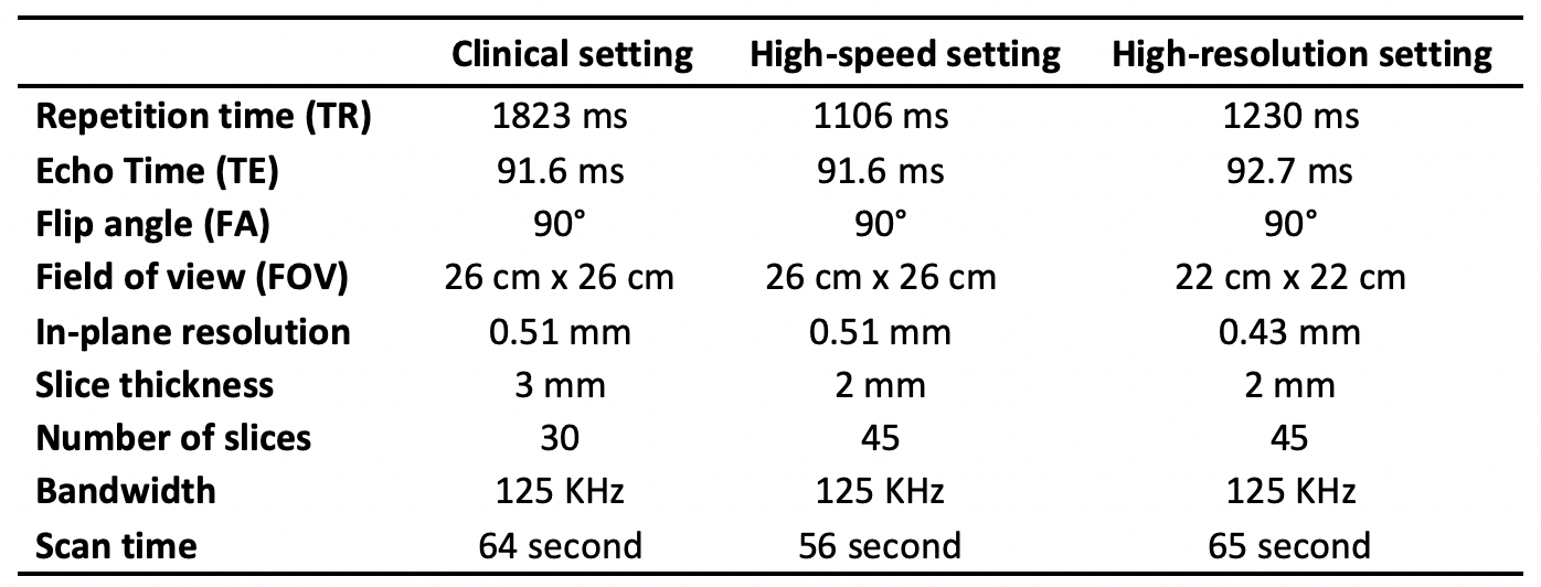
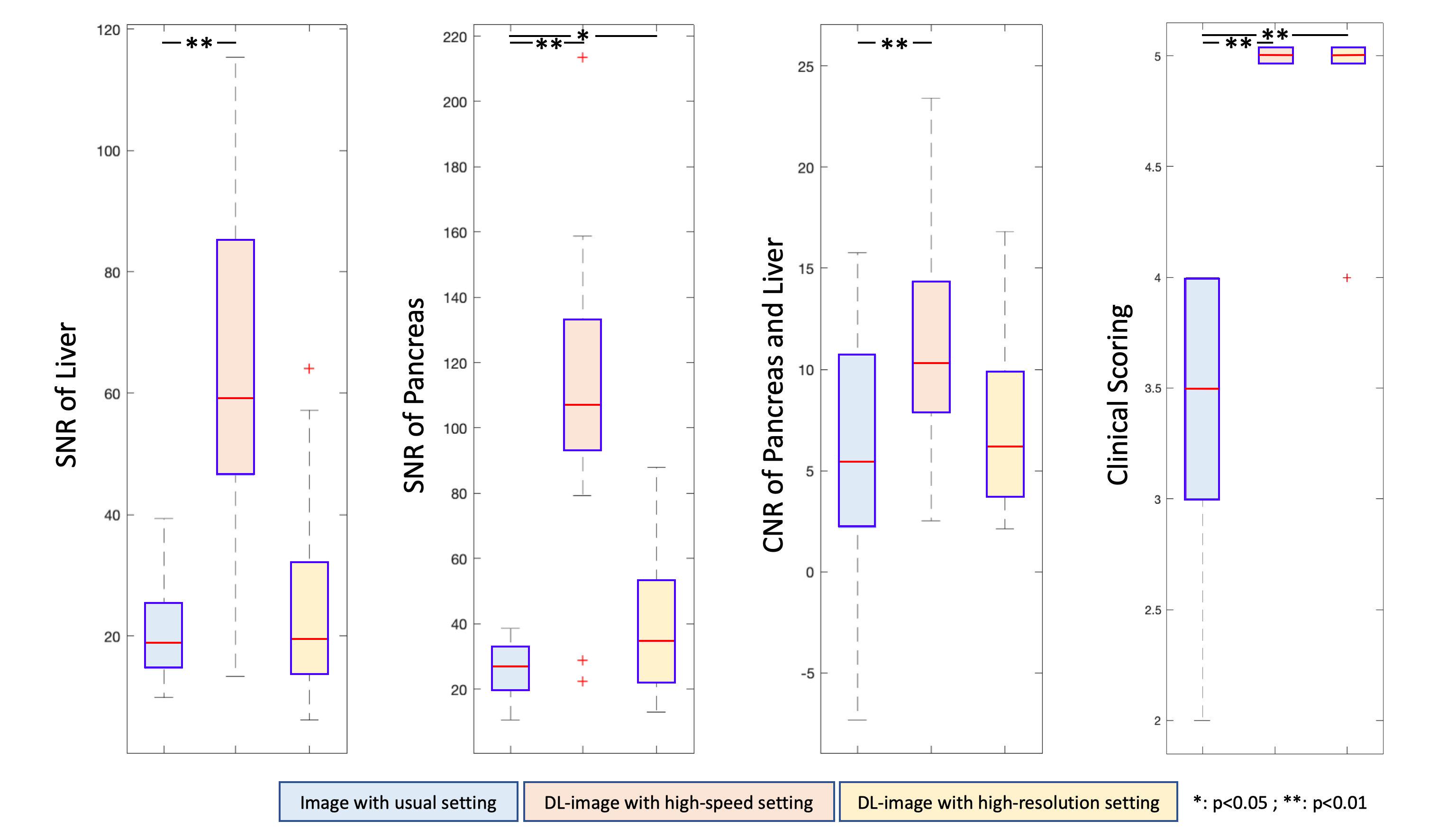
Twenty participants were enrolled retrospectively in this study. All the datasets including axial T2-weighted single-shot fast spin-echo (SSFSE) abdominal imaging was performed on a 3T clinical scanner (Signa Architect; GE Healthcare, Milwaukee, USA) equipped with an adaptive AIR™ Coil for signal detection and a whole-body coil for radio-frequency excitation. Those images were reconstructed by conventional pathway (non-DL) and by AIR™ Recon DL (DLRecon) process. The acquisition parameters for T2-weighted SSFSE images with clinical setting, high-speed setting, and high-resolution setting are listed in the Table 1. To compare the image quality of conventional MR images with images reconstructed by DLRecon technique, ROIs of pancreas (ball shape with 6-mm radius), liver (ball shape with 12-mm radius), and background noise (ball shape with 12-mm radius) were selected manually by two experienced radiology technologists, and the SNR and CNR were assessed based on the signal intensity of those ROIs (Figure 1). The quality of images in clinical diagnosis in terms of perceived SNR, perceived sharpness, presence of artefacts, and overall quality is evaluated from one experienced radiologist by a 5-point score:(1, very bad; 2, bad; 3, fair; 4, good; 5, very good).RESULTS and DISCUSSION
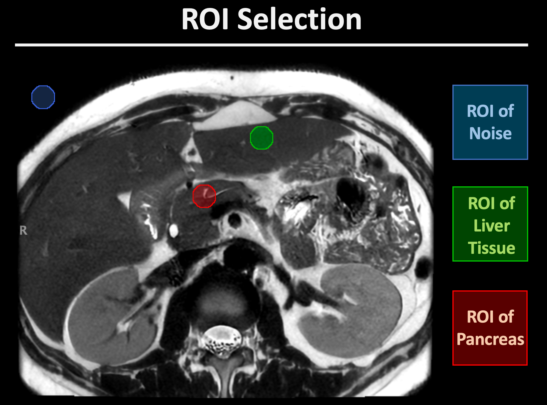
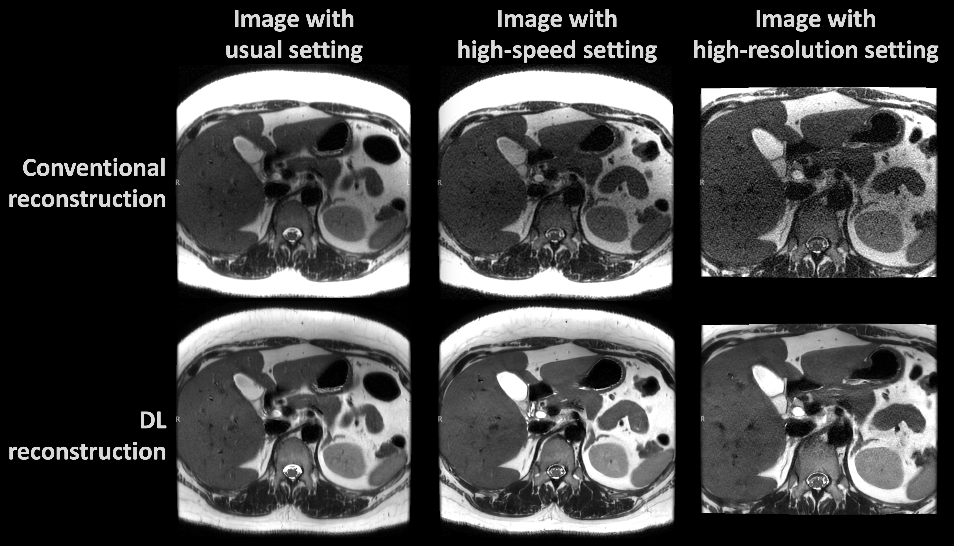
A representative abdominal imaging with DL and non-DLRecon was demonstrated in the Figure 2, and the image quality of images by DLRecon was obviously superior to the images by non-DLRecon. The SNR, CNR, and clinical scoring of images with high-speed setting and DLRecon were all more improved than that of the images with usual setting and conventional reconstruction (Figure 3). The images with high-resolution setting and DLRecon showed equal or slightly better SNR and CNR than that of the images with usual setting with non-DLRecon; moreover, the diagnostic performance of the images with high-resolution setting and DLRecon showed significantly higher clinical score than that of images with usual setting with non-DLRecon.CONCLUSION
In this study, abdominal MRI images in three different parameter settings with and without DLRecon were compared in subjective and objective image quality indices. With the DLRecon technique, temporal resolution can be improved by decreasing breath holding from 1 minute to 40 seconds, while maintaining good image quality. The results showed that DLRecon could make more efficient use of the acquired data to increase SNR, produce detailed structure images and reduce acquisition time and, consequently, improve the workflow in clinical practice.Acknowledgements
No acknowledgement found.References
1. Chen, F., Taviani, V., Malkiel, I., Cheng, J. Y., Tamir, J. I., Shaikh, J., Chang, S. T., Hardy, C. J., Pauly, J. M., & Vasanawala, S. S. (2018). Variable-density single-shot fast spin-echo MRI with deep learning reconstruction by using variational networks. Radiology, 289(2), 366–373.
2. Higaki, T., Nakamura, Y., Tatsugami, F., Nakaura, T., & Awai, K. (2019). Improvement of image quality at CT and MRI using deep learning. Japanese Journal of Radiology, 37(1), 73–80.
3. Kim, M., Kim, H.S., Kim, H.J., Park, J.E., Park, S.Y., Kim, Y.H., Kim, S.J., Lee, J., Lebel, M.R. (2021), Thin-Slice Pituitary MRI with Deep Learning-based Reconstruction: Diagnostic Performance in a Postoperative Setting. Radiology. Jan;298(1):114-122.
4. Litjens, G., Kooi, T., Bejnordi, B.E., Setio, A.A.A., Ciompi, F., Ghafoorian, M., van der Laak, J.A.W.M., van Ginneken, B., Sánchez, C.I. (2017), A survey on deep learning in medical image analysis, Med. Image Anal. 42, 60–88.
5. van der Velde, N., Hassing, H.C., Bakker, B.J., Wielopolski, P.A., Lebel, R.M., Janich, M.A., Kardys, I., Budde, R.P.J., Hirsch, A. (2021), Improvement of late gadolinium enhancement image quality using a deep learning-based reconstruction algorithm and its influence on myocardial scar quantification. Eur Radiol. Jun;31(6):3846-3855.
6. Xie, D., Li, Y., Yang, H., Bai, L., Wang, T., Zhou, F., Zhang, L., & Wang, Z. (2020). Denoising arterial spin labeling perfusion MRI with deep machine learning. Magnetic Resonance Imaging, 68(January), 95–105.
7. Zhang S, Wang X, Schuler FW, et al. Deep Learning Reconstruction improves CEST MRI. Proc. Of ISMRM 2020, 3100.
8. Chen, B-T., Yeh, C-Y., Chen, Y-C., Li, C-W., Shieh, C-C., Lin, C-Y., Liu, K-L. (2022). Deep Learning Reconstruction for Abdomen Diagnosis: Improvement of Image Quality and Diagnostic Performance.International Symposium of Magnetic Resonance in Medicine 2022 Conference.
Figures