3146
Evaluating chronic hepatitis B-related liver fibrosis with a continuous-time random-walk diffusion model1Department of Medical Imaging Center, Nanfang Hospital, Southern Medical University, Guangzhou, China, 2Shenzhen United Imaging Research Institute of Innovative Medical Equipment, Shenzhen, China, 3MR Collaboration, Central Research Institute, United Imaging Healthcare, Shanghai, China
Synopsis
Keywords: Liver, Diffusion/other diffusion imaging techniques
Early characterization of liver fibrosis has great clinical significance as it allows monitoring of treatment response to antifibrotic agents in patients with chronic liver disease. Most recently, high b-values diffusion-weighted imaging (DWI) based on the continuous-time random-walk (CTRW) model has been introduced. Compared to the healthy control group, the mean D, α, and β values were significantly lower in the hepatic fibrosis group. Spatial diffusion heterogeneity β showed the best diagnostic performance. This non-invasive method has the potential to realize efficient evaluation and improve clinical treatment decision-making.
Introduction
Viral hepatitis is the main cause of liver fibrosis and cirrhosis in patients in China, among which chronic hepatitis B is the most common [1]. Early characterization of liver fibrosis has great clinical significance as it allows monitoring of treatment response to antifibrotic agents in patients with chronic liver disease [2]. While liver biopsy was limited by invasiveness, sampling errors, and interobserver variability, there has been an increasing demand for non-invasive tools to evaluate liver fibrosis [3].Most recently, high b-values diffusion-weighted imaging (DWI) based on the continuous-time random-walk (CTRW) model has been introduced [4]. It offers novel quantitative parameters including diffusion coefficient (D), temporal diffusion heterogeneity (α), and spatial diffusion heterogeneity (β), with capabilities of not only providing an accurate characterization of tissue cellularity but also directly reflecting intravoxel structural heterogeneity by introducing temporal and spatial diffusion parameters α and β, which is related to tissue complexity and microenvironment [5, 6]. With regard to the conventional Gaussian DWI model and other non-Gaussian DWI models, the CTRW model relaxes a priori distributions of waiting times and distance increments of water molecular diffusion, providing a realistic description of the complex structures of biological tissues in more detail and in-depth [7, 8].
Several recent studies have provided strong evidence that the CTRW has great potential for more accurate information on water diffusion in complex living tissues on glioma [5, 7], Parkinson’s disease [6], and breast cancer [8]. However, no study of CTRW has been reported in assessing liver diseases. Thus, the purpose of this study was to investigate the value of CTRW-DWI in characterizing liver fibrosis in patients with chronic hepatitis B.
Methods
A cohort of 20 adult patients with histologically confirmed liver fibrosis and 20 healthy subjects were included in this study. All the participants underwent magnetic resonance imaging (MRI) examination on a 3.0T scanner (uMR780, United Imaging Healthcare, Shanghai, China) with a 12-channel body coil. Multi-b-values DWI was acquired using echo planar imaging (EPI) sequence with the respiratory trigger in the axial plane (repetition time (TR) /echo time (TE): 4000 ms/78 ms, flip angle (FA): 90°, slice thickness: 5 mm, field of view (FOV): 300 × 356 mm2, matrix: 242 × 288, b-values: 0, 200, 400, 600, 800, 1,000, 2,000 s/mm2). The CTRW model was calculated through the following fitting formula:$$$S/S_0=E_α(-(bD_m)^β)$$$
Where S0 is the signal intensity when b-value = 0 s/mm2
For each subject, a total of six circular ROIs (mean area, 299.8mm2) were drawn within the right lobe of the liver on the central two continuous sections (three ROIs per section), avoiding large intrahepatic vessels and focal hepatic lesions. The left lobe and right liver dome were avoided as measurements could be unreliable from cardiac motion artifacts.
Mann-Whitney U test was used to assess differences in CTRW-derived parameters between the liver fibrosis group and the healthy control group. The diagnostic performance of different CTRW-derived parameters for characterizing liver fibrosis was assessed by receiver operating characteristic (ROC) analyses. The Youden index was used to determine the cutoff value, along with the area under the curve (AUC), sensitivity, specificity, and accuracy. P < 0.05 was considered statistically significant.
Results
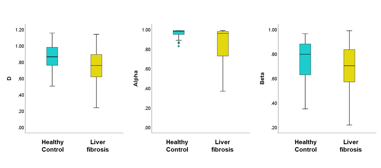
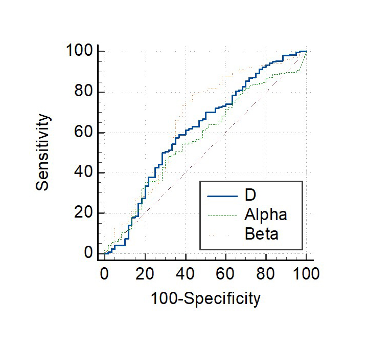
As shown in Figure 1, compared to the healthy control group, the mean D, α, and β values were significantly lower in the hepatic fibrosis group (P < 0.001 for D and β, P = 0.007 for α). As shown in Figure 2, all the CTRW-derived parameters could be used to differentiate the hepatic fibrosis group and the healthy control group (D: sensitivity = 58.5%, specificity = 69.1%, accuracy = 63.8%, AUC = 0.66; α: sensitivity = 45.6%, specificity = 76.4%, accuracy = 61.0%, AUC = 0.62; β: sensitivity = 73%, specificity = 67.3%, accuracy = 70.2%, AUC = 0.70). Among all the CTRW-derived parameters, β showed the best diagnostic performance with the largest AUC and the highest sensitivity and accuracy.Discussion
This study utilized parameters derived from the CTRW model to characterize liver fibrosis in patients with chronic hepatitis B. Unlike the mono-exponential DWI, the CTRW model provides new parameters, it is able to not only characterize the cellular density but also reflect different aspects of intravoxel tissue structural heterogeneity by using two fractional-order parameters, α, and β, with respect to time and space, respectively [4]. Compared to the healthy control group, the mean D, α, and β values were significantly lower in the hepatic fibrosis group. These results were in concordance with previous findings that the accumulation of extracellular matrix and restricted water diffusion in fibrotic tissues [9-11].Conclusion
This study showed that the CTRW model provided a non-invasive method to evaluate liver fibrosis in patients with chronic hepatitis B. CTRW-derived parameter β showed the best diagnostic performance. This non-invasive method has the potential to realize efficient evaluation and improve clinical treatment decision-making.Acknowledgements
No acknowledgement found.References
[1] Liaw Y, Kao J, Piratvisuth T, et al. Erratum to: Asian-Pacific consensus statement on the management of chronic hepatitis B: a 2012 update. Hepatology international,2012,6(4):809-810.
[2] Friedman S L. Liver fibrosis – from bench to bedside. Journal of hepatology,2003,38:38-53.
[3] De Robertis R, D'Onofrio M, Demozzi E, et al. Noninvasive diagnosis of cirrhosis:A review of different imaging modalities. World journal of gastroenterology : WJG,2014,20(23):7231-7241.
[4] Karaman M M, Sui Y, Wang H, et al. Differentiating low- and high-grade pediatric brain tumors using a continuous-time random-walk diffusion model at high b-values. Magn Reson Med,2016,76(4):1149-1157.
[5] Sui Y, Xiong Y, Jiang J, et al. Differentiation of Low- and High-Grade Gliomas Using High b-Value Diffusion Imaging with a Non-Gaussian Diffusion Model. AJNR Am J Neuroradiol,2016,37(9):1643-1649.
[6] Zhong Z, Merkitch D, Karaman M M, et al. High-Spatial-Resolution Diffusion MRI in Parkinson Disease: Lateral Asymmetry of the Substantia Nigra. Radiology,2019,291(1):149-157.
[7] Karaman M M, Zhang J, Xie K L, et al. Quartile histogram assessment of glioma malignancy using high b-value diffusion MRI with a continuous-time random-walk model. NMR Biomed,2021,34(4):e4485.
[8] Qin Y, Tang C, Hu Q, et al. Assessment of Prognostic Factors and Molecular Subtypes of Breast Cancer With a Continuous-Time Random-Walk MR Diffusion Model: Using Whole Tumor Histogram Analysis. Journal of magnetic resonance imaging : JMRI,2022.
[9] Leitão H S, Doblas S, Garteiser P, et al. Hepatic Fibrosis, Inflammation, and Steatosis: Influence on the MR Viscoelastic and Diffusion Parameters in Patients with Chronic Liver Disease. Radiology,2017,283(1):98-107.
[10] Jiang H, Chen J, Gao R, et al. Liver fibrosis staging with diffusion-weighted imaging: a systematic review and meta-analysis. Abdom Radiol (NY),2017,42(2):490-501.
[11] Fujimoto K, Tonan T, Azuma S, et al. Evaluation of the mean and entropy of apparent diffusion coefficient values in chronic hepatitis C: correlation with pathologic fibrosis stage and inflammatory activity grade. Radiology,2011,258(3):739-748.
Figures