3120
Automatic Localization of EEG Electrodes from MR Structural Images1Department of Biomedical Engineering, University of Calgary, Calgary, AB, Canada, 2Department of Radiology, University of Calgary, Calgary, AB, Canada
Synopsis
Keywords: Data Analysis, Segmentation, EEG Electrodes Localization, T1-weighted MRI
The localization of EEG sources is one of the fundamental approaches to facilitate the interpretation of EEG data. However, the accuracy of source localization depends on the exact knowledge of the position of the electrodes on the scalp, which currently requires time-consuming and/or expensive approaches. Here, an automatic method is proposed that retrieves the electrode positions by localizing the curvature changes in T1-weighted MRI images caused by electrodes. The results show an average detection sensitivity of ~96.4%, with an average position error of 4.23 mm for all subjects.Purpose
Recent developments in EEG source imaging have made it possible to localize brain generators using the information on the electrical field recorded on the surface of the head. There are many approaches for EEG source localization, but precise localization depends on accurate knowledge of the positions of the EEG electrodes. By considering that each electrode induces artifacts that imprint relatively higher curvature than the surrounding scalp in anatomical MRI scans, we propose a novel, fully automatic approach based on T1-weighted MRI images.Material &methods
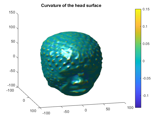
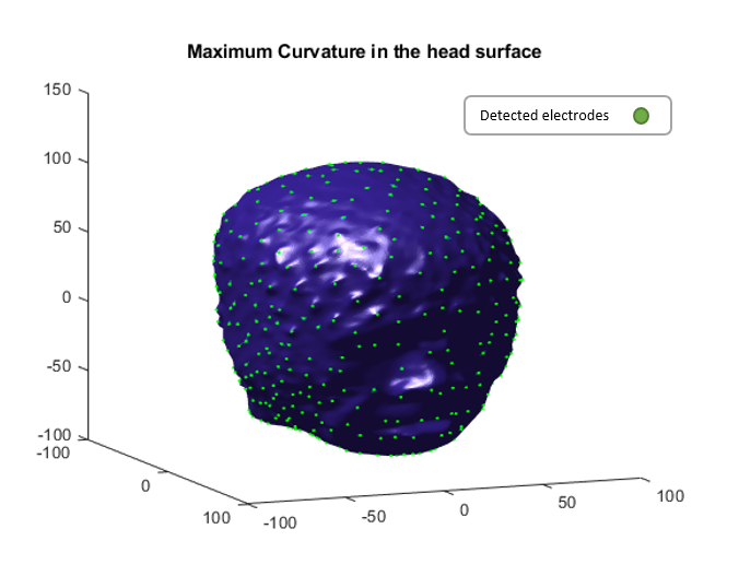
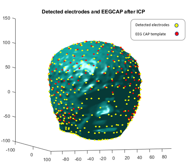
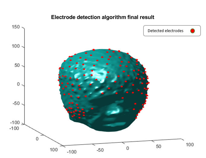
A T1w sequence was acquired in five healthy volunteers who wore a 256-channel MR-compatible cap (Magstim EGI) during the MRI scan. Localization of the EEG electrodes proceeded in two steps: first, we extract the head surface and its curvature, and second, we perform an Iterative Closest Point (ICP) 1 registration to match the curvature peaks with a template of standard EEG electrode positions. In the first stage, uniformity correction is applied to the anatomical T1 image using FSL’s FAST algorithm 2. Then, we extract the head surface and the curvatures with the Brainstorm Toolbox 3 (using the global threshold segmentation method). Figure 1 shows the head surface and its curvature. Then we detect the local maxima of the surface curvature, which are induced by the electrodes on the MRI images (figure 2). Finally, we should determine which of the detected curvature peaks correspond to real electrodes and which detections are false positives. So, we register the detections to a template of standard EEG electrode positions 4 using the ICP algorithm (Figure 3). Each template position is matched to the closest detection within 10 mm, and the remaining detections are considered as false positives. Figure 4 shows the results of the detected electrodes.Results
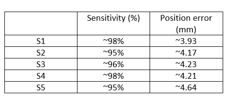
The algorithm detected a large number of curvature peaks (695 on average), of which many are likely to be false positives (Figure 2). An average of 247 detections could be matched with the template electrode locations, therefore the number of false positives averaged 448. However, as seen in figure 3, the false positives were mostly distributed on the face and the bottom of the head (e.g., neck). These detections are far from the template locations, so they can be easily ignored. By discarding detections with a distance greater than 10 mm from the template locations, corresponding to the diameter of an electrode cup, the average number of false detections was reduced to 137. Most of the remaining false detections were located at the bottom part of the head, where the high-density EEG net includes several electrodes that are, however, less relevant for source localization. When ignoring detections below the ears, the number of false detections was further reduced to 27 on average. The final detected electrodes after applying our algorithm are shown in Figure 4, with the sensitivity and position error for all subjects shown in figure 5. On average, 96% of the electrodes could be successfully localized.Discussion/Conclusion
We have demonstrated that our algorithm could accurately and automatically detect the position of high-density EEG electrodes using a common T1-weighted sequence. Also, the method does not require any modification of the electrodes. This is in contrast to previous approaches that required other non-conventional MRI sequences or modified electrodes to make them easier to identify in MRI images 5, 6. This technique has multiple applications for EEG source localization, particularly for EEG-fMRI studies in which T1-weighted scans are commonly acquired.Acknowledgements
This work was supported by NSERC Discovery Grant RGPIN-2021-02797 and CIHR grant PJT-183825.References
1. Besl PJ, McKay ND. Method for registration of 3-D shapes. InSensor fusion IV: control paradigms and data structures 1992.
2. Jenkinson M, Beckmann CF, Behrens TE, Woolrich MW, Smith SM. Fsl. Neuroimage. 2012.
3. Tadel F, Baillet S, Mosher JC, Pantazis D, Leahy RM. Brainstorm: a user-friendly application for MEG/EEG analysis. Computational intelligence and neuroscience. 2011.
4. Geodesics E. Geodesic Sensor Net Technical Manual. Electrical Geodesics: Eugene, OR, USA. 2007.
5. Fleury M, Barillot C, Mano M, Bannier E, Maurel P. Automated electrodes detection during simultaneous EEG/fMRI. Frontiers in ICT. 2019.
6. Pinte C, Fleury M, Maurel P. Deep learning-based localization of EEG electrodes within MRI acquisitions. Frontiers in Neurology. 2021.
Figures