3105
Improved diagnostic efficacy on structural abnormalities of knee using high-resolution deep learning-based 2D FSE images
Xiaxia Wu 1, Weiyin Vivian Liu2, and Yunfei Zha1
1Renmin Hospital of Wuhan University, Wuhan, China, 2GE Healthcare,MR Reaearch China,Beijing, Wuhan, China
1Renmin Hospital of Wuhan University, Wuhan, China, 2GE Healthcare,MR Reaearch China,Beijing, Wuhan, China
Synopsis
Keywords: Image Reconstruction, Cartilage
Diagnostic performance was limited to image resolution and contrast between target tissues and surrounding tissues. A rapid knee imaging has been perused but no loss of image quality is critical. This study proposed a rapid knee imaging based on two-dimensional fast spin echo sequence and examined the reliability and diagnostic performance of deep learning-based reconstruction T1-, T2- and PD- weighted images on knee joint pathology via comparison of images with and without deep-learning reconstruction algorithm (DLR). Diagnostic efficacy on knee structural abnormalities of 2D DLR FSE sequence elevated using knee arthroscopy results as the gold standard.Introduction and Purpose
Reference standards for knee MRI are proton density (PD)– and T1–weighted fast spin-echo (FSE) sequences due to the excellent tissue contrast and high in-plane spatial resolution with good assessment of meniscal, ligamentous, and cartilaginous injuries. Limited to scan time, images often have lower resolution and poor image quality. Early detection of knee osteoarthritis is difficult because the contrast between the articular cartilage and the surrounding tissues is low. Deep learning reconstruction (DLR) algorithm has showed benefits for clinical imaging field for better image quality, less noise, and shorter scan time[1]. Our study aimed to propose a rapid deep learning reconstruction-based knee imaging protocol and ensured its feasibility in clinical knee imaging via evaluation of the image quality and diagnostic performance of 2D fast spin echo (FSE) sequences on knee.Materials and methods
With approval of the Institutional Review Board, a total of 92 patients in supine position underwent knee routine MRI with a protocol including 2D FSE-based T1-, T2-, and PD-weighted images using 18-channel knee coils (Table 1). Among 92 patients, the arthroscopic findings of 22 patients in this prospective study were used as the reference standard for diagnosis. Two datasets of original images (FSEO) and DLR images (FSEDL) were automatically generated. Diagnostic performance and image quality of FSEO and FSEDL was independently evaluated by radiologists with 3 to 9 years of experience in interpreting musculoskeletal MRI, according to International Cartilage Regeneration&Joint Preservation Society (ICRS) [2]and the 5-point Likert scale. Objective evaluation of image quality such as SNR and CNR were also obtained. Assessment of pathologies and internal derangement were conducted by the same two radiologists and included the evaluation of the medial and lateral menisci; anterior and posterior cruciate ligaments; and cartilage defects of the medial and lateral femur trochlea, the medial tibia plateau, the trochlear groove, and the retropatellar cartilage. Structural abnormal-ities were graded as 0 = normal, 1 = altered (degenerative, postoperative), and 2 = tear. Areas of bone marrow edema (femoral, patellar, tibial), as well as fractures and joint effusion, were evaluated being present or absent. If there were discrepancies between the readers, a consensus reading was enclosed to define false-positive and false-negative findings. Evaluation was performed repeatedly with an interval of at least 2 weeks. Assessment was recorded and analyzed, in particular the correlation with arthroscopic findings using SPSS (version 25.0, IBM Corp). P<0.05 was considered statistical difference.Results
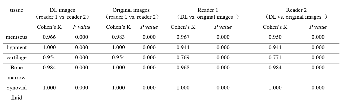
The overall image quality, sharpness and diagnostic confidence for FSEDL were higher compared to FSEO, showing significantly improved sharpness (p < 0.05). Inter- and intra-reader agreement was substantial to almost perfect (ICC=0.710 -0.898) (Table 2). In objective evaluation, SNR and CNR of PDWIDL and T1WIDL images were significantly higher than that of PDWIO and T1WIO images (p < 0.05) (Table 3). Two radiologists assessed the sequences regarding structural abnormalities of the knee based on FSEO and FSEDL (Table 4). Inter- and intra-reader agreement were moderate-excellent consistent (κ = 0.792-1.000) for the detection of internal derangement. Intra-reader agreement was substantial to almost perfect (κ=0.769-0.771) for the assessment of cartilage defects and almost perfect (κ = 0.944-1.000) for the assessment of meniscal, ligament. When we classified the reference diagnoses as normal and abnormal, the sensitivities of FSEDL ranged from 92.3% to 100% compared with 66.7% to 100% for FSEO, and the specificities of both FSEDL and FSEO ranged from88.9% to 100%. The accuracy of FSEDL ranged from 83.3% to 100% compared with 81.8 to 100% for FSEO. There were no significant differences in sensitivity, specificity and accuracy between FSEDL and FSEO for diagnostic performance (P = 0.309–1.000). A total of 10 cartilage lesions were detected by reader 1 on FSEDL compared with 8 lesions were detected on FSEO,and 11 lesions were detected on FSEDL compared with 10 lesions were detected on FSEO by reader 2. The diagnostic accuracy of cartilage injury on FSEDL is higher than FSEO. However for two readers, there was no statistically significant difference between the two images for the detection of these structural abnormalities. (all, P > 0.05).Discussion and conclusions
In this study, deep learning technology is embedded in the stage of MRI reconstruction of original data, and then the noise of MRI is effectively removed in the stage of original data acquisition, and finally pure MR Signal is achieved. Therefore, high signal-to-noise ratio and high-resolution images can be achieved at faster scanning speed. In terms of cartilage defect classification, the diagnostic accuracy of cartilage injury on FSEDL is higher than FSEO. This may attribute to DL-based image reconstruction, leading to improved image quality and increased contrast between cartilage and neighboring structure. The lesion clarity elevated. 2D FSE sequences of the knee using deep-learning reconstruction are clinically feasible, showing excellent image quality and improving diagnostic efficacy compared to the original images.Acknowledgements
The author would like to thank Guangnan Quan for technical assistance.References
[1] HERRMANN J, KELLER G, GASSENMAIER S, et al. Feasibility of an accelerated 2D-multi-contrast knee MRI protocol using deep-learning image reconstruction: a prospective intraindividual comparison with a standard MRI protocol [J]. Eur Radiol, 2022.
[2] MAINIL-VARLET P, AIGNER T, BRITTBERG M, et al. Histological assessment of cartilage repair: a report by the Histology Endpoint Committee of the International Cartilage Repair Society (ICRS) [J]. J bone joint surg am, 2003, 85-A Suppl 2(null): 45-57.
Figures
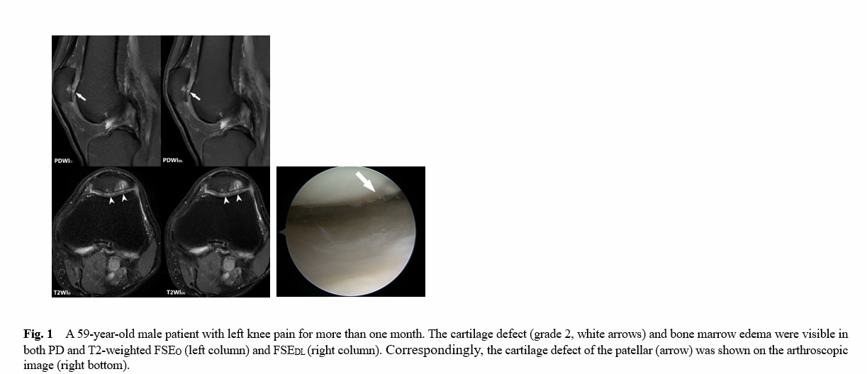
Fig. 1 A 59-year-old male patient with left knee pain
for more than one month. The cartilage defect (grade 2, white arrows) and bone
marrow edema were visible in both PD and T2-weighted FSEO (left
column) and FSEDL (right column). Correspondingly,
the cartilage defect of the
patellar (arrow) was shown on the arthroscopic image (right bottom).
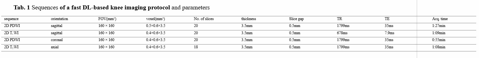
Table 1 Sequences of a fast DL-based knee imaging protocol and
parameters
Table
3 Comparison of SNR and CNR in different tissues
between PDWIO, T1WIO images and PDWIDL, T1WIDL
images.
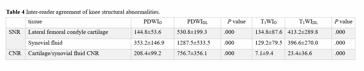
Table 4 Inter-reader agreement of knee
structural abnormalities.
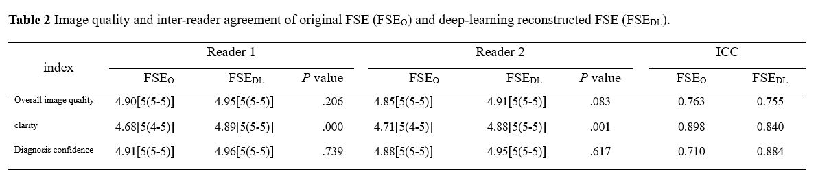
Table 2 Image quality and inter-reader
agreement of original FSE (FSEO) and deep-learning reconstructed FSE
(FSEDL).
DOI: https://doi.org/10.58530/2023/3105