3095
Slab-selection in free-running cardiac and respiratory motion-resolved bSSFP 5D whole-heart MRI
Robin Ferincz1, Ludovica Romanin1, Jérôme Yerly2, Davide Piccini1,3,4, Matthias Stuber2, and Christopher William Roy1
1Department of Radiology, Lausanne University Hospital (CHUV) and University of Lausanne (UNIL), Lausanne, Switzerland, 2Center for Biomedical Imaging (CIBM), Lausanne, Switzerland, 3Advanced Clinical Imaging Technology (ACIT), Siemens Healthineers International AG, Lausanne, Switzerland, 4LTS5, École Polytechnique Fédérale de Lausanne (EPFL), Lausanne, Switzerland
1Department of Radiology, Lausanne University Hospital (CHUV) and University of Lausanne (UNIL), Lausanne, Switzerland, 2Center for Biomedical Imaging (CIBM), Lausanne, Switzerland, 3Advanced Clinical Imaging Technology (ACIT), Siemens Healthineers International AG, Lausanne, Switzerland, 4LTS5, École Polytechnique Fédérale de Lausanne (EPFL), Lausanne, Switzerland
Synopsis
Keywords: Data Acquisition, Artifacts
Recent advances have enabled high resolution cardiac and respiratory motion-resolved 5D whole-heart MRI using non-contrast enhanced non-selective 3D radial bSSFP. However, the nature of bSSFP, the subject dependent anatomy, as well as the underlying sparse reconstruction can lead to banding and streaking artifacts, which degrade image quality and reduce diagnostic utility. In this work, the impact of slab-selective RF pulses in a programmable orientation that is independent of the k-space trajectory is assessed in a cohort of healthy volunteers. Preliminary results suggest that a subject-specific slab orientation can reduce artifacts and improve image quality.Introduction
The free-running framework (FRF) provides a simplified workflow for 3D dynamic cardiac MRI by acquiring 3D whole-heart data continuously within a fixed scan time and then retrospectively reconstructing fully self-gated cardiac and respiratory motion-resolved (5D) images 1. In previous work, free-running acquisitions have leveraged bSSFP acquisitions with non-selective RF pulses 1,2. The use of bSSFP sequences holds many advantages including high SNR and blood-to-myocardium contrast. However, sensitivity to B0 inhomogeneities, banding artifacts, and subject-specific hyper-intensity regions (e.g. fat) can lead to streaking artifacts which degrade image quality in 3D radial whole-heart acquisitions. In this work, we explore the impact of spatially selective (slab) excitations in free-running 3D radial bSSFP data. We test the hypothesis that by varying the orientation of the spatially confined volume, signals from specific body-regions outside the field-of-view (FOV) can be reduced, thus improving image quality. We programmed our sequence such that the slab orientation is decoupled from the orientation of the radial trajectory, allowing us to maintain a repeated readout orientated along the superior-inferior direction for self-gating, and qualitatively and quantitatively compare axial, sagittal, and coronal orientation in back-to-back scans from four healthy volunteers.Methods
Free-running 3D radial bSSFP data with a Phyllotaxis trajectory were acquired using a research sequence in 4 healthy volunteers (2 male, ages 25-26 years) on a 1.5T clinical MRI scanner (MAGNETOM Sola, Siemens Healthcare, Erlangen, Germany). The body mass index (BMI) was recorded for each volunteer. Relevant scan parameters included: RF excitation angle: 60°, resolution: (1.38 mm)3, FOV (220 mm)3 with two-fold oversampling in all directions, TE/TR: 1.53/3.05 ms, readout bandwidth: 1002 Hz/pixel. To examine the influence of the slab-orientation on the image quality, for each volunteer three back-to-back scans were performed using axial, sagittal, and coronal orientations of the slab selective gradient and its rewinder while keeping the k-space trajectory unchanged to allow for retrospective self-gating of cardiac and respiratory motion 1. As a reference a non-selective acquisition also has been performed on each volunteer. For each orientation, the width of the slab was set to the size of the cubic, non-oversampled FOV (220mm). The center of the slabs was positioned at isocenter for a fair comparison between each orientation. For each orientation and volunteer, a 3D static reconstruction of the oversampled FOV using all acquired data was performed to visualize and compare the slab profiles. Additionally, fully self-gated cardiac and respiratory motion resolved 5D whole heart images were reconstructed as previously described for each acquisition across all volunteers 1. The resulting 5D images for each volunteer were inspected for artifacts by an expert with over 10 years of experience in cardiac MRI and assigned a grade. Image grades for acquisitions of each volunteer with respective axial, sagittal and coronal slab-orientation are listed in Tab. 1.Results
3D and 5D image reconstructions were successfully obtained from all 4 volunteers and across all 3 orientations. Fig. 1 shows the 3D static reconstructions from volunteers 1 and 2 while Fig. 2 shows the same from volunteers 3 and 4. Overall, there is a visible influence of the slab-selection, compared to the non-selective cases, in the amount of artifacts. There is a clear reduction in signal in the upper and lower quarters of the axial slabs, reduction in the left and right quarters of the sagittal slabs, while minimal signal reduction is apparent in the coronal slab due to the majority of the bright anatomy remaining inside the excited volume. In each case, a non-ideal slab profile is observed. Fig. 3 contains animations of the cardiac and respiratory motion-resolved 5D image reconstructions from volunteers 1 and 2, while Fig. 4 displays the same from volunteers 3 and 4. The axial orientation provides relatively good suppression of streaking artifacts originating in the lower abdomen as indicated by green arrows in Fig. 3b (Vol. 2 BMI: 25.2) and Fig. 4a (Vol. 3 BMI: 24.8). Conversely, for artifacts originating from the arms and shoulders, the sagittal orientation clearly reduces streaking as shown by blue arrows in Fig. 4b (Vol. 4 BMI: 22.9). Finally, the coronal orientation tends to retain the most artifacts across all volunteers as the slab does not suppress signal from the chest wall and relatively little artifact originates from the spine.Discussion and Conclusion
A free-running 3D radial bSSFP sequence was modified to include slab-selective excitations with a programable orientation that is decoupled from the orientation of the k-space trajectory. In this preliminary study, visual comparison of axial, sagittal, and coronal slab orientations yielded varying degrees of artifact reduction that appear linked to the subject’s BMI. Future work is needed to optimize the slab profile including the duration of the RF pulse and slab width. Furthermore, a larger cohort with varied body sizes may yield additional insight into the link between potential improvement in image quality and a subject-specific orientation.Acknowledgements
MS is the PI on the Swiss National Science Foundation grants 320030_173129 and 201292 that funded part of this research. CWR is the PI on Swiss National Science Foundation Grant PZ00P3_202140 that funded part of this research.References
1. Di Sopra L, Piccini D, Coppo S, Stuber M, Yerly J. An automated approach to fully self-gated free-running cardiac and respiratory motion-resolved 5D whole-heart MRI. Magn Reson Med. 2019 Dec;82(6):2118-2132.
2. Masala N, Bastiaansen JAM, Di Sopra L, et al. Free-running 5D coronary MR angiography at 1.5T using LIBRE water excitation pulses. Magn Reson Med. 2020;84:1470–1485.
3. Ahmad R, Ding Y, Simonetti OP. Edge Sharpness Assessment by Parametric Modeling: Application to Magnetic Resonance Imaging. Concepts Magn Reson Part A Bridg Educ Res. 2015 May;44(3):138-149.
Figures
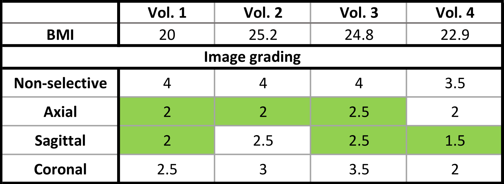
Tab. 1:
Listing of visually assigned image grading from 0 (low quality) to 5 (high
quality) for all four volunteers with respective body mass indices (BMI). Green cells are highlighting the lowest grade of each volunteer.
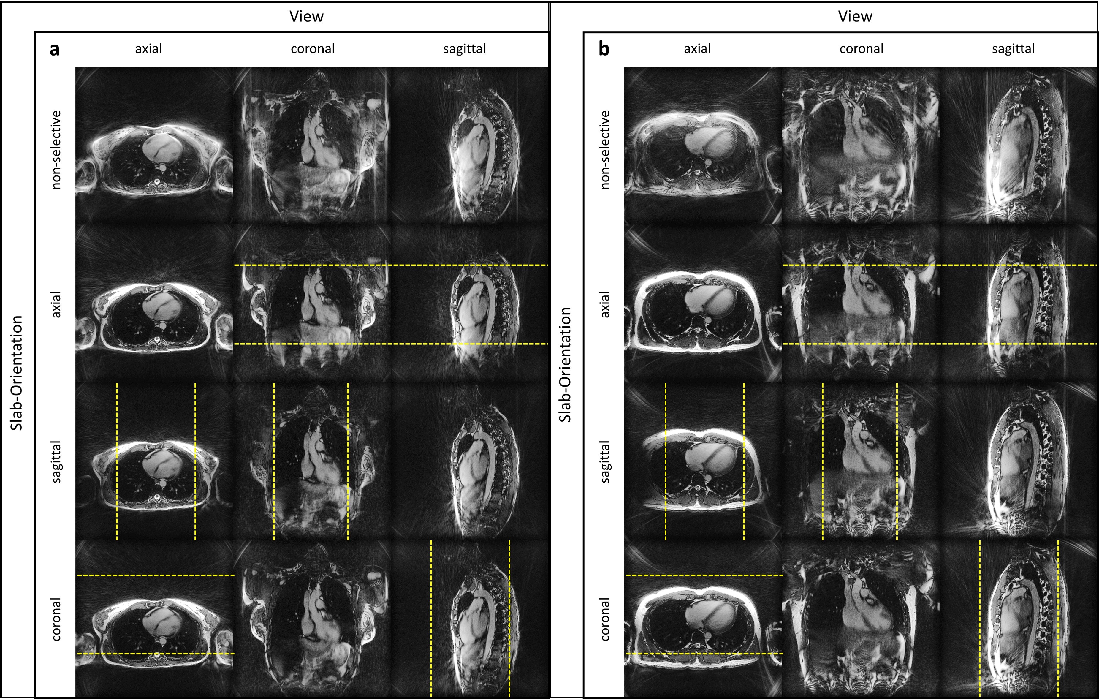
Fig. 1: Comparison of static reconstructions of
free-running 3D radial bSSFP data with slab-selective RF excitations in three
orientations for volunteer 1 (a) and 2 (b). For each slab orientation, the
oversampled FOV is shown with the prescribed slab denoted by yellow dashed
lines.
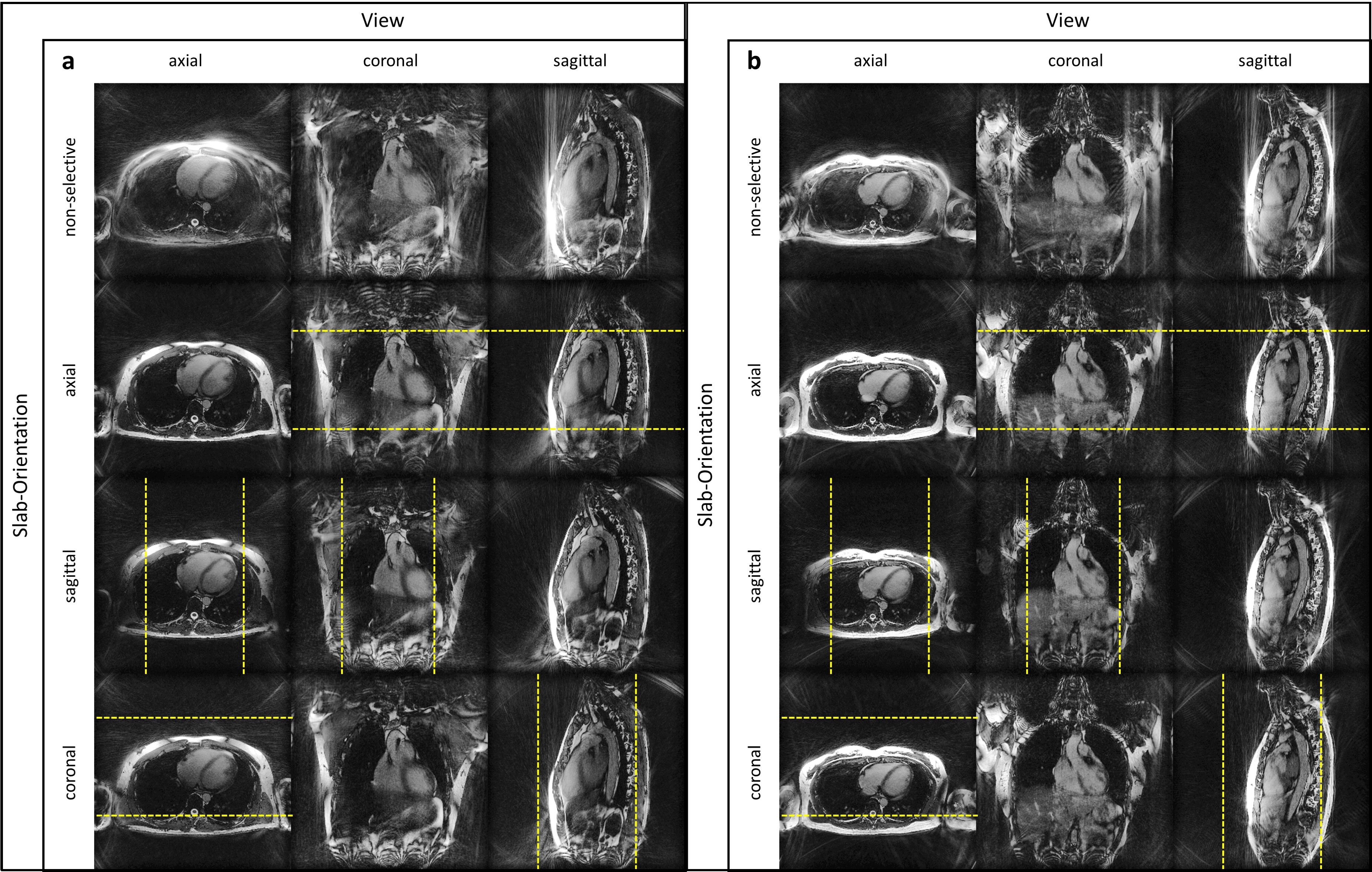
Fig. 2: Comparison of static reconstructions of
free-running 3D radial bSSFP data with slab-selective RF excitations in three
orientations for volunteer 3 (a) and 4 (b). For each slab orientation, the
oversampled FOV is shown with the prescribed slab denoted by yellow dashed
lines.

Fig. 3: Animated comparison of cardiac
motion-resolved reconstructions of free-running 3D radial bSSFP data with
slab-selective RF excitations in three orientations for volunteer 1 (a) and 2
(b). Green arrows denote the location of streaking artifact from the lower
abdomen which is relatively suppressed using the axial slab orientation.
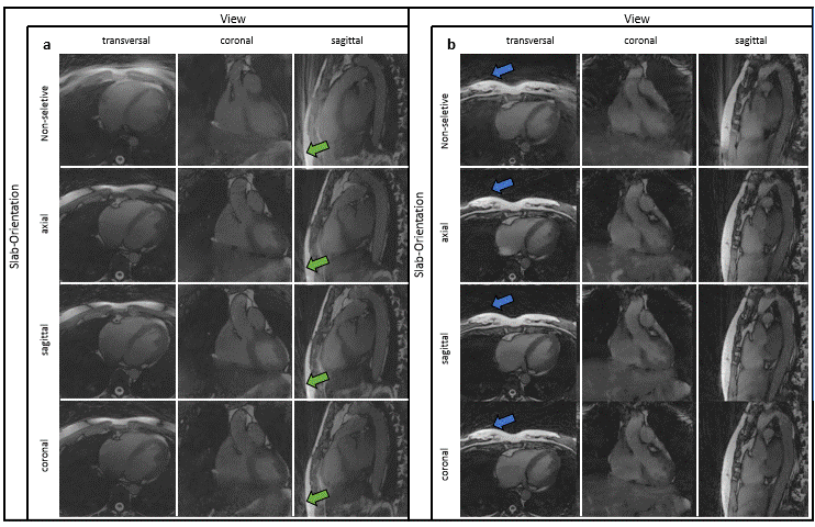
Fig. 4: Animated comparison of cardiac
motion-resolved reconstructions of free-running 3D radial bSSFP data with
slab-selective RF excitations in three orientations for volunteer 3 (a) and 4
(b). Green arrows denote the location of streaking artifact from the lower
abdomen which is relatively suppressed using the axial slab-orientation, while
blue arrows highlight streaking originating from the arms and shoulders which
is better suppressed by the sagittal slab orientation.
DOI: https://doi.org/10.58530/2023/3095