3092
Improvement of the time-resolved 4DMRI image quality through super-resolution reconstruction using compressed sensing1Medical Physics, Memorial Sloan Kettering Cancer Center, New York, NY, United States
Synopsis
Keywords: Image Reconstruction, Radiotherapy, TR-4DMRI, multi-breathing cycles, breathing irregularities, image quality, compressed sensing
Since the development of respiratory-correlated four-dimensional computed tomography (RC-4DCT) [1] and magnetic resonance imaging (RC-4DMRI) [2,3], patient-specific respiratory-induced tumor motion has been incorporated in radiotherapy for treating mobile tumors, such as lung, liver and pancreatic cancer. However, the RC-based snapshot 4D imaging only provides one breathing cycle, often contains severe binning motion artifacts, and may not represent tumor motion over 20-minute treatment, affecting treatment outcomes. Therefore, respiratory motion irregularities remain a challenge in radiotherapy. In this study, we report an improved time-resolved 4DMRI technique that captures multi-breath and can be used clinically and quantifies tumor motion irregularities.Introduction
Dynamic magnetic resonance imaging (MRI) has been increasingly applied in radiotherapy for patient motion simulation and real-time motion monitoring, such as 2D cine MRI in sagittal, coronal, or even beam eye’s view (BEV) for MRI simulator or MR-integrated linear accelerator (MRL). Respiratory-correlated (RC) 4DMRI is also developed to provide volumetric images within a single-breathing cycle through the retrospective binning technique, similar to RC 4D computed tomography (4DCT)[1-3]. However, RC-4DMRI has some limitations that prevent it from further clinical applications, for example, it can only be reconstructed retrospectively, contains only one-breathing-cycle motion, and may suffer from binning artifacts. Therefore, prospective or time-resolved (TR) 4DMRI techniques have been reported to overcome these limitations, including the 2D-cine-guided reconstruction based on the deformable image registration (DIR) among RC-4DMRI, as well as the super-resolution reconstruction based on DIR between low-resolution 3D cine in free-breathing (FB) and high-resolution breath-hold (BH) MR images [4-7]. The two dynamic volumetric TR-4DMRI techniques have been reviewed recently [8] and the super-resolution reconstructed TR-4DMRI approach is independent of RC-4DMRI. Since the first publication of super-resolution reconstructed TR-4DMRI, this technique has been further developed and optimized for improved image quality, including an enhanced deformation range of Daemon from 2cm to 6cm [5], a 2-step DIR to boost the alignment of low-contrast tissue [6], and a hybrid strategy to minimize the sliding-motion artifact. On the other hand, the delineation of lung tumors and five nearby organs at risk (OARs) based on 4DMRI has been explored and compared among different MR contrasts, such as T1w and T2w. Therefore, the TR-4DMRI method has been established and is ready for clinical implementation. The purpose of this work was to further improve TR-4DMRI image quality using compressed sensing and super-resolution reconstructions.Methods
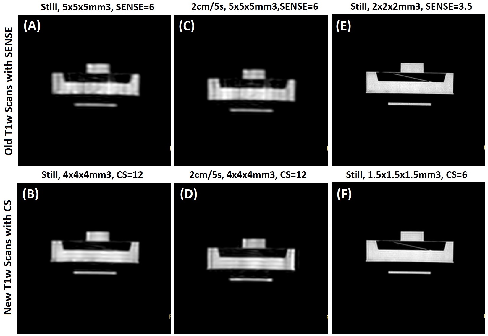
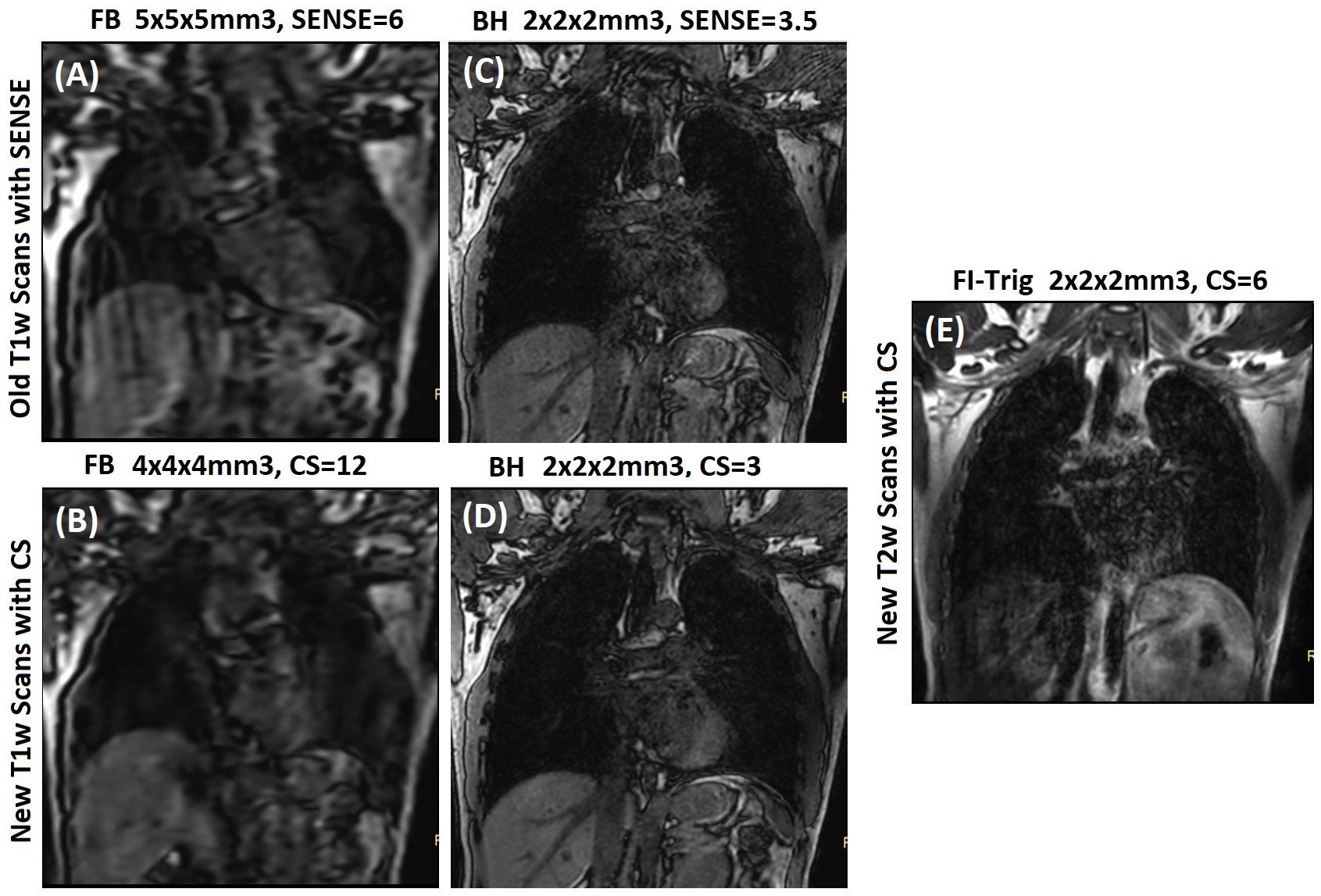
In this study, we employed the newly developed compressed sensing (CS) cartesian acquisition and golden-angle radial acquisition on a 3T MR scanner (Ingenia Elition, Philips Healthcare) to scan FB and BH MRI images with improved quality for the reconstruction of TR-4DMRI. The sequence parameters for T1w scans were TE/TR=1.1-1.3ms/2.6-2.9ms and flip angle = 15˚. The T2w scans were acquired with navigator gating to expiration at about 40% efficiency with TE/TR=134/2040ms, voxel size=2×2×2mm3, and flip angle = 90˚. The SENSE or compressed sensing (CS) was applied to create the old and new scans for comparison. All scans were performed in the coronal view with the same field of view (330´330´252cm3). The BH scans were performed with a high spatial resolution (2×2×2mm3) while the FB Cine T1w scans were performed with the same temporal resolution (2Hz) at two different spatial resolutions (old scan with SENSE: 5×5×5mm3, new scan with CS: 4×4×4mm3). A motion phantom was used to test the image quality of the sequences. A Philips QA phantom (PIQT) was placed on a wheeled cart, which can be moved by a stick driven by a sinusoidal mobile platform that was placed outside the bore in the experiment. The motion range (±10mm in the superior-inferior direction) and period (5 seconds) can be controlled by the controller. In addition, a healthy volunteer (male, 30 years old) was recruited to test the performance of the sequences in humans. The study was approved by our institutional review board.Results and Discussion
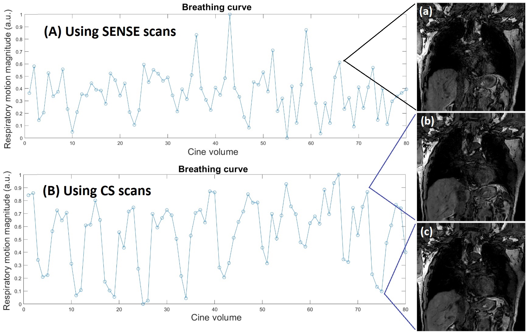
The image quality of the high- and low-resolution MRI images has been improved, as illustrated in motion-free and mobile phantom and human images in Figs. 1 and 2. In the phantom experiment, the image quality between the motion-free and moving phantom at low resolution is similar (Fig.1A-D), suggesting the low image quality is mostly resulted from the low resolution and acquisition parameters, rather than from motion. Comparing the old with new parallel acquisitions, the CS produces higher image quality than the SENSE. At high resolution with the voxel size of either 2x2x2 mm3 or 1.5x1.5x1.5mm3, the image quality is substantially improved as the lower SENSE and CS factors are applied. For human subjects, the image quality is shown in Fig. 2, in which the necessary images were provided using the old and new scanning protocols. Both FB and BH image qualities are higher in the CS scan, showing few artifacts in FB and higher signal-to-noise ratio and spatial resolution. Fig. 3 shows the improved overall quality of the new TR-4DMRI, as well as breathing waveforms with irregularities.Conclusion
The image quality of the multi-breath TR-4DMRI has been improved by using compressed sensing (CS) in FB and BH image acquisitions, replacing previously used SENSE. The improved TR-4DMRI technique with enhanced image quality is ready for clinical implementation.Acknowledgements
This research is in part supported by the MSK Cancer Center Support Grant/Core Grant (P30 CA008748).References
1. Keall PJ, et al. The management of respiratory motion in radiation oncology report of AAPM Task Group 76. Med Phys 2006;33(10):3874-900.
2. Cai J, et al. Improving Tumor-to-Tissue CNR of 4D-MRI Using Deformable Image Registration. Pract Radiat Oncol 2013;3(2 Suppl 1):S8-9.
3. Liu Y, et al. Accuracy of respiratory motion measurement of 4D-MRI: A comparison between cine and sequential acquisition. Med Phys 2016;43(1):179.
4. Li G, et al. Novel Super-Resolution Approach to Time-Resolved Volumetric 4-Dimensional Magnetic Resonance Imaging With High Spatiotemporal Resolution for Multi-Breathing Cycle Motion Assessment. Int J Radiat Oncol Biol Phys 2017;98(2):454-62.
5. Li G, et al. Introduction of a pseudo demons force to enhance deformation range for robust reconstruction of super-resolution time-resolved 4DMRI. Med Phys 2018;45(11):5197-207.
6. Nie X, et al. Enhanced super-resolution reconstruction of T1w time-resolved 4DMRI in low-contrast tissue using 2-step hybrid deformable image registration. J Appl Clin Med Phys 2020;21(10):25-39.
7. Nie X, et al. A super-resolution framework for the reconstruction of T2-weighted (T2w) time-resolved (TR) 4DMRI using T1w TR-4DMRI as the guidance. Med Phys 2020;47(7):3091-102.
8. Li G, et al. Respiratory-Correlated (RC) vs. Time-Resolved (TR) Four-Dimensional Magnetic Resonance Imaging (4DMRI) for Radiotherapy of Thoracic and Abdominal Cancer. Frontiers in oncology 2019;9(9):1024.
Figures