3085
Towards motion-resolved interleaved radial 23Na/1H magnetic resonance imaging of the human heart at 7 Tesla1Fraunhofer Institute for Digital Medicine MEVIS, Bremen, Germany, 2Institute of Radiology, University Hospital Erlangen, Erlangen, Germany, 3Friedrich-Alexander-Universität Erlangen-Nürnberg (FAU), Erlangen, Germany, Erlangen, Germany, 4mediri GmbH, Heidelberg, Germany, 5Faculty 1 (Physics/Electrical Engineering), University of Bremen, Bremen, Germany, 6Division of Medical Physics in Radiology, German Cancer Research Center (DKFZ), Heidelberg, Germany, Erlangen, Germany
Synopsis
Keywords: Image Reconstruction, Myocardium
Motion correction in interleaved 23Na/1H MRI of the human heart is important to improve the diagnostic reliability of reconstructed images and derived quantitative parameters. Therefore, this work demonstrates and compares the application of different reconstruction techniques to undersampled and motion-gated 23Na/1H MRI data at 7 T.Introduction
23Na concentration can be an important indicator for cell viability, making 23Na MRI a promising technique for providing additional diagnostic information about dysfunctional myocardium1,2. However, low signal-to-noise ratio (SNR) requires additional high-resolution 1H images for segmentation of cardiac compartments and partial volume correction which prolongs scan time. This limitation can be overcome by an interleaved density-adapted radial 23Na/1H sequence scheme which enables the acquisition of cardiac 23Na and 1H images within a single scan. However, since typical scan times are in the range of minutes, interleaved 23Na/1H imaging still requires robust compensation of respiratory and cardiac motion prior to further analysis. Motion compensation can be accomplished by retrospective gating of acquired 23Na/1H projections. Reconstructed images from gated data however often suffer from low quality due to streaking artifacts arising from high undersampling factors. The BART toolbox3 offers a variety of algorithms, designed for artifact-free reconstruction of undersampled MRI data. To this aim, we evaluate different reconstruction techniques applied to motion-gated 23Na/1H MRI data to derive the optimal setup for future applications.Methods
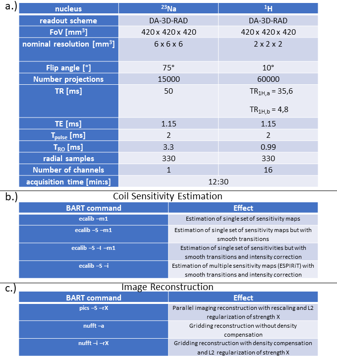
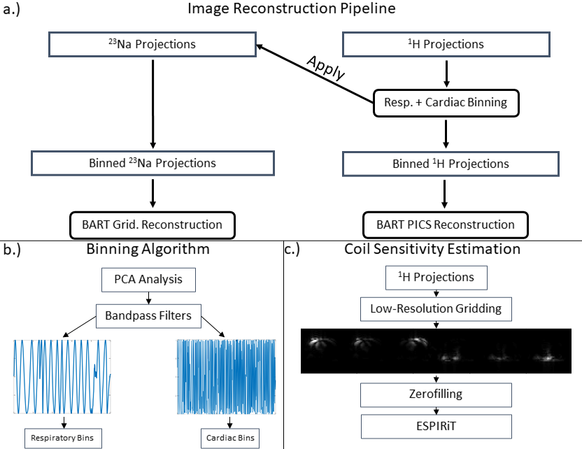
23Na and 1H images were reconstructed from data which were acquired using a dual-nuclear interleaved sequence with a density-adapted 3D radial readout scheme4,5. The interleaved sequence scheme allows to acquire four 1H projections within a single 23Na TR, thus being especially time efficient. Related sequence parameters are given in Fig. 1. In the given scenario, 15,000 23Na and 60,000 1H projections were acquired following a 3D golden angle scheme, which enables respiratory and cardiac gating. 1H data were acquired using a sixteen channel receive coil, enabling parallel imaging reconstruction while 23Na data were acquired using a single channel only. Acquired 23Na/1H data were reconstructed according to Figure 2a. 1H projections were used for retrospective respiratory and cardiac gating. Two motion states were defined for each type of motion, resulting in a total number of four motion bins. PCA-based analysis of the k-space center signal of every fourth 1H projection was used to define the motion states of the three preceding 1H projections and the preceding 23Na projection (cf. Fig. 2b). To evaluate optimal image reconstruction technique, different reconstruction mechanisms from the BART toolbox were applied to the gated 23Na/1H data. First, gridding reconstruction of motion-gated 23Na/1H images was applied in combination with pre-/post-density compensation, followed by a sum-of-squares coil combination (in case of multi-channel 1H data). Furthermore, parallel imaging reconstruction techniques were tested on 1H data due to the multi-channel readout. Therefore, all non-gated 1H projections were first used to reconstruct 3D low-resolution images (40x40x40 voxels) of individual coils which were used to estimate coil sensitivity profiles using different algorithms (cf. Fig. 2c). The different calibration commands are given in Fig. 1b and the calibration technique, yielding the best image quality, was further used to assess the effect of different regularization terms during the image reconstruction process.Results
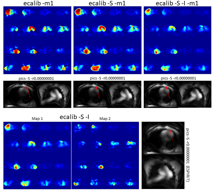
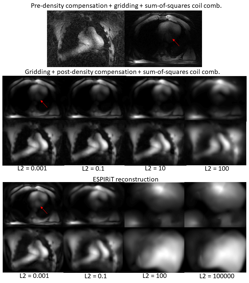
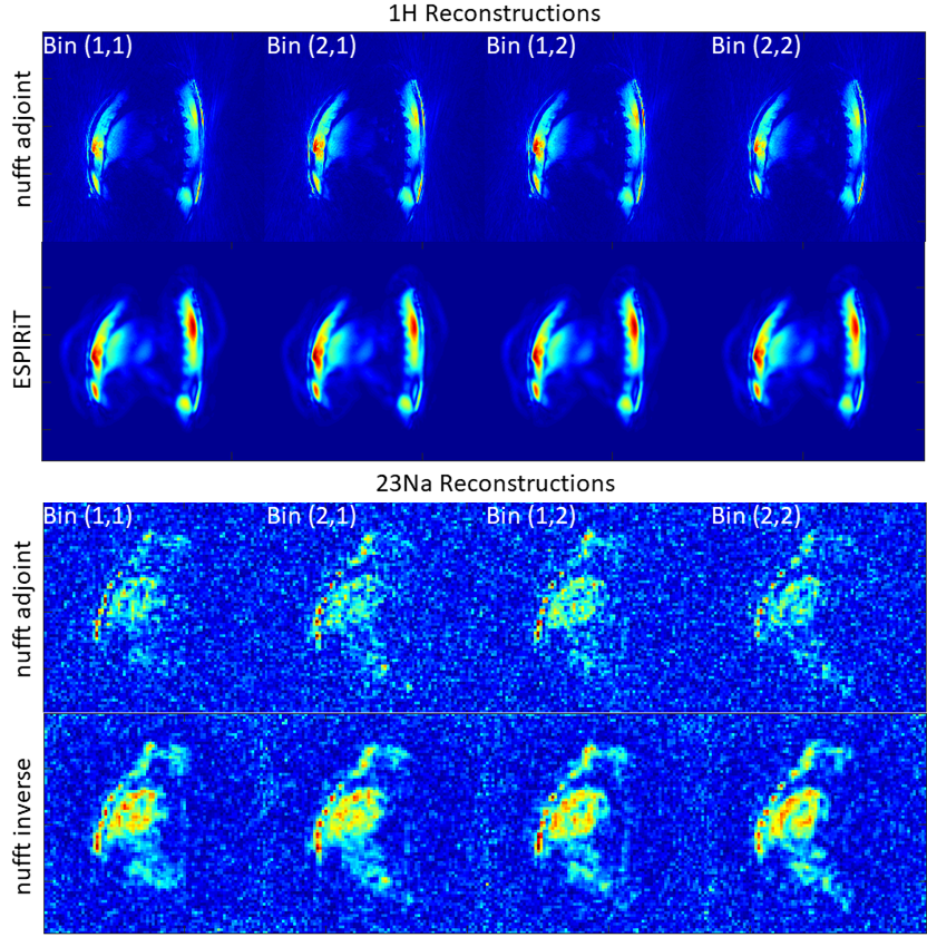
Estimated sensitivity profiles and their respective effects on 1H image reconstruction are shown in Fig. 3. Individual reconstructed 1H images using ESPIRiT reconstruction6 with different L2 regularization weights are compared to standard gridding reconstructions in Figure 4. Figure 5 finally compares reconstructed motion-gated 23Na/1H images using gridding only (23Na)/gridding and ESPIRiT reconstructions (1H).Discussion
The calibration of 1H coil sensitivity maps showed to have a direct impact on the resulting image quality. Here, the ESPIRiT calibration showed a reduction of artifacts (cf. arrows in Fig. 3) when compared to the other methods and was thus applied during the following parallel imaging reconstructions. The standard gridding reconstruction with pre-density compensation resulted in sharp 1H images with residual streaking artifacts, appearing in a noise-like fashion (cf. Fig. 4). Post-density compensation appears to reduce noise-like streaking artifacts at the cost of reduced sharpness. Strong L2 regularization resulted in strong image blurring and is thus not recommended. In comparison, the ESPIRiT reconstruction further enhances the signal in the heart region (cf. Fig. 4 arrows) but also shows a loss in detail structures, especially at higher L2 regularization factors. In general, it appears that L2 regularization has no benefits for the image reconstruction applied in this work. The same trend shows when comparing gridding and ESPIRiT reconstructions for 1H data in Fig. 5. In case of 23Na data, the gridding routine with post-density compensation seems to be beneficial for reduction of noise-like artifacts. However, it should be investigated whether the same can be achieved using additional filter operations (e.g., Hanning) in case of gridding with pre-density compensation. It should also be mentioned that the presented data suffers from severe B1 inhomogeneity which results in signal loss in structures distant to the transmit coils. Parallel transmit techniques could therefore be beneficial to homogenize the signal intensity.Conclusion
This work evaluated reconstruction algorithms from the BART toolbox in combination with interleaved and motion-gated 1H/23Na MRI data. A combination of ESPIRiT reconstruction of 1H data and gridding reconstruction with post-density compensation of 23Na data can be recommended to reduce the noise in final images. However, future work must investigate, whether similar or better results can be achieved using gridding alone with additional filter operations or other reconstruction frameworks such as XD-GRASP7.Acknowledgements
This project was funded by the Deutsche Forschungsgemeinschaft (DFG) under project number 449552397.References
1. Joern J W Sandstede, Hanns Hillenbrand, Meinrad Beer et al., Time course of 23Na signal intensity after myocardial infarction in humans. Magn Reson Med . 2004 Sep;52(3):545-51
2. Johanna Lott, Tanja Platt, Sebastian C Niesporek et al., Corrections of myocardial tissue sodium concentration measurements in human cardiac 23Na MRI at 7 Tesla. Magn Reson Med . 2019 Jul;82(1):159-173
3. Uecker et al., The BART Toolbox for Computational Magnetic Resonance Imaging, Proc Intl Soc Magn Reson in Med, 2016
4. Armin M Nagel, Frederik B Laun, Marc-André Weber et al., Sodium MRI using a density-adapted 3D radial acquisition technique, Magn Reson Med . 2009 Dec;62(6):1565-73
5. Ruck et al., Interleaved 23Na/1H MRI of the human heart at 7 Tesla, Proc Intl Soc Magn Reson in Med, 2022
6. Martin Uecker, Peng Lai, Mark J Murphy et al., ESPIRiT--an eigenvalue approach to autocalibrating parallel MRI: where SENSE meets GRAPPA, Magn Reson Med . 2014 Mar;71(3):990-1001
7. Li Feng, Leon Axel, Hersh Chandarana et al., XD-GRASP: Golden-angle radial MRI with reconstruction of extra motion-state dimensions using compressed sensing, Magn Reson Med . 2016 Feb;75(2):775-88
Figures