3082
Non-contrast high-resolution 4D-peripheral MR angiography using retrospective echo planar imaging with Compressed SENSE
Yasuhiro Goto1, Michinobu Nagao2, Masami Yoneyama3, Johannes M Peeters4, Yasutomo Katsumata3, Isao Shiina1, Kazuo Kodaira1, Yutaka Hamatani1, Takumi Ogawa1, Mana Kato1, and Shuji Sakai2
1Department of Radiological Services, Tokyo Women's Medical University, Tokyo, Japan, 2Department of Diagnostic imaging & Nuclear Medicine, Tokyo Women's Medical University, Tokyo, Japan, 3Philips Japan, Tokyo, Japan, 4Philips Healthcare, Best, Netherlands
1Department of Radiological Services, Tokyo Women's Medical University, Tokyo, Japan, 2Department of Diagnostic imaging & Nuclear Medicine, Tokyo Women's Medical University, Tokyo, Japan, 3Philips Japan, Tokyo, Japan, 4Philips Healthcare, Best, Netherlands
Synopsis
Keywords: Image Reconstruction, Blood vessels
In this study demonstrated that the higher reduction factor (R=10.0) with a one-minute scan still provided sufficient image quality with significantly faster scan time compared with conventional REPI-SENSE. REPIX would be useful for further assessment of PAD pathology even with multiple VENC acquisitions still in a clinically feasible scan time.REPIX 4D-MRA well depicted peripheral arteries clearly in a significantly short time thanks to Compressed SENSE, compared with conventional SENSE.Introduction
Peripheral artery disease (PAD) is an atherosclerotic vascular disease, which is associated with significant morbidity and mortality [1,2]. Early detection and diagnosis are important to prevent complications without aggravating PAD. Diagnosing the degree of occlusion in peripheral arteries is very important, especially for protecting the toes and ankles [3]. Non-contrast enhanced (NCE) MRA studies are crucial because PAD patients often have renal impairment [4], but current NCE-MRA techniques have limitations in terms of image quality and acquisition time when applied to foot MRA [5]. To solve these problems, we focused on peripheral pulse (PPU)-triggered retrospective turbo field-echo echo planar imaging (TFEPI, REPI) 4D-FLOW based on quantitative flow sequencing and demonstrated the utility of REPI [6.7]. Since REPI sequence is based on EPI, it can reduce scan times compared to traditional 4D FLOW sequences [6]. (Figure 1.) To further accelerate the acquisition time in addition to combining EPI readout, sensitivity encoding (SENSE) is most frequently used. However, SENSE with high reduction factor often causes significant degradation of signal-to-noise ratio (SNR), depending on the coil geometry factor (g-factor) [8.9]. Therefore, REPI 4D-MRA used SENSE with conservative reduction factor to prevent the image quality deterioration, which results in difficulty to further shorten the scan time. Recently, compressed sensing with sensitivity encoding (Compressed SENSE) [10] has been extended to allow combining with 3D EPI sequence, including 3D TFEPI/REPI. We hypothesized that the combination of REPI and Compressed SENSE (REPIX) can further shorten the acquisition time without g-factor related SNR penalty unlike the SENSE. The purpose of this study was to investigate whether REPIX enables to further reduce the scan time and improve the image quality of high-resolution non-contrast 4D peripheral MRA.Methods
[Subjects] A total of six volunteers (4 men, 2-woman, age range 22 to 47) were examined on a 3.0T MRI (Ingenia, Philips Healthcare). The study was approved by the local IRB, and written informed consent was obtained from all subjects. [Imaging quality assessment] To examine the usefulness of foot REPI 4D-MRA with SENSE (REPI-SENSE) and with Compressed SENSE (REPIX), the evaluation of proximal to distal vessels was segmented with joints as the border. (Figure 2.) The blood vessels of the peripheral arteries of the foot were evaluated on a 5-point scale. Three radiologists visually evaluated the blood vessels in the foot. [Statistical analysis] Statistical analysis was performed with Wilcoxon signed-rank test and judged the difference as significant at p<0.05. [scan parameter] REPI 4D-MRA: FOV (mm) = 180×160, spatial resolution (mm) = 1.0×1.0×2.0, TR/TE (ms) = 12/7.2, Flip angle (°) = 10, TFE-factor=3, EPI-factor=2, Recon heart phase = 8, VENC (cm/sec) = 10, Number of slices = 80, acquisition time = 1m53s. REPI-SENSE: SENSE factor = 10, REPIX: C-SENSE factor = 10.Results and Discussion
REPIX showed significantly higher visual assessment scores in all four areas compared to REPI-SENSE. (Figure 3.) Comparing REPIX and REPI-SENSE, REPIX showed higher vascular imaging capability with shorter acquisition time. (Figure 4.) In REPIX reconstruction, Compressed SENSE reconstruction framework, including wavelet denoising, worked well to reduce g-factor related image noise effectively. Further advanced Compressed SENSE technology (SmartSpeed) [11] including deep learning constrained reconstruction would be promising to further accelerate the san time of 4D REPI in future.In this study demonstrated that the higher reduction factor (R=10.0) with a one-minute scan still provided sufficient image quality with significantly faster scan time compared with conventional REPI-SENSE. REPIX would be useful for further assessment of PAD pathology even with multiple VENC acquisitions still in a clinically feasible scan time.Conclusion
REPIX 4D-MRA well depicted peripheral arteries clearly in a significantly short time thanks to Compressed SENSE, compared with conventional SENSE. (Figure 5.) It might be extended for other body parts such as abdominal MRA.Acknowledgements
No acknowledgement found.
References
1. Kiernan, T. J., Hynes, B. G., Ruggiero, N. J., Yan, B. P., & Jaff, M. R. (2010). Comprehensive evaluation and medical management of infrainguinal peripheral artery disease: "when to treat, when not to treat". Techniques in vascular and interventional radiology, 13(1), 2–10. https://doi.org/10.1053/j.tvir.2009.10.002. 2. Parikh, S. V., Saya, S., Divanji, P., Banerjee, S., Selzer, F., Abbott, J. D., Naidu, S. S., Wilensky, R. L., Faxon, D. P., Jacobs, A. K., & Holper, E. M. (2011). Risk of death and myocardial infarction in patients with peripheral arterial disease undergoing percutaneous coronary intervention (from the National Heart, Lung, and Blood Institute Dynamic Registry). The American journal of cardiology, 107(7), 959–964. https://doi.org/10.1016/j.amjcard.2010.11.019. 3. Forsythe, R. O., Brownrigg, J., & Hinchliffe, R. J. (2015). Peripheral arterial disease and revascularization of the diabetic foot. Diabetes, obesity & metabolism, 17(5), 435–444. https://doi.org/10.1111/dom.12422. 4. Schubert, T., Takes, M., Aschwanden, M., Klarhoefer, M., Haas, T., Jacob, A. L., Liu, D., Gutzeit, A., & Kos, S. (2016). Non-enhanced, ECG-gated MR angiography of the pedal vasculature: comparison with contrast-enhanced MR angiography and digital subtraction angiography in peripheral arterial occlusive disease. European radiology, 26(8), 2705–2713. https://doi.org/10.1007/s00330-015-4068-6. 5. Reimer, P., & Boos, M. (1999). Phase-contrast MR angiography of peripheral arteries: technique and clinical application. European radiology, 9(1), 122–127. https://doi.org/10.1007/s003300050642. 6. Goto Y, et al. Non-contrast high-resolution 4D-peripheral MRA using Retrospective EPI. Proc. ISMRM. 2021:1622. 7. Goto Y, et al. Non-contrast 4D Dynamic Coronary MRA using Retrospective EPI (REPI) 4D-Flow Sequence. Proc. ISMRM. 2021:2072. 8. Yoneyama M, et al. Noise Reduction in Prostate Single-Shot DW-EPI utilizing Compressed SENSE Framework. Proc. ISMRM. 2019:1634. 9. Shiina I, et al. Improvement of multi-echo gradient-spin-echp (mGraSE) myocardial T2 mapping utilizing Compressed SENSE reconstruction framework. Proc. ISMRM. 2021:3603. 10. Geerts-Ossevoort L, et al. Compressed SENSE Speed done right. Every time. The Netherlands: Philips Healthcare; 2018 Jan. Report No: 4522 991 31821. https://www.philips.de/content/dam/b2bhc/de/resourcecatalog/landingpages/ingeniaelition/White_Paper_Compressed_SENSE-opt.pdf 11. Peeters H, Chung H, Valvano G, Yakisikli D, van Gemert J, de Weerdt E, van de Ven K. Philips SmartSpeed, No compromise Image quality and speed at your fingertip. (2022) Available via https://www.philips.com/c-dam/b2bhc/master/landing-pages/smartspeed/philips-smart-speed-brochure.pdfFigures
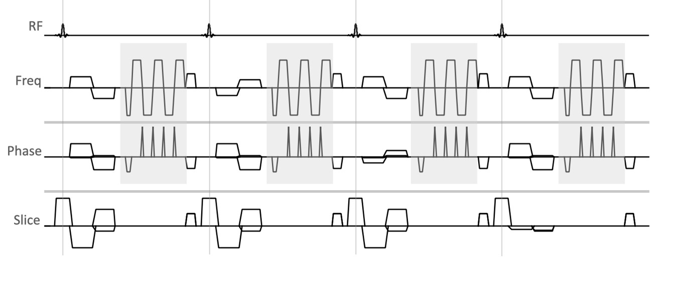
Figure 1. Scheme of the REPI 4D-MRA sequence is shown in Figure 1.
TFEPI sequence is being combined with EPI readout, it is capable of scan time
acceleration and improves SNR.
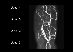
Figure 2. Visual evaluation. The visual
evaluation was divided into four areas with the joint as a boundary, and each
was evaluated for the depiction of blood vessels. Area 1 : Chopard joint -
Lisfranc joint, Area 2 : Lisfranc joint - MP joint, Area 3 : MP joint - IP
joint, PIP joint, Area 4 : IP joint, PIP joint - Distal.
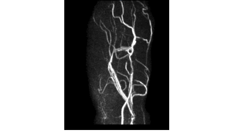
Figure 3. REPI continuously images all cardiac cycles, it collects
8 images with 1R-R. Therefore, by replaying all continuously images, arteries
and veins can be distinguished and diagnosed.
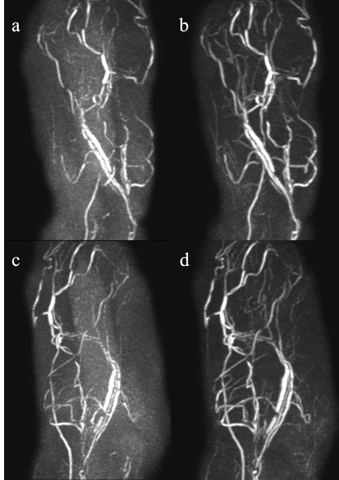
Figure 4. We show images of REPI-SENSE (a,c) and REPIX (b,d) used for comparison. REPIX significantly improves
vascular structure in ALL area compared to the REPI-SENSE.
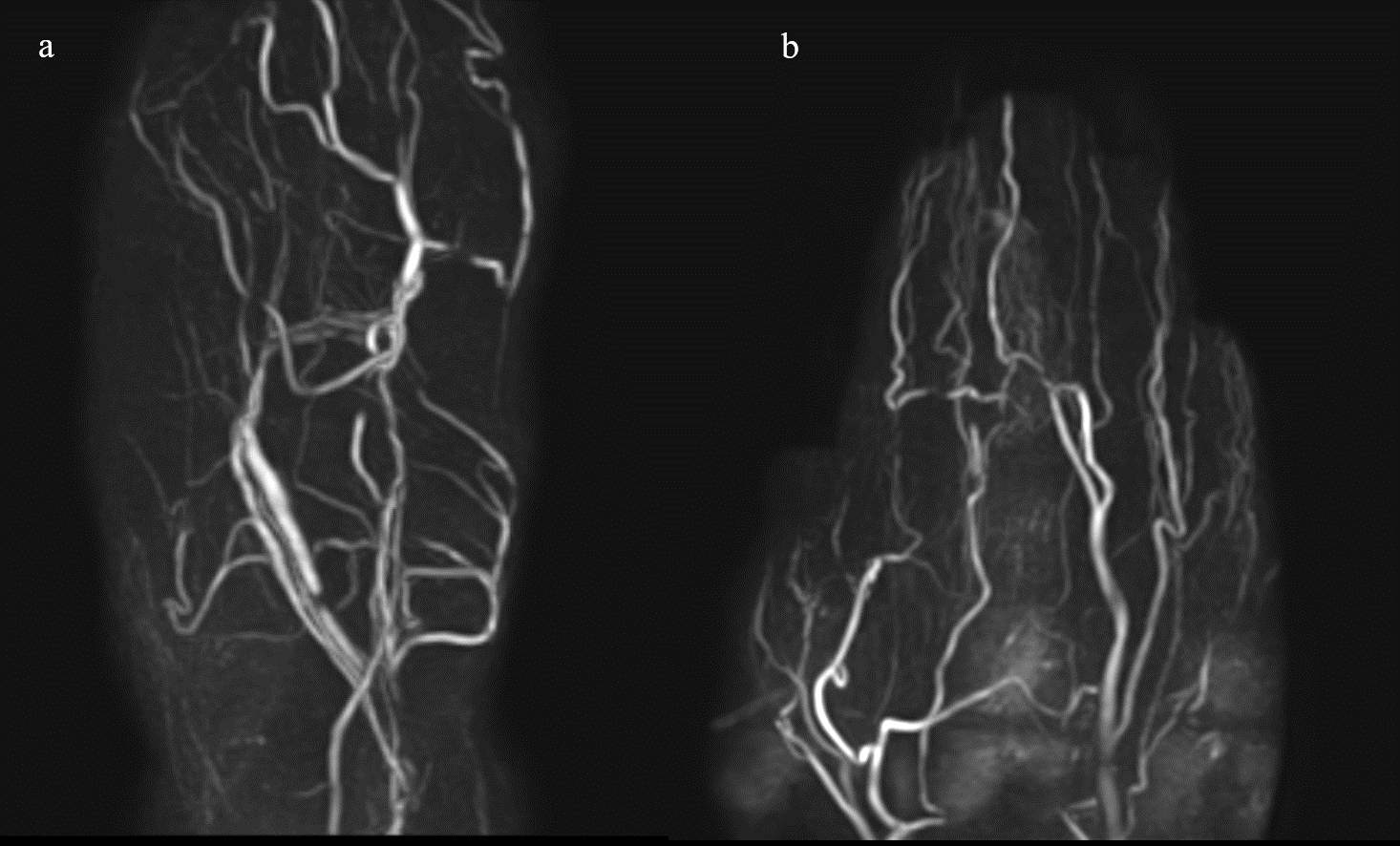
Figure 5. Representative REPIX 4D-MRA of the foot (a). REPIX 4D-MRA could visualize blood vessels from proximal to
distal with high robustness. In addition, hands can be imaged with REPIX 4D-MRA as well as feet (b).
DOI: https://doi.org/10.58530/2023/3082